


Culturing and morphological development of Galumna zachvatkini Grishina, 1982 (Acari, Oribatida, Galumnidae)
Ermilov, Sergey G.  1
1
1✉ University of Tyumen, X-BIO Institute, Tyumen, Russia.
2026 - Volume: 66 Issue: 4 pages: 929-945
https://doi.org/10.24349/set9-edpjZooBank LSID: C9BD5C38-AA97-4E0A-928F-FCB0C9BA14CA

Original research
Keywords
Abstract
Introduction
Galumna zachvatkini Grishina, 1982 (Acari, Oribatida, Galumnidae) was described by Grishina (1982) based on specimens collected from Ukraine. It is characterized by distinct sexual dimorphism in the shape of the A3 porose areas (strongly elongate in males vs. oval or slightly elongate in females). The currently known distribution is in the southern Palaearctic region; it inhabits litter, soil, ant nests, and decaying stumps (Grishina 1982; Shtanchaeva and Subías 2010; Vladimirova 2018).
The main goals of the present study were to: provide a supplementary description of the adult of G. zachvatkini based on material from Dagestan (southwestern Russia) and summarize its key morphological traits; obtain (by laboratory culturing) all active juvenile instars (larva, proto-, deuto-, and tritonymph); document the duration of development of each juvenile instar and the subsequent pre-ecdysis resting periods; and to describe the juvenile instars in detail. Additionally, the synonymy of Galumna bimorpha Mahunka, 1987 is discussed.
The original description of the adult G. zachvatkini was incomplete; in particular, it lacked information on the gnathosoma, legs, epimeral region, and lateral body features, and setae measurements were lacking (Grishina 1982). Data on the culturing of Galumna Heyden, 1826 are available for seven identified species, but remain unknown for G. zachvatkini (Norton and Ermilov 2014, 2024; Ermilov and Mashkov 2026; Ermilov and Mzeek 2026b). Data on the morphology of juvenile instars of Galumna have been reported for 10 identified species and one subspecies (Norton and Ermilov 2014, 2024; Ermilov and Mashkov 2026; Ermilov and Mzeek 2026b). Grishina (1982) described the juveniles of G. zachvatkini, but her data are brief; only figures of the larva and deutonymph were provided, with no information on the gnathosoma and legs. In the present study, comprehensive data on the juvenile morphology of G. zachvatkini are provided.
Material and methods
Material
Nine adults of G. zachvatkini: Russia, southern Dagestan, near the village of Nyugdi, bank of the Gyulgerychay River, 41°51′20″N, 48°26′30″E, sifted litter under black alder trees, 5 January 2026, collected by O.L. Makarova and K.V. Makarov. Nine adults of G. zachvatkini: Russia, southern Dagestan, 2 km east of the village of Mollakent, 41°55′21″N, 48°24′37″E, damp Elaeagnus sp. trees between the dunes, litter of blackberries, 6 January 2026, collected by O.L. Makarova and K.V. Makarov.
Culturing
The culturing method in this study was previously described in detail by Ermilov and Mzeek (2026a). Adults (males and females) of G. zachvatkini were placed in a group of 18 individuals into a single glass jar with a screw-on lid. As soon as juveniles were present, they were maintained separately, in individual plastic chambers. The jar and chambers were filled with a mix of Plaster-of-Paris and activated charcoal (PPC). The mites were offered the following foods of plant and animal origin: algae (Pleurococcus sp.) from linden bark, lichen (Cladonia sp.) and dry tree leaves from a mixed forest, fungi (Alternaria sp., Fusarium solani, and Penicillium janczéwskii) from cultured media, freeze-dried nematodes (Panagrellus redivivus) from cultured source, and dry baker's and brewer's yeast. Food was supplied ad libitum to determine dietary preferences. Food was available to the mites in abundance. All culture vessels were maintained at a constant room temperature (≈25ºС) under high humidity conditions, stored in a closed box without access to light. A Zeiss Stemi 305 stereomicroscope was used to observe the developmental instars. The adults were observed every 48 hours, while juveniles were observed daily. The identity of the emerged adults was confirmed via a taxonomic identification, using a light microscope and temporary slides with water, or glycerine, or lactic acid. Mite culturing was conducted between January 2026 and April 2026.
Morphology
For measurement and illustration, specimens were mounted in lactic acid on temporary cavity slides. Body length was measured in lateral view, from the tip of the rostrum to the posterior edge of gastronotum. Maximum body width was measured in dorsal aspect. Setae were measured from the side to avoid foreshortening. All measurements are in micrometers (µm). Formulas for leg setation are given in parentheses according to the sequence trochanter-femur-genu-tibia-tarsus (famulus included). Formulas for leg solenidia are given in square brackets, according to the sequence genu-tibia-tarsus. Drawings were made using a camera lucida attached to the Leica DM 2500 transmission light microscope. Images were obtained with an AxioCam ICc3 camera using a Carl Zeiss transmission light microscope Axio Lab.A. Image stacks were combined using the Helicon Focus Pro (v. 8.0) suite.
Terminology
Morphological terminology used in this paper follows that of Grandjean: see Travé and Vachon (1975) for references, Ermilov and Klimov (2017), and Norton and Ermilov (2017) for overviews.
Abbreviations
Prodorsum: rl = rostral ledge; L = lamellar line; S = sublamellar line; N = prodorsal leg niche; E, T = lateral ridges of body; ro, le, in, bs, ex = rostral, lamellar, interlamellar, bothridial, and exobothridial setae, respectively; Ad = dorsosejugal porose area; D = dorsophragma; P = pleurophragma; ps = prodorsal macrosclerite. Gastronotum: Aa, A1, A2, A3 = porose areas; c-, d-, l-, h-, p- = series of alveoli/setae; mp = median pore; ia, im, ip, ih, ips = lyrifissures/cupules; gla = opisthonotal gland opening; es = excentrosclerite; gs = gastronotic macrosclerite; gf = gastronotic furrow; gb = gastronotic band; δ = ecdysial cleavage line (line of dehiscence); tt = microtuberculate tubercle; als = anterolateral sclerite; ls = lateral sclerite; vs = ventral sclerite; pvs = posteroventral sclerite; PS = segment; pss = sclerite anterior to PS segment. Gnathosoma: SG = subcapitular gena; SM = subcapitular mentum; a, m, h = subcapitular setae; or = adoral seta; sup, inf, d, cm, acm, ul, su, vt, lt = palp setae; as = axillary saccule; ω = palp solenidion; cha, chb = cheliceral setae; Tg = Trägårdh organ. Epimeral and lateral podosomal regions: ep3, ep4 = epimeres III and IV, respectively; 1a, 1b, 1c, 2a, 3a, 3b, 4a, 4b = epimeral setae; PdI, PdII = pedotecta I, II, respectively; z = aperture of supracoxal gland; dis = discidium; cir = circumpedal carina; sc = scale; Cl = Claparède's organ; oh = humeral organ. Anogenital region: g, ag, an, ad = genital, aggenital, anal, and adanal setae, respectively; iad = adanal lyrifissure/cupule; Ap = postanal porose area; po = preanal organ; cgs = circumgenital macrosclerite; ads = adanal sclerite. Legs: Tr, Fe, Ge, Ti, Ta = trochanter, femur, genu, tibia, and tarsus, respectively; ACII, ACIII = acetabula II, III, respectively; pa = porose area; ω, φ, σ = solenidia; e = famulus; d, l, v, bv, ev, ft, tc, it, p, u, a, s, pv, pl = setae. Instars: LA, PN, DN, TN, AD = larva, protonymph, deutonymph, tritonymph, and adult, respectively.
Results
Culturing
The adult mites put into the jar on January 17, 2026, displayed high locomotor activity, moving rapidly within it, pausing rarely to feed (on Cladonia sp., Alternaria sp., and Pleurococcus sp.).
No spermatophores and eggs were found, but several larvae were present in the jar on February 3. Subsequently, new larvae were constantly being born. It is possible that this species has extended internal development (e.g., viviparity), or that eggs were laid deep in food substrates or cracks in the PPC, making them difficult to detect. Subsequent examinations of the females revealed no eggs or prelarvae inside them. Similar observations have been made for some other galumnid mites (Ermilov and Mashkov 2026; Ermilov and Tolstikov 2026). Juvenile instars were active within chambers and fed on Alternaria sp. and Pleurococcus sp. Pre-ecdysis resting periods occurred on the surface of food substrates, in irregularities (cracks, pits) of the PPC, and on the chamber walls.
A total of 11 individuals successfully developed from larvae to adults. The development took 26–32 (≈29) days at ≈25ºC (Table 1). Among active instars, the larval and protonymphal instars were the shortest (≈3.5 and ≈3.4 days, respectively), while the deutonymphal and tritonymphal instars were longer (≈4.1 and ≈4.9 days, respectively). The durations of the pre-ecdysis resting periods differed slightly following the larval, protonymphal, and deutonymphal instars (they took from ≈2.8, ≈2.4, and ≈3.0 days, respectively), while the period was longest after the tritonymph (≈4.5 days) (Table 1).
Download as Note: Active instars, not including immobile prelarva, which was not observed. Min – minimum. Max – maximum. Ave – Average. SEM – Standard Error of the Mean. n – number of individuals.
Instar
Min
Max
Ave ± SEM
n
Larva
3
4
3.5±0.1
11
Pre-ecdysis resting period
2
4
2.8±0.1
11
Protonymph
3
5
3.4±0.1
13
Pre-ecdysis resting period
2
3
2.4±0.1
13
Deutonymph
4
5
4.1±0.1
16
Pre-ecdysis resting period
2
4
3.0±0.1
16
Tritonymph
4
7
4.9±0.3
16
Pre-ecdysis resting period
4
6
4.5±0.1
16
Total development larva–adult
26
32
29.0±0.5
11
Morphology
Galumna zachvatkini Grishina, 1982
(Figures 1–8)
Supplementary description of adult
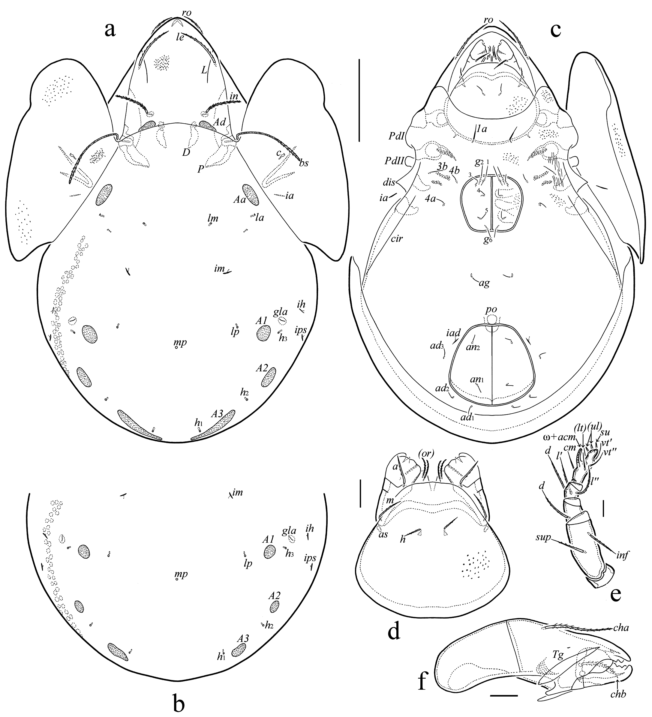
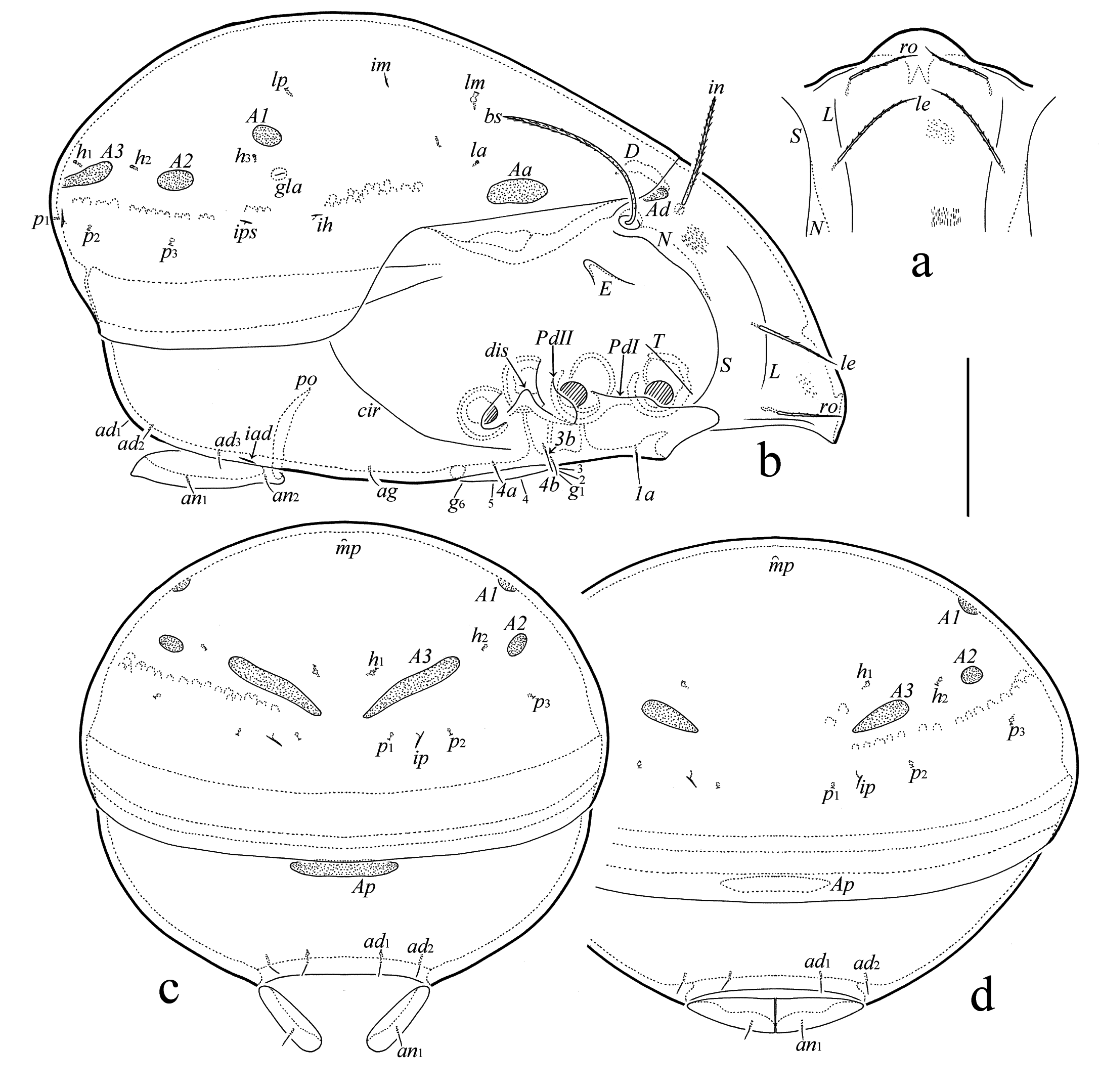
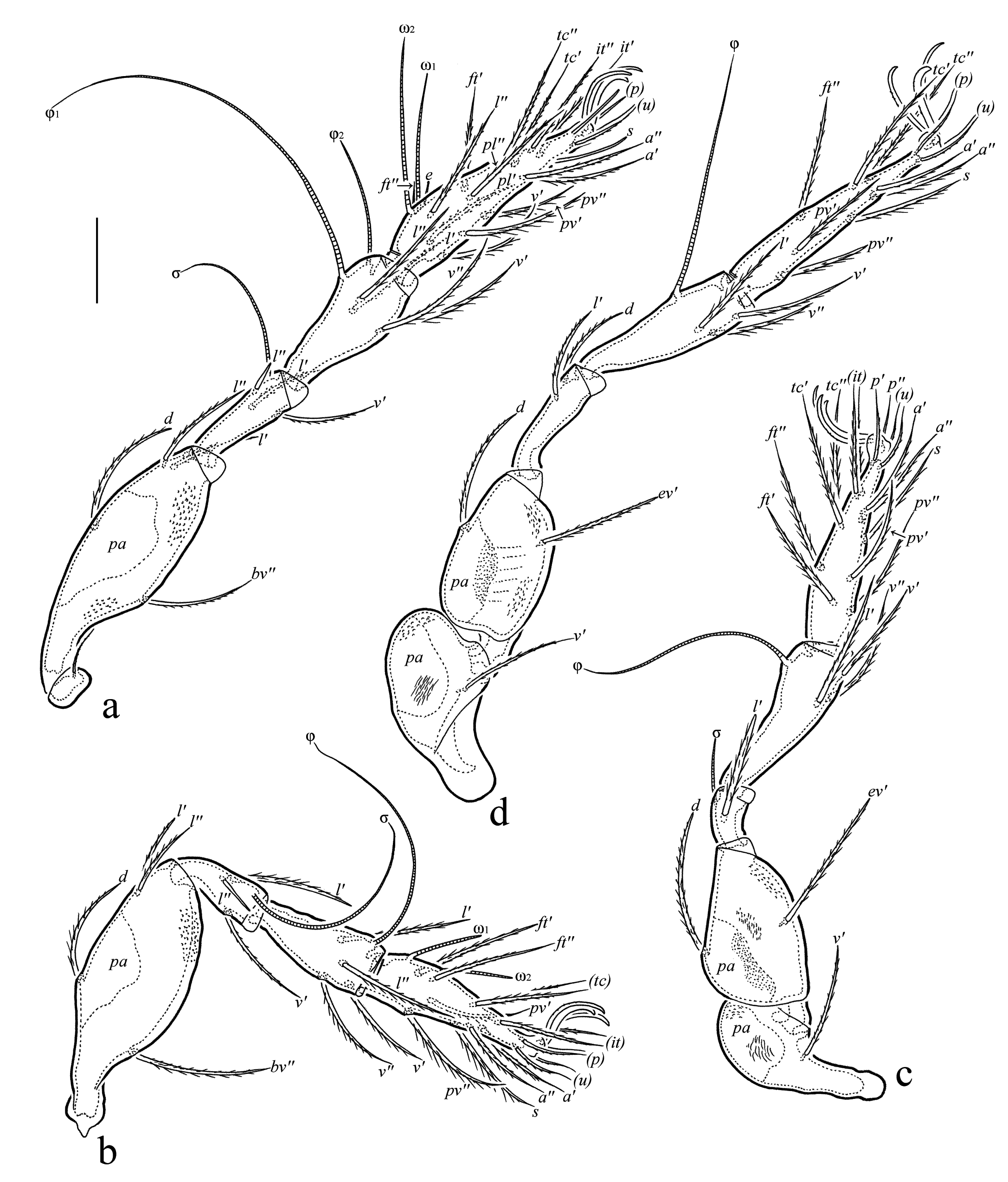
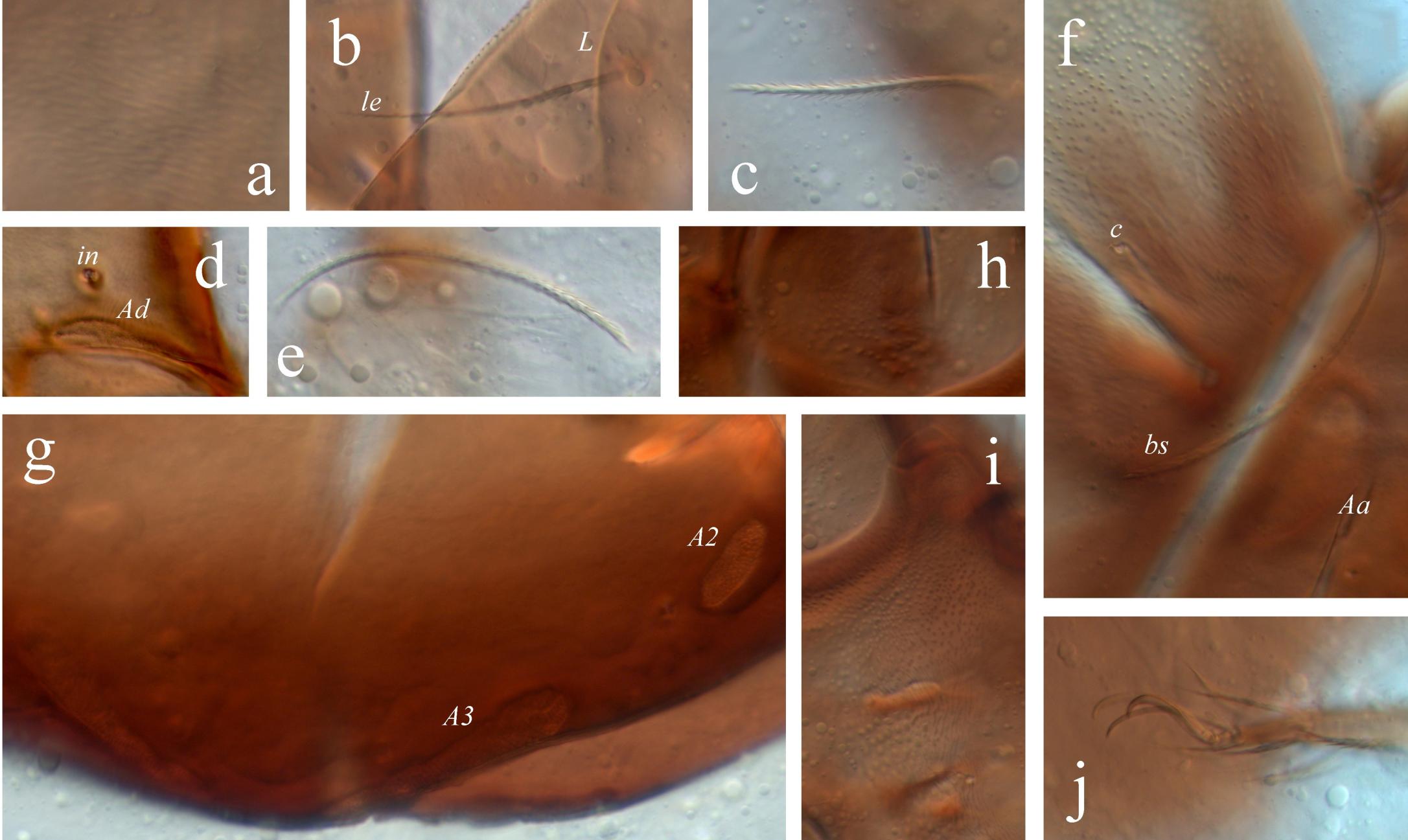
Measurements – Body length 495–555 (n=10). Notogaster width 345–360 (n=10). No difference between males and females in body size.
Integument (Figs 4a, f, h, i) – Body color brown. Body surface microtuberculate sculpturing. Microtubercles simple or elongate, clearer on prodorsum, pteromorphs, subcapitular mentum, antiaxial side of leg femora I–IV, trochanters III, IV, and in epimeral region. Additionally, prodorsum, epimeral region, and leg trochanters III, IV partially with short, thin, raised lines.
Prodorsum (Figs 1a, 2a, b, 4b–f) – Rostrum rounded. Sublamellar line thin, complete, curving backward at end. Lamellar line thin, incomplete, with reduced distal part. Rostral (56–64) and lamellar (60–67) setae setiform, barbed. Insertion of lamellar seta distinctly lateral to lamellar line. Interlamellar seta (71–79) stiff, heavily barbed. Bothridial seta (123–131) slightly expanded in distal half (sometimes vaguely lanceolate), barbed, directed posterolaterad. Exobothridial seta not observed. Dorsosejugal porose area oval (26–34 × 7–11), located posterior to insertion of interlamellar seta. Dorsophragma medium-sized, elongate longitudinally.
Notogaster (Figs 1a, b, 2b–d, 4f, g) – Dorsosejugal suture complete, slightly convex medially. All notogastral setae represented by setal alveoli. Four pairs of porose areas present: Aa oval (30–37 × 11–19), longitudinally oriented, located close to pteromorphal hinge; A1 circular (17–22); A2 circular (19–22) or oval (19–26 × 17–22); A3 strongly elongate in males (64–75 × 15–19) versus oval or slightly elongate in females (30–41 × 11–19). Median pore present in both genders, located between A1. Opisthonotal gland opening located close and lateral or anterolateral to A1. Lyrifissure im located between setal alveoli lm and lp, ip between p1 and p2.
Gnathosoma (Figs 1d–f, 4h) – Subcapitulum size 127–135 × 105–116. Subcapitular setae a (22) and m (22) setiform, slightly barbed, h (15) setiform, roughened. Seta a thickest, h thinnest. Adoral seta (15) setiform, barbed. Palp length 97–101. Palp setation 0-2-1-3-9(+ω). Postpalpal seta (6) spiniform. Chelicera length 150–157. Cheliceral setae (cha 49–52; chb 32–34) setiform, barbed.
Epimeral and lateral podosomal regions (Figs 1c, 2b, 4i) – Epimeral setal formula 1-0-1-2. Setae (1a 22; 3b 17–19; 4a, 4b 11) setiform, thin, nearly smooth. Circumpedal carina medium-sized, directed to insertion of 3b, but distinctly not reaching it.
Anogenital region (Figs 1c, 2b–d) – Anogenital setal formula 6-1-2-3. Genital (g1, g2 17–19; g3–g6 11), aggenital (11), anal (11), and adanal (11) setae setiform, thin, nearly smooth. Anterior edge of genital plate with two setae (g1, g2), but g3 inserted close to anterior edge. Aggenital seta inserted between genital and anal apertures, equally distanced from them. Adanal setae ad1 and ad2 posterior, ad3 lateral to anal plate. Distance ad1–ad2 shorter than ad2–ad3. Adanal lyrifissure located close, parallel or slightly oblique to anterolateral part of anal plate. Postanal porose area elongate (71–75 × 15–19).
Legs (Figs 3a–d, 4j) – Median claw slightly thicker than lateral claws. All claws slightly barbed on dorsal side. Each lateral claw with small tubercle ventrodistally. Porose area on femora I–IV and on trochanters III, IV distinct. Proximoventral porose area of tarsi I–IV and distoventral porose area of tibiae I–IV absent. Formulas of leg setation and solenidia: I (1-4-3-4-20) [1-2-2], II (1-4-3-4-15) [1-1-2], III (1-2-1-3-15) [1-1-0], IV (1-2-2-3-12) [0-1-0]. Homology of setae and solenidia indicated in Table 2. Famulus straight, inserted anterior to solenidion ω1. Seta s of tarsus I eupathidial. Solenidion of tibia IV inserted in anterior part of dorsal side of segment.
Download as Notes: Roman letters refer to normal setae, Greek letters refer to solenidia. Single prime (‘) marks setae on the anterior and double prime (’’) setae on the posterior side of a given leg segment. Parentheses refer to a pair of setae. Setae are listed only for the instar in which they first appear.
Instar
Tr
Fe
Ge
Ti
Ta
Leg I
LA
–
d, bv’’
(l), σ
(l), v’, φ1
(ft), (tc), (p), (u), (a), s, (pv), (pl), e, ω1
PN
–
–
–
–
ω2
DN
–
(l)
–
φ2
–
TN
v’
–
– / v’
v’’
(it)
AD
–
–
– / v’
–
l’‘, v’
Leg II
LA
–
d, bv’’
(l), σ
l’, v’, φ
(ft), (tc), (p), (u), (a), s, (pv), ω1
PN
–
–
–
–
–
DN
–
(l)
–
l’’
ω2
TN
v’
–
– / v’
v’’
(it)
AD
–
–
– / v’
–
–
Leg III
LA
–
d, ev’
l’, σ
v’, φ
(ft), (tc), (p), (u), (a), s, (pv)
PN
–
–
–
–
–
DN
v’
–
–
l’
–
TN
–
–
–
v’’
(it)
AD
–
–
–
–
–
Leg IV
PN
–
–
–
–
ft’’, (p), (u), (pv)
DN
–
d, ev’
d, l’
v’, φ
(tc), (a), s
TN
v’
–
–
l’, v’’
–
AD
–
–
–
–
–
Supplementary description of juvenile instars
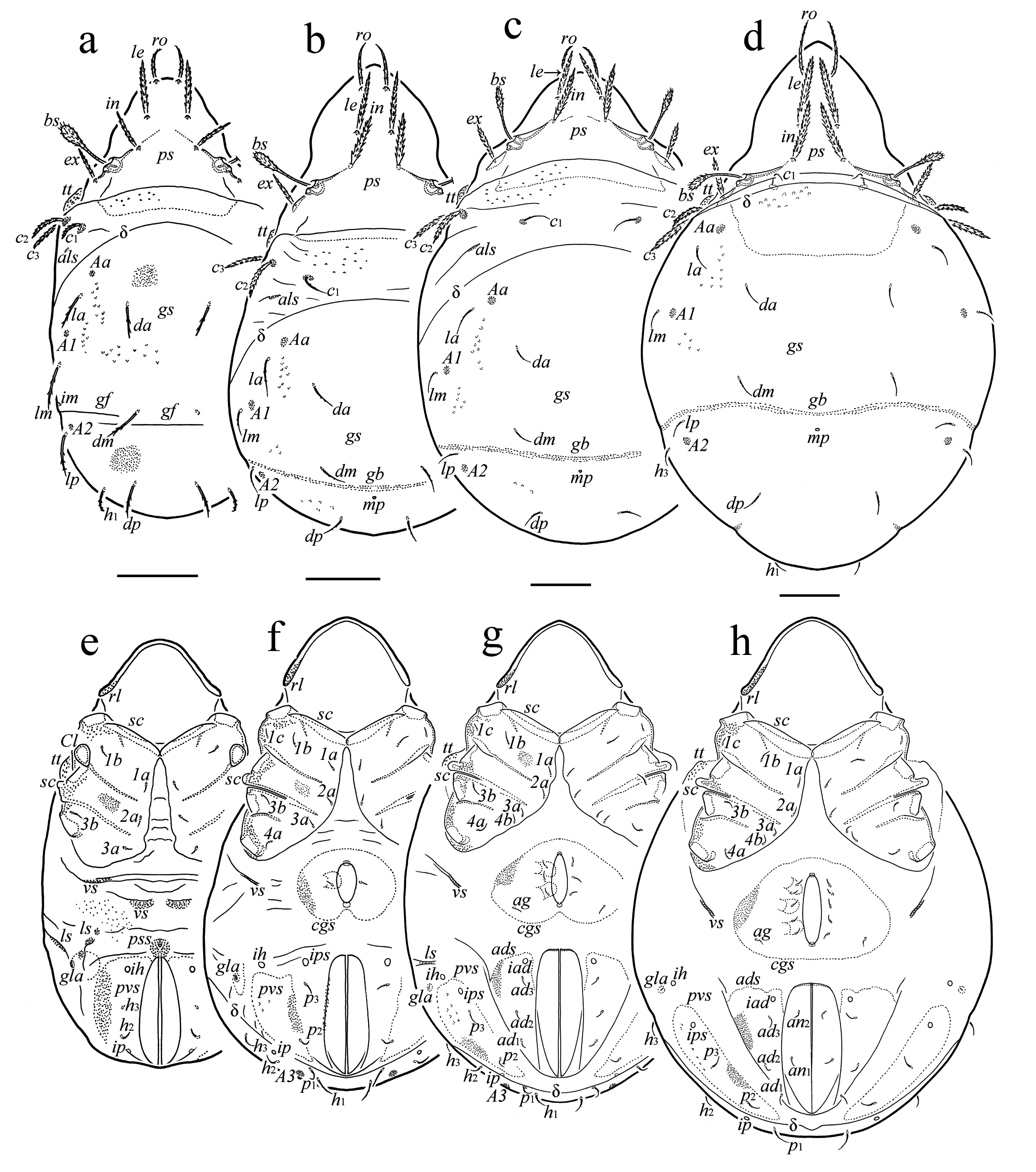
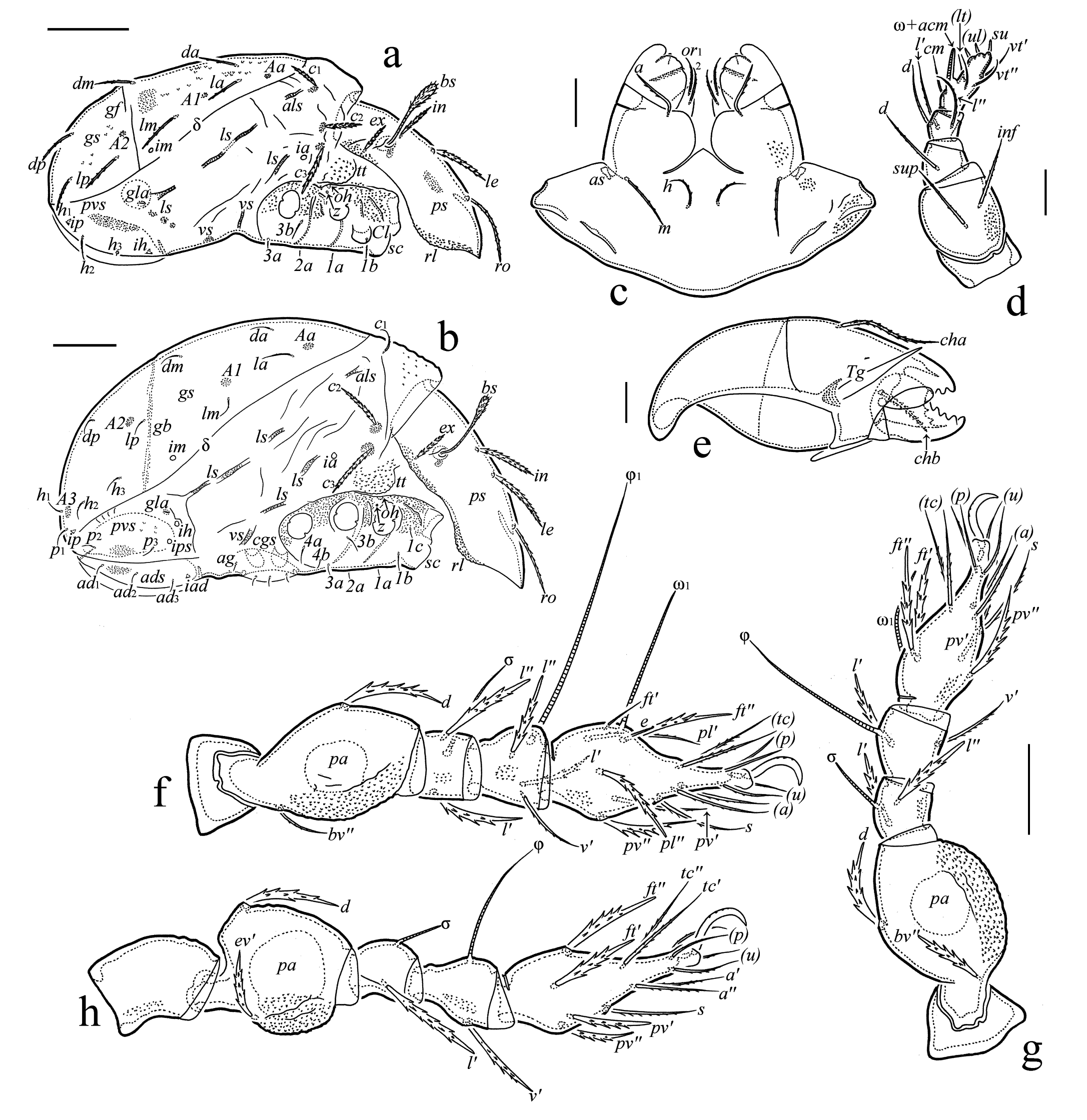
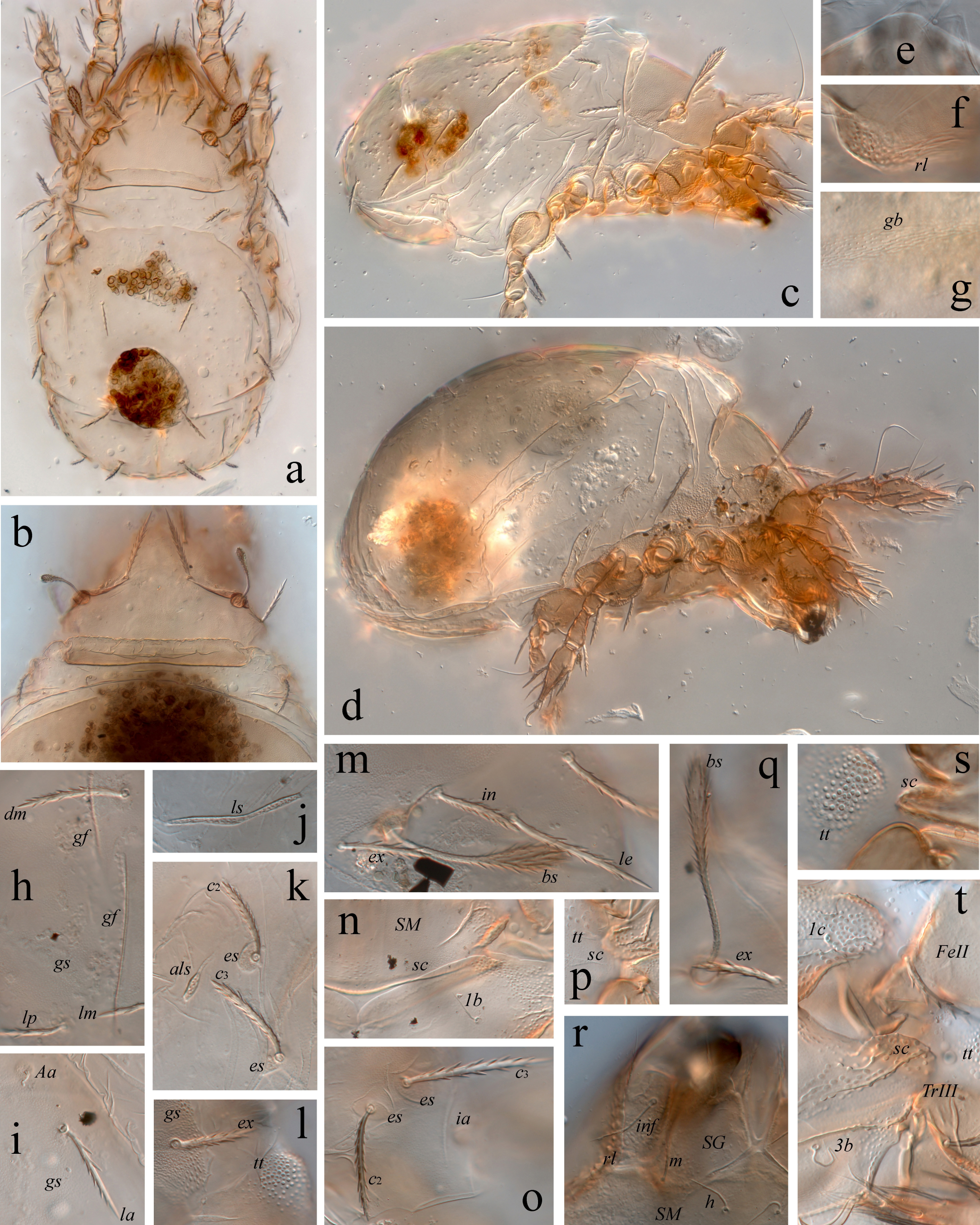
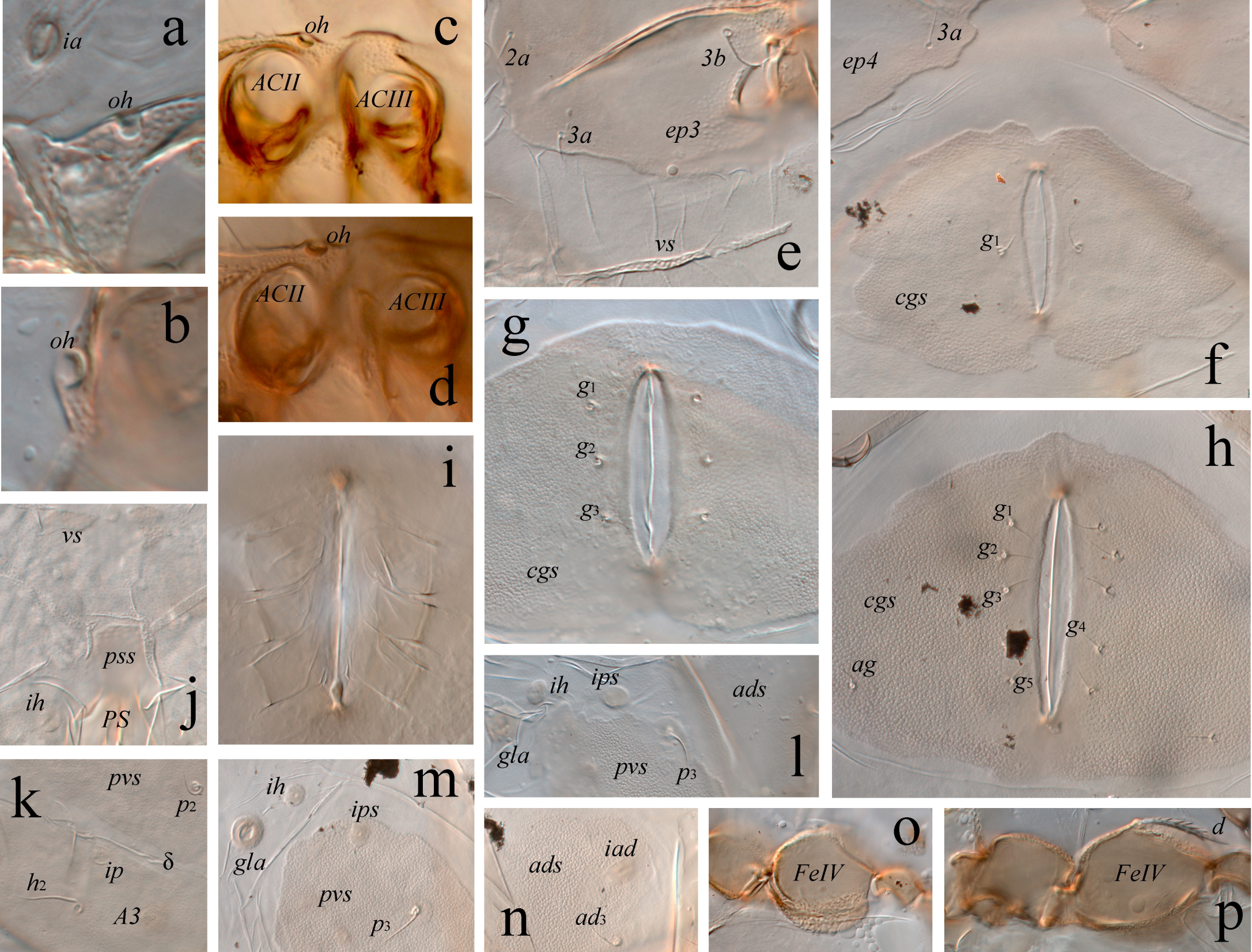
Measurements – Total length of LA 270–285 (n=5), PN 300–345 (n=5), DN 405–420 (n=4), TN 435–480 (n=4). Maximum width of LA 150–157 (n=5), PN 180–210 (n=5), DN 255–285 (n=4), TN 300–330 (n=4).
Integument (Figs 5a–h, 6a–d, f– h, 7f, l, p, s, t, 8a, f–h, m, n, o) – Body light brown, but sclerites, gnathosoma, epimeres, and legs darker. Cuticle mostly densely microporose. Lateral ledge of prodorsum, large tubercle between seta c3 and acetabulum II, podosomal region, lateral parts of epimeres, subcapitular mentum, palp femur, and antiaxial side of leg segments (partially) densely microtuberculate. Anterior part of gastronotum before macrosclerite with sparse tubercles. Gastronotic macrosclerite and posteroventral sclerites partially with sparse, vaguely developed foveolae. In LA, ventral side partially with microtubercles. Cuticle of gastronotum and ventral side outside sclerites with some plications.
Prodorsum (Figs 5a–d, 6a, b, 7a–f, l, m, q) – Relatively short, about 1/2 (LA) to 2/5 (PN, DN, TN) length of gastronotum (in lateral aspect). Rostrum rounded. Lateral part of prodorsum with distinct ledge. Prodorsal macrosclerite well defined. Rostral seta (LA 45–49; PN 52–60; DN 60–67; TN 64–71) setiform, barbed. Lamellar (LA 41; PN 49–60; DN 60–64; TN 64–71), interlamellar (LA 26–30; PN 37–41; DN 52–56; TN 56–64), and exobothridial (LA 22–24; PN 30–34; DN 34–37; TN 41–45) setae thick, heavily barbed. Bothridial seta (LA 52–56; PN 60–64; DN, TN 67–71) clavate, with long, roughened stalk and shorter, barbed head.
Gastronotic region (Figs 5a–h, 6a, b, 7a–d, g–l, o, s, 8a–d, j–m) – Gastronotic macrosclerite well defined; its boundaries coincide with ecdysial cleavage line (line of dehiscence). In LA, gastronotic macrosclerite with transverse furrow (behind setae dm and cupules im), which is twice interrupted, forming three parts. In PN, DN, and TN, gastronotic macrosclerite with transverse band represented by slight, dense, raised lines located anterior to median pore. Porose areas (three pairs in LA, four pairs in PN, DN, and TN) circular (7–11), located on macrosclerite. Larva with 12 pairs of setae. Of these, setae c3 (34–39), c2 (24–30), and c1 (19–26) thick, heavily barbed; la, lm, dp, lp, h1 (all 24–28) and da, dm (all 19–22) stiff, barbed; h2 (13–15) setiform, thin, roughened; and h3 (1) vestigial. Nymphs with 15 pairs of setae. Of these, setae c3 (PN 41–49; DN 60–64; TN 56–64) and c2 (PN 34–37; DN 45–53; TN 45–49), thick, heavily barbed; c1 (PN 22–26; DN, TN 26–30) setiform, thin, slightly barbed; la (PN, DN, TN 26–30) and da, dm, lm, dp, lp (PN 19–22; DN, TN 26–30) setiform, thin, roughened; h1–h3, p1 (PN 11–13; DN, TN 19–26) and p2, p3 (PN, DN, TN 11–13) setiform, thin, nearly smooth. Setae c1–c3 (in all instars) with excentrosclerite at their bases. Small median pore slightly observed in nymphs. Cupules ia, im, and ip well visible. One large tubercle located between seta c3 and acetabulum II. Anterolateral part of gastronotum with small sclerite posterolateral to seta c1. Lateral part of gastronotum with four to five elongate sclerites between ia and gla. One pair of large posteroventral sclerites present in all instars. Ventral side with one pair of elongate sclerite. Larva with additional one pair of oval ventral sclerites between epimeres and segment PS, one circular sclerite close and anterior to segment PS, and several small sclerites anteromedial to gla.
Gnathosoma (Figs 6c–e, 7r) – Subcapitulum size of LA 56–64 × 64–67; PN 75–79 × 79–82; DN 94 × 97–101; TN 105 × 109–112. Subcapitular setae a (LA 13; PN 15; DN 17; TN 19–22), m (LA 15; PN 17; DN 19; TN 22–24), h (LA 11; PN 13; DN 15; TN 17–19), and two pairs of adoral setae (LA 9–11; PN, DN 11–13; TN 15) setiform, thin, roughened. Chelicera length of LA 71–79; PN 94–97; DN 116–124; TN 131–135. Cheliceral setae (cha of LA 26–30; PN 30–34; DN, TN 41–45; chb of LA 19–22; PN 22–26; DN, TN 26–30) setiform, barbed. Palp length of LA 52–56; PN 60–64; DN, TN 77–86. Palp setal formula in all instars 0-2-1-3-9(+1ω). Length of postpalpal seta of LA 2; PN, DN, TN 4.
Epimeral and lateral podosomal regions (Figs 5e–h, 6a, b, 7c, d, n, p, s, t, 8e, f) – Epimeral region well bordered, with defined elongate triangular sternal region. Anterior margin of coxisternum with one pair of elongate scales overlapping medially. Epimeral setal formula (for successive epimeres) of LA 3-1-2 (1c as typical scale covering Claparède's organ); PN 3-1-2-1; DN, TN 3-1-2-2. Epimeral setae acicular; setae 1b, 3b (LA 11; PN 15; DN, TN 17–19), and 4a (DN, TN 17–19) roughened, while others (LA 7; PN, DN, TN 7–9) nearly smooth. Humeral organ present but faint. Small scale (well visible in ventral view) located posterior to acetabulum II.
Anogenital region (Figs 5e–h, 6a, b, 7c, d, 8f–i, l, n) – Circumgenital macrosclerite distinct in nymphs, large, concave posteriorly in PN and DN. One pair of large adanal macrosclerites present in DN and TN. Ontogeny of genital (7–9), aggenital (7–9), anal (11), and adanal (11) setal formulas, from PN to TN: 1-3-5, 0-1-1, 0-0-2, 0-3-3, respectively. All setae acicular, without excentrosclerite at their bases. Paraproctal valves of LA (segment PS), PN (segment AD), and DN (segment AN) glabrous (without setae and alveoli). Cupules ih, ips, iad, and opisthonotal gland opening well visible.
Legs (Figs 6f–h, 7a, c, d, 8o, p) – Claw of each leg slightly barbed on dorsal side. Paraxial porose area on all femora well visible. Leg setal formulas: LA: I (0-2-2-3-16) [1-1-1], II (0-2-2-2-13) [1-1-1], III (0-2-1-1-13) [1-1-0]; PN: I (0-2-2-3-16) [1-1-2], II (0-2-2-2-13) [1-1-1], III (0-2-1-1-13) [1-1-0], IV (0-0-0-0-7) [0-0-0]; DN: I (0-4-2-3-16) [1-2-2], II (0-4-2-3-13) [1-1-2], III (1-2-1-2-13) [1-1-0], IV (0-2-2-1-12) [0-1-0]; TN: I (1-4-2/3-4-18) [1-2-2], II (1-4-2/3-4-15) [1-1-2], III (1-2-1-3-15) [1-1-0], IV (1-2-2-3-12) [0-1-0]. Homology of setae and solenidia indicated in Table 2. Some setae of segments thick, heavily barbed.
General remarks
Synonymy
The sexually dimorphic species Galumna bimorpha was described by Mahunka (1987) from Hungary. He compared the new species with G. berlesei Oudemans, 1917 and G. rossica Sellnick, 1926, but not with G. zachvatkini. Galumna bimorpha and G. zachvatkini are similar in all morphological traits (particularly in the specific shape of the notogastral porose areas A3 in males and females) and no clear differences exist between them. Therefore, G. bimorpha is a junior synonym of G. zachvatkini: Galumna zachvatkini Grishina, 1982 (=Galumna bimorpha Mahunka, 1987 n. syn.).
Adult diagnosis of G. zachvatkini
Based on the supplementary descriptions (Mahunka 1987 – as G. bimorpha; and present data above) and on the original description (Grishina 1982), the following diagnostic morphological traits for G. zachvatkini are proposed: Body length 475–575. Body surface microtuberculate sculpturing. Rostrum rounded. Lamellar line with reduced distal part. Rostral and lamellar setae long, setiform, barbed. Interlamellar seta long, stiff, heavily barbed. Bothridial seta longer than ro, le, and in, slightly expanded in distal half, barbed. Dorsosejugal porose area, dorsosejugal suture, and median pore present. Four pairs of porose areas present (Aa oval, longitudinally oriented, located close to pteromorphal hinge; A1 circular; A2 circular or oval; A3 strongly elongate in males versus oval or slightly elongate in females). Lyrifissure im located between setal alveoli lm and lp. Epimeral setal formula 1-0-1-2. Epimeral and anogenital setae short, simple. Circumpedal carina medium-sized. Postanal porose area elongate. Leg famulus inserted anterior to solenidion ω1. Leg solenidion of tibia IV inserted in anterior part of dorsal side of segment.
Juvenile diagnosis of G. zachvatkini
Based on the supplementary description (present data above) and on the original description (Grishina 1982), the following diagnostic morphological traits for G. zachvatkini are proposed: Rostrum rounded. Lateral part of prodorsum with tuberculate ledge. Prodorsal setae well developed: rostral seta setiform, barbed; lamellar, interlamellar, and exobothridial setae thick, heavily barbed; bothridial seta clavate, barbed. In LA, gastronotic macrosclerite with transverse furrow, which is twice interrupted, forming three parts versus in PN, DN, and TN, gastronotic macrosclerite with transverse band represented by slight, dense, raised lined. In LA, gastronotic setae c1–c3 medium-sized, thick, heavily barbed; da, la, dm, lm, dp, lp, h1 medium-sized, stiff, barbed; h2 shorter, setiform, thin, roughened; and h3 vestigial. In PN, DN, and TN, setae c2 and c3 medium-sized, thick, heavily barbed; other setae short, setiform, slightly barbed to smooth. Setae c1–c3 (in all instars) with excentrosclerite at their bases. Median pore present in nymphs. Large tubercle, covering dense microtubercles, located between seta c3 and acetabulum II. Gastronotum with one pair of anterolateral sclerites, four to five pairs of lateral sclerites, one pair of ventral sclerites, and one pair of large posteroventral sclerites. LA with additional one pair of oval ventral sclerites, one circular sclerite close and anterior to segment PS, and several small sclerites anteromedial to gla. Anterior margin of coxisternum with one pair of elongate scales overlapping medially. Epimeral setal formula in DN and TN 3-1-2-2. Humeral organ faint. Scale located posterior to acetabulum II. Circumgenital macrosclerite in nymphs large, concave posteriorly in PN and DN. One pair of large adanal macrosclerites present in DN and TN. Thick, heavily barbed setae present on leg segments.
Development
The development of G. zachvatkini from larva to adult is characterized by ≈29 days at ≈25ºC. Within Galumna, relatively similar data are known for G. flabellifera Hammer, 1958 (25 days at ≈25ºC, see Ermilov and Tolstikov 2026), G. flagellata aegyptica Afifi and Abdel-Halim, 1989 (≈32 days at ≈25ºC, see Afifi and Abdel-Halim 1989), G. parva Woodring, 1965 (≈29 days at ≈23ºC, see Woodring 1965), G. tarsipennata Oudemans, 1914 (≈27 or ≈28 days at ≈25ºC depending on the diet, see Shereef et al. 1989), G. triquetra Aoki, 1965 (≈25 days at ≈27ºC, see Shereef and Haq 1992), and Galumna sp. (≈26 days at ≈28ºC, see Haq and Shereef 1992). Other known data for Galumna show shorter (e.g., G. tarsipennata depending on the diet) or longer (e.g., G. ithacensis (Jacot, 1929)) development from larva to adult (Norton and Ermilov 2014, 2024; Ermilov and Mashkov 2026; Ermilov and Mzeek 2026b). However, objective comparison of development durations across species is difficult due to varying culturing conditions.
Juvenile morphology
In a comparison of larval morphology within Galumna, the larva of G. zachvatkini can be easily distinguished from those of G. alata (Hermann, 1804) (see Seniczak et al. 2012) and G. curvifamulus Ermilov, Hugo-Coetzee, Khaustov and Theron, 2017 (see Ermilov et al. 2017) by the presence of thick and heavily barbed lamellar and interlamellar setae, and gastronotic setae c1–c3 (vs. setiform and barbed); medium-sized, stiff, barbed gastronotic setae on the macrosclerite (vs. short and acicular), a transverse gastronotic furrow behind dm and ip (vs. absent), a faint humeral organ (vs. well-developed), and one large tubercle (covering microtubercles) between gastronotic seta c3 and acetabulum II (vs. tubercle absent). The larval instars of G. flabellifera, G. flagellata aegyptica, and G. ithaensis were only briefly described (Sengbusch 1954; Reddy et al. 1978; Afifi and Abdel-Halim 1989), therefore, their comparison with G. zachvatkini is problematic.
In a comparison of nymphal morphology within Galumna, the nymphs of G. zachvatkini can be easily distinguished from those of G. acutirostrum Ermilov and Anichkin, 2010 (see Ermilov and Mashkov 2026), G. alata (Hermann, 1804) (see Seniczak et al. 2012), G. curvifamulus Ermilov, Hugo-Coetzee, Khaustov and Theron, 2017 (see Ermilov et al. 2017), and G. tarsipennata Oudemans, 1914 (see Travé 1970) by the presence of thick and heavily barbed lamellar and interlamellar setae, and gastronotic setae c1–c3 (vs. setiform and barbed); longer, setiform gastronotic setae on the macrosclerite (vs. short, acicular); a faint humeral organ (versus well developed), and one large tubercle (covering microtubercles) between gastronotic seta c3 and acetabulum II (vs. tubercle absent). The nymphs of G. elimata, G. flabellifera, Galumna flagellata aegyptica, G. ithaensis, and G. parva were only briefly described (Oudemans 1914; Sengbusch 1954; Woodring 1965; Reddy et al. 1978; Afifi and Abdel-Halim 1989), therefore, their comparison to those of G. zachvatkini is problematic.
Seniczak et al. (2021) provided a supplementary description of the adult of Galumna flagellata flagellata Willmann, 1925 and the description of its active juvenile instars. They noted the similarity between the juveniles of G. flagellata flagellata and G. zachvatkini. According to their data, the juvenile instars of these species differ from each other by the morphology of the humeral organ (typical organ absent vs. present), the length of the interlamellar seta in the tritonymph (as long as the lamellar seta vs. shorter), and the length of the gastronotic seta c2 in the larva (longer than c3 vs. shorter). However, these distinctions appear inaccurate. In both species, the humeral organ is present but faint (see Figs 11a, b in Seniczak et al. 2021 vs. Figs 6a, b above). The lengths of the lamellar and interlamellar setae in the tritonymph do not differ significantly, and the larval seta c2 appears shorter than c3 (see Fig. 8a in Seniczak et al. 2021). Thus, there are no clear differences between the juvenile instars of G. flagellata flagellata (from Seniczak et al. 2021) and G. zachvatkini (present data), except for the absence (Figs 14a, b in Seniczak et al. 2021) vs. presence (vs. Figs 5g, h, 6b above) of the large adanal sclerite bearing adanal setae in the deutonymph and tritonymph; however, the authors (as well as Grishina in the original description, 1982) might have overlooked it. Furthermore, the bothridial seta in the adult of G. flagellata flagellata is typically setiform, attenuate, and very long (Willmann 1925; Pérez-Íñigo 1993; Weigmann 2006), whereas in the description by Seniczak et al. (2021), this seta is clearly shorter and expanded in the distal half. Consequently, the description of the ontogenetic instars of G. flagellata flagellata in Seniczak et al. (2021) strongly resembles that of G. zachvatkini. However, the authors explicitly state that their adult specimens of G. flagellata flagellata lacked sexual dimorphism in the shape of porose areas A3, which is a key diagnostic feature of G. zachvatkini. It is possible that the juveniles of these two species are indeed very similar, and the different morphology of the bothridial seta in G. flagellata flagellata represents intraspecific variation. Alternatively, G. flagellata flagellata and G. zachvatkini might be synonymous, representing a single species with dimorphic males—a hypothesis that could be tested with molecular data in the future.
Acknowledgements
I cordially thank Dr. Julia Baumann (Institute of Biology, University of Graz, Graz, Austria) and two anonymous reviewers for valuable comments, Dr. Olga L. Makarova (Severtsov Institute of Ecology and Evolution, Russian Academy of Sciences, Moscow, Russia) and Prof. Dr. Kirill V. Makarov (Moscow Pedagogical State University, Moscow, Russia) for oribatid mite sampling, and Dr. Artyom V. Stepanov (University of Tyumen, Tyumen, Russia) for providing fungi from the cultured media. This study was supported by the Ministry of Science and Higher Education of the Russian Federation within the framework of the Carbon Measurement Test Area in Tyumen′ Region (FEWZ-2024-0016).
References
- Afifi A.M., Abdel-Halim S.M. 1989. Description of developmental stages of the new subspecies Pergalumna flagellata aegypticus, with notes on its biology (Acari: Oribatida: Galumnidae). Bul. Soc. Ent. d'Égypte, 68: 123-130.
- Aoki J. 1965. Oribatiden (Acarina) Thailands. I. Nature and Life in Southeast Asia, Kyoto, 4: 129-193.
- Ermilov S.G., Anichkin A.E. 2010. Three new species of Galumnidae (Acari: Oribatida) from Cat Tien National Park, southern Vietnam. Zootaxa, 2681: 20-34. https://doi.org/10.11646/zootaxa.2681.1.2
- Ermilov S.G., Klimov P.B. 2017. Generic revision of the large-winged mite superfamily Galumnoidea (Acari, Oribatida) of the world. Zootaxa, 4357: 1-72. https://doi.org/10.11646/zootaxa.4357.1.1
- Ermilov S.G., Mashkov K.A. 2026. On the biology of the oribatid mite Galumna acutirostrum Ermilov & Anichkin, 2010 (Acari, Oribatida, Galumnidae). Acta Phyt. Ent. Hung., 61: 3-13. https://doi.org/10.1556/038.2026.00287
- Ermilov S.G., Mzeek H. 2026a. Culturing and descriptions of the juvenile and adult instars of Scheloribates (Scheloribates) milleri Jacot, 1936 (Acari, Oribatida, Scheloribatidae). Zool. Zh., 105: 33-48. https://doi.org/10.7868/S3034545626030035
- Ermilov S.G., Mzeek H. 2026b. Unusual juvenile morphology in Galumnidae (Acari, Oribatida): culturing and development of Trichogalumna ohkuboi from Russia. Zootaxa, 5802: 83-100. https://doi.org/10.11646/zootaxa.5802.1.4
- Ermilov S.G., Tolstikov A.V. 2026. Culturing of three species of Galumnidae (Acari: Oribatida). Acarina, 34: 15-19. https://doi.org/10.21684/0132-8077-2026-34-1-15-19
- Ermilov S.G., Hugo-Coetzee E.A., Khaustov A.A., Theron P.D. 2017. Juvenile instars of Galumna curvifamulus (Acari, Oribatida, Galumnidae). Acarina, 25: 87-95. https://doi.org/10.21684/0132-8077-2017-25-2-87-95
- Grishina L.G. 1982. A new species of the genus Galumna (Acariformes, Oribatei). Zool. Zh., 61: 146-149.
- Hammer M. 1958. Investigations on the oribatid fauna of the Andes Mountains. I. The Argentine and Bolivia. Det Kong. Dansk. Vidensk. Selsk. Biol. Skr., 10: 1-129.
- Hermann J.F. 1804. Memoire apterologique. Strassbourg: F.G. Levrault. 144 pp. https://doi.org/10.5962/bhl.title.137910
- Jacot A.P. 1929. American oribatid mites of the subfamily Galumninae. Bul. Mus. Comp. Zool. Harvard Coll., 69: 1-37.
- Mahunka S. 1987. A survey of the oribatids of the Kiskunság National Park (Acari: Oribatida). In: Mahunka S. (Ed.). Fauna of Kiskunság National Park, vol. 2. Budapest: Akadémiai Kiadó. pp. 346-397.
- Norton R.A., Ermilov S.G. 2014. Catalogue and historical overview of juvenile instars of oribatid mites (Acari: Oribatida). Zootaxa, 3833: 1-132. https://doi.org/10.11646/zootaxa.3833.1.1
- Norton R.А., Ermilov S.G. 2017. Identity of the oribatid mite Oribata curva and transfer to Trichogalumna (Acari, Oribatida, Galumnidae), with discussion of nomenclatural and biogeographical issues in the ′curva′ species-group. Zootaxa, 4272: 551-564. https://doi.org/10.11646/zootaxa.4272.4.4
- Norton R.A., Ermilov S.G. 2024. Catalogue of juvenile instars of oribatid mites (Acari: Oribatida)-the next decade (2014-2023). Zootaxa, 5419: 451-494. https://doi.org/10.11646/zootaxa.5419.4.1
- Oudemans A.C. 1914. Acarologisches aus Maulwurfsnestern [second continuation and conclusion]. Arch. Natur., Abteilung A, 79: 1-69.
- Oudemans A.C. 1917. Notizen über Acari, 26. Reihe (Oribatoidea) (Gruppe der Galumnae). Arch. Natur. (A). 83: 1-84.
- Pérez-Íñigo C. 1993. Acari. Oribatei, Poronota. In: Ramos M.A. et al. (Ed.). Fauna Iberica, Vol. 3. Madrid: Museo Nacional de Ciencias Naturales Press, 3. 320 pp.
- Reddy A.S., Kumar R., Mathur S.N. 1978. Studies on the biology of Galumna flabellifera Hammer, 1958 and its role as fungal spore carrier. In: Edwards C.A., Veeresh G.K. (Eds.). Soil Biology and Ecology in India. Hebbal, Bangalore: University of Agricultural Sciences, 22. pp. 134-143.
- Sellnick M. 1926. New Russian Oribatidae. Bul. Biol. Res. Ins. Perm State Univ., 4: 339-342. (In Russian)
- Sengbusch H.G. 1954. Studies on the life history of three oribatoid mites with observations on other species. Ann. Ent. Soc. America, 47: 646-667. https://doi.org/10.1093/aesa/47.4.646
- Seniczak A., Seniczak S., Rodríguez-Fernández S., Ondoño E.F. 2021. Morphological ontogeny of Galumna flagellata Willmann (Acari: Oribatida: Galumnidae). Int. Journ. Acarol., 47: 199-217. https://doi.org/10.1080/01647954.2021.1880478
- Seniczak S., Iturrondobeitia J.C., Seniczak A. 2012. The ontogeny of morphological traits in three species of Galumnidae (Acari: Oribatida). Int. Journ. Acarol., 38: 612-638. https://doi.org/10.1080/01647954.2012.709276
- Shereef E.A.M., Haq M.A. 1992. Biologia of Galumna triquetra Aoki, 1965, a vector mite of cestode parasites. In: Ramachandran Nair C.G. (Ed.). Proceedings of the 4th Kerala Science Congress, Thrissur. pp. 238-240.
- Shereef G.M., Afifi A.M., Abdel-Halim S.M. 1989. Life history of three oribatid mites in Egypt. Bul. Ent. Soc. Egypt, 68: 115-122.
- Shtanchaeva U.Ya., Subías L.S. 2010. Catalogue of oribatid mites of the Caucasus. Makhachkala: Dagestan Sci. Center, Rus. Acad. Sci. 276 pp. (in Russian)
- Travé J. 1970. Les stases immatures du genre Neoribates (Parakalumnidae, Oribates). Parakalumnidae et Galumnidae. Acarologia, 12: 208-215.
- Travé J., Vachon M. 1975. François Grandjean. 1882-1975 (Notice biographique et bibliographique). Acarologia, 17: 1-19.
- Vladimirova N.V. 2018. Oribatid mites (Acari: Oribatida) of the North-Eastern Altai, Russia. Euroasian Ent. Journ., 15: 715-720. (In Russian)
- Weigmann G. 2006. Hornmilben (Oribatida). Die Tierwelt Deutschlands. Teil 76. Keltern: Goecke and Evers. 520 pp.
- Willmann C. 1925. Neue und seltene Oribatiden. Jah. Ent. Vereins, Bremen, 13: 7-11.
- Woodring J.P. 1965. The biology of five new species of oribatids from Louisiana. Acarologia, 7: 564-576.
to post any comment or correction.


2026-05-11
Date accepted:
2026-10-07
Date published:
2026-10-09
Edited by:
Baumann, Julia

This work is licensed under a Creative Commons Attribution 4.0 International License
2026 Ermilov, Sergey G.
 Download article Download low definition
Download the citation
RIS with abstract
(Zotero, Endnote, Reference Manager, ProCite, RefWorks, Mendeley)
RIS without abstract
BIB
(Zotero, BibTeX)
TXT
(PubMed, Txt)