


Spermatogenesis and sperm structure in astigmatic mites Glycyphagus domesticus (De Geer) and Diplaegidia columbae (Buchholz), with some comments on the actual structure of the testis (Acariformes: Astigmata)
Witaliński, Wojciech  1
and Podkowa, Dagmara
2
1
and Podkowa, Dagmara
2
1✉ Department of Comparative Anatomy, Institute of Zoology and Biomedical Research, Jagiellonian University, Gronostajowa 9, 30-387 Kraków, Poland.
2Department of Comparative Anatomy, Institute of Zoology and Biomedical Research, Jagiellonian University, Gronostajowa 9, 30-387 Kraków, Poland.
2026 - Volume: 66 Issue: 4 pages: 868-889
https://doi.org/10.24349/2xva-xiwfOriginal research
Keywords
Abstract
Introduction
Overall knowledge of spermatogenesis and the resultant sperm structure in astigmatic mites had been boosted by several studies conducted over the years (Alberti 1980b; Witaliński et al. 1986; Witaliński and Afzelius 1987; Witaliński 1988; Witaliński et al. 1990), which eventually contributed to a review paper (Liana and Witaliński 2005). Most recently, two species belonging to Hemisarcoptoidea, one of the ten superfamilies of Astigmata, were studied with respect to sperm structure and spermiogenesis, i.e. a pest of stored food Carpoglyphus lactis (Carpoglyphidae) (Florek and Witaliński 2010), and a parasite of solitary bees Chaetodactylus osmiae (Chaetodactylidae) (Rożej-Pabijan and Witaliński 2018).
All studies evidenced concomitant striking similarities supporting monophyly of Astigmata. At least three characters of sperm cells were considered as autapomorphies, i.e. 1) the multiform shape of the sperm, 2) the sperm chromatin structure – chromatin threads embedded in cytoplasm rather than in a compact nucleus delimited with nuclear envelope, and 3) the lack of an acrosome. That said, despite the apomorphic characters, some substantial differences in sperm organization were also noted among the species (Liana and Witaliński 2005).
This study provides a detailed description of spermatogenesis and sperm structure in the two phylogenetically and ecologically distant astigmatan species, i.e. the haplo-diploid stored food pest Glycyphagus domesticus (Glycyphagoidea: Glycyphagidae) and the diplo-diploid feather mite Diplaegidia columbae (Analgoidea: Analgidae). Several preliminary ultrastructural data on these species has also been reported previously (Witaliński and Liana 2010; Witaliński 2014).
In both species, spermatozoa are the pleomorphic cells, whereas the acrosome is absent. Nevertheless, chromatin organization of the sperms is granular, thus being atypical for Astigmata. Other peculiar structures emerging during spermatogenesis were also observed and addressed accordingly.
Material and methods
Glycyphagus domesticus (De Geer, 1778) males were obtained from a laboratory culture, kindly provided by Dr. Roberto Nannelli, Istituto Sperimentale per la Zoologia Agraria, Florence, Italy, whereas Diplaegidia columbae (Buchholz, 1869) males were collected from the covering plumage of city pigeons in Kraków, Poland, and expertly identified by Prof. Jacek Dabert, A. Mickiewicz University, Poznań, Poland.
The males of both species were immersed one by one into a droplet of Karnowsky's fixative (Karnovsky 1965), a mixture containing 2% formaldehyde and 2.5% glutaraldehyde in 0.1 M cacodylate buffer, pH 7.2, on a Parafilm-coated microscopic slide. Then the anterior part of the body was cut off with a fine razor blade for better penetration of the fixative. The remaining rear parts containing the testes were transferred into fresh fixative for 24h at 4o C. After fixation, the material was rinsed four times for 15 min in 0.1 M cacodylate buffer containing 8% sucrose and postfixed overnight at 4o C in 1.4% osmium tetroxide in 8% sucrose. Dehydration was carried out in a graded ethanol series, then in propylene oxide. The material was finally embedded in EponTM 812 substitute (Sigma-Aldrich).
Semithin sections (ca. 0.3 µm thick) on microscopic slides were stained with an Azur II and methylene blue (1:1) mixture, whereas the ultrathin sections (ca. 80 nm thick) were collected on the formvar-coated grids, contrasted with uranyl acetate and lead citrate, according to standard protocols (Venable and Coggeshall 1965), and subsequently examined with transmission electron microscope JEOL JEM 100SX (JEOL Ltd., Tokyo, Japan) at 80 kV, in the Department of Cell Biology and Imaging, Institute of Zoology and Biomedical Research, Jagiellonian University, Krakow, Poland.
Results
Since the male reproductive systems in both species under study differ significantly, they are going to be described separately.
Reproductive system and structure of the testis in Glycyphagus
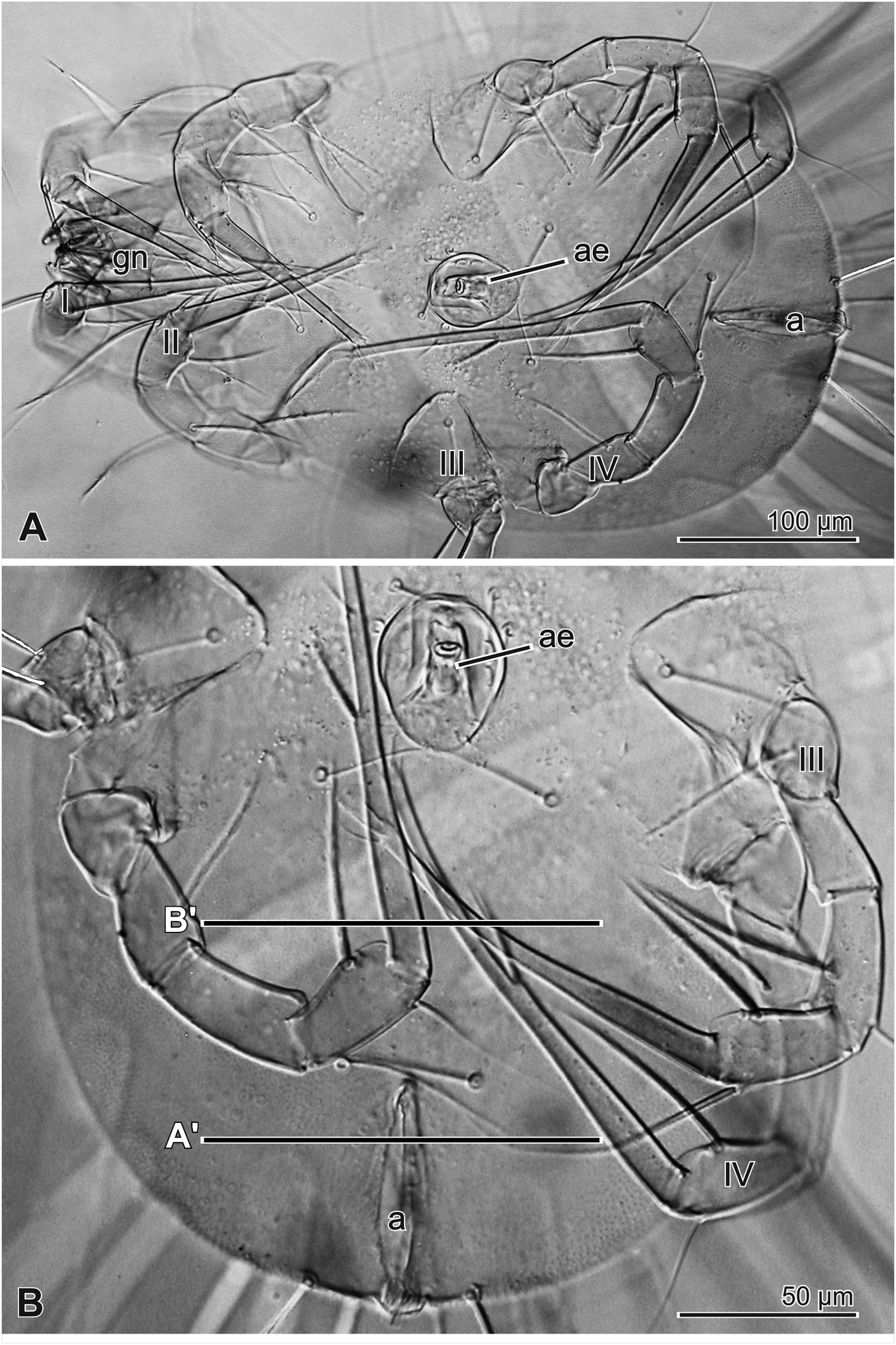
In Glycyphagus domesticus male (Fig. 1), the reproductive system consists of the paired large and roundish testes, located at the posterior end of the body on both sides of anal atrium (Fig. 2A′). More anteriorly, however, the left testis is terminating earlier than the right one, being replaced with a male accessory gland (Fig. 2B′). In the dorsal part of each testis, a compact germarium containing spermatogonia may be encountered, whereas the spermatocytes and spermatids are more spread out ventrally (Fig. 2A′). In the lower parts of the testes, the assemblages of spermatozoa occur in deferent ducts (DD in Fig. 2A′ as well as RDD and LDD in Fig. 2B′).
Each of the paired deferent ducts leaves a mid-ventral surface of the testis and runs anteriorly along the ventral cuticle. The diameter of the left testis gradually diminishes, being ultimately replaced with an accessory gland (Fig. 2B′).
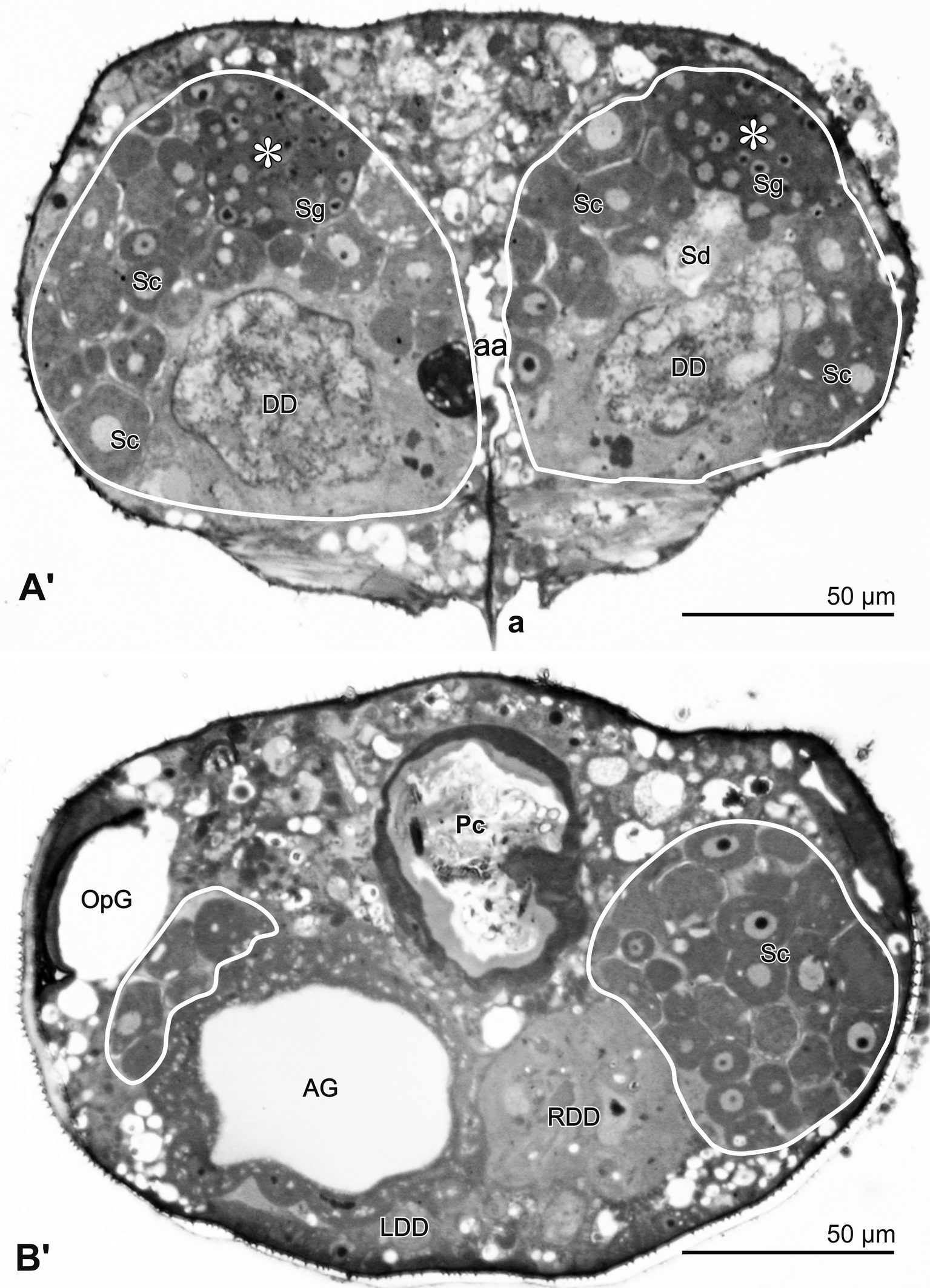
A thick wall of the unpaired accessory gland surrounds an ''empty'' lumen (Fig. 2B′). The duct of the gland and both deferent ducts join together into a cuticle-lined ejaculatory duct, which is S-shaped in the axial plane and enters into the basis of the aedeagus, to finally terminate at its tip. The aedeagus is located on the male ventral side, just behind the fourth pair of legs (Fig. 1).
Spermatocytogenesis in Glycyphagus
The spermatogonia in the germarium (Figs 2A′ and 3A) are tightly packed polygonal cells (ca. 4–7 μm in diameter; n=5). They contain a relatively large nucleus with a prominent nucleolus.
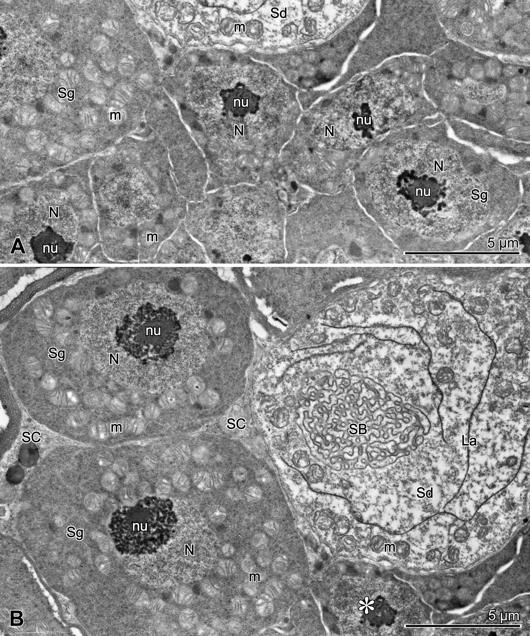
The spermatogonia which enter spermatocyte stage increase in diameter, separate from the ventral or ventro-lateral surface of germarium and become roundish. Such spermatogonia (Fig. 3B) (ca. 7.8–11.5 μm in diameter; n=5) show a moderately electron-dense cytoplasm containing a lot of free ribosomes. Central roundish nucleus (4.2–5.8 μm; n=5) contains a prominent central nucleolus. Around the nucleus, numerous spherical mitochondria with lamellar cristae are spread out.
Similar to spermatogonia, spermatocytes have an increased diameter (Fig. 2A′, B′) (ca. 16x17 up to 20x29 μm in diameter; n=5) and a spongy layer, formed by anastomosing membranes (Fig. 4A), appears on their surface. This layer allegedly produced via the increase in cisterns of superficially located Golgi bodies (Fig. 4A). During spermatocyte development, the thickness of spongy layer increases (Fig. 4A), roundish nucleus gradually enlarges and round nucleolus disappears. Finally, the nuclear envelope also disappears and is no longer discernible. Meiotic division has not been observed.
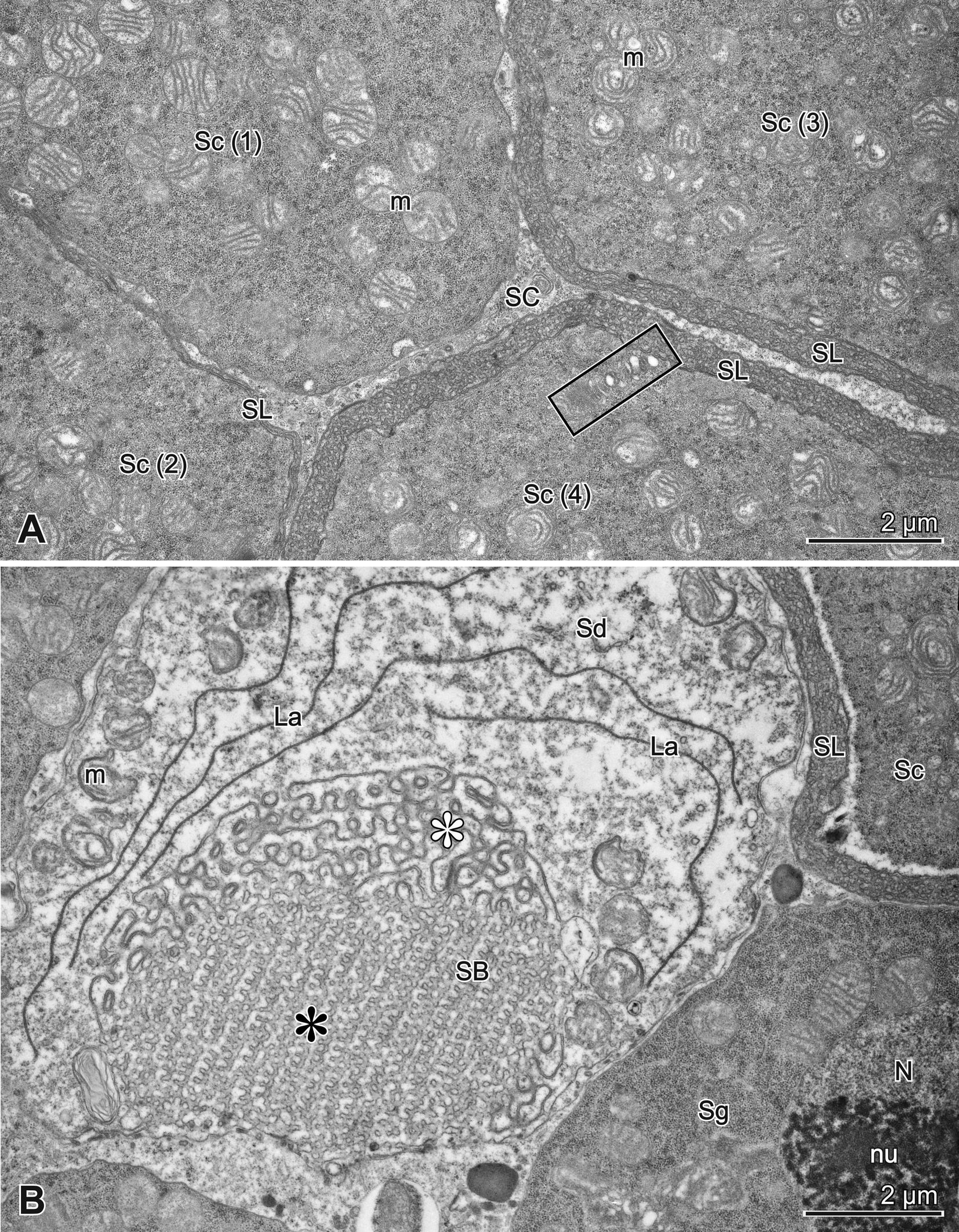
Like in other Astigmata, the spaces between the germinal cells located outside the germarium are filled with the processes of scarce somatic stroma cells (Figs 3B, 4A).
Spermiogenesis and sperm structure in Glycyphagus
Spermatids (Figs 3, 4B and 5A) are the singular, roundish cells (13–15 μm in diameter; n=5); their electron-lucent cytoplasm contains several profiles of electron-dense lamellae, irregularly shaped mitochondria, and conspicuous, large spongy body (ca. 7 μm in diameter; n=4), formed by the anastomosing membranes. The spongy body – representing a structure termed OSER (Organized Smooth Endoplasmic Reticulum) (Anderson et al. 1983; Snapp et al. 2003) – is derived from spermatocyte the superficial spongy layer, which becomes internalized in one place of cell periphery, hence being no longer visible on the spermatid surface.
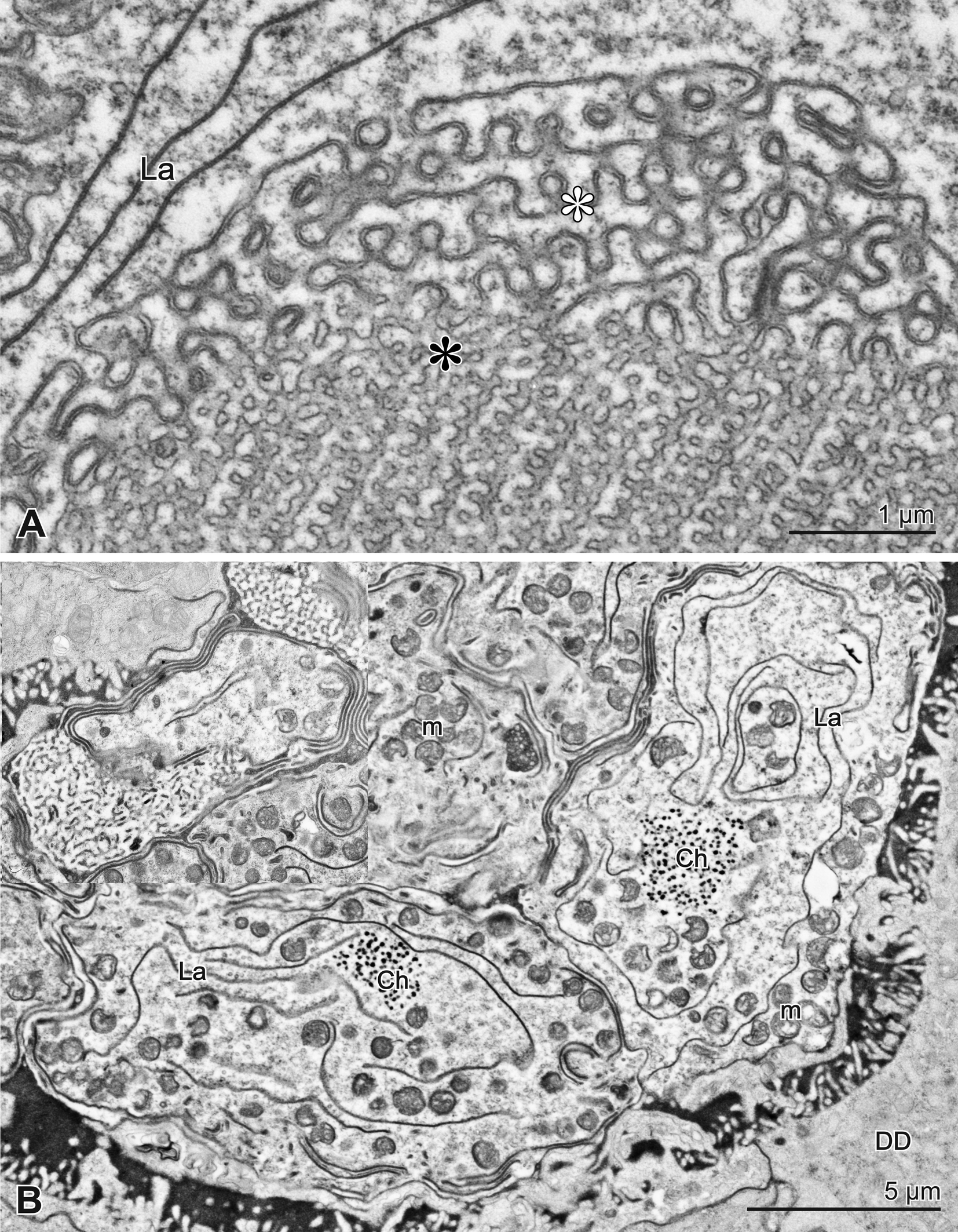
Such the spongy body comprises two compartments, i.e. a peripheral layer with sinusoidal, anastomosing double membranes, and a more central core containing a regular pattern of membranes (Figs 4B, 5A).
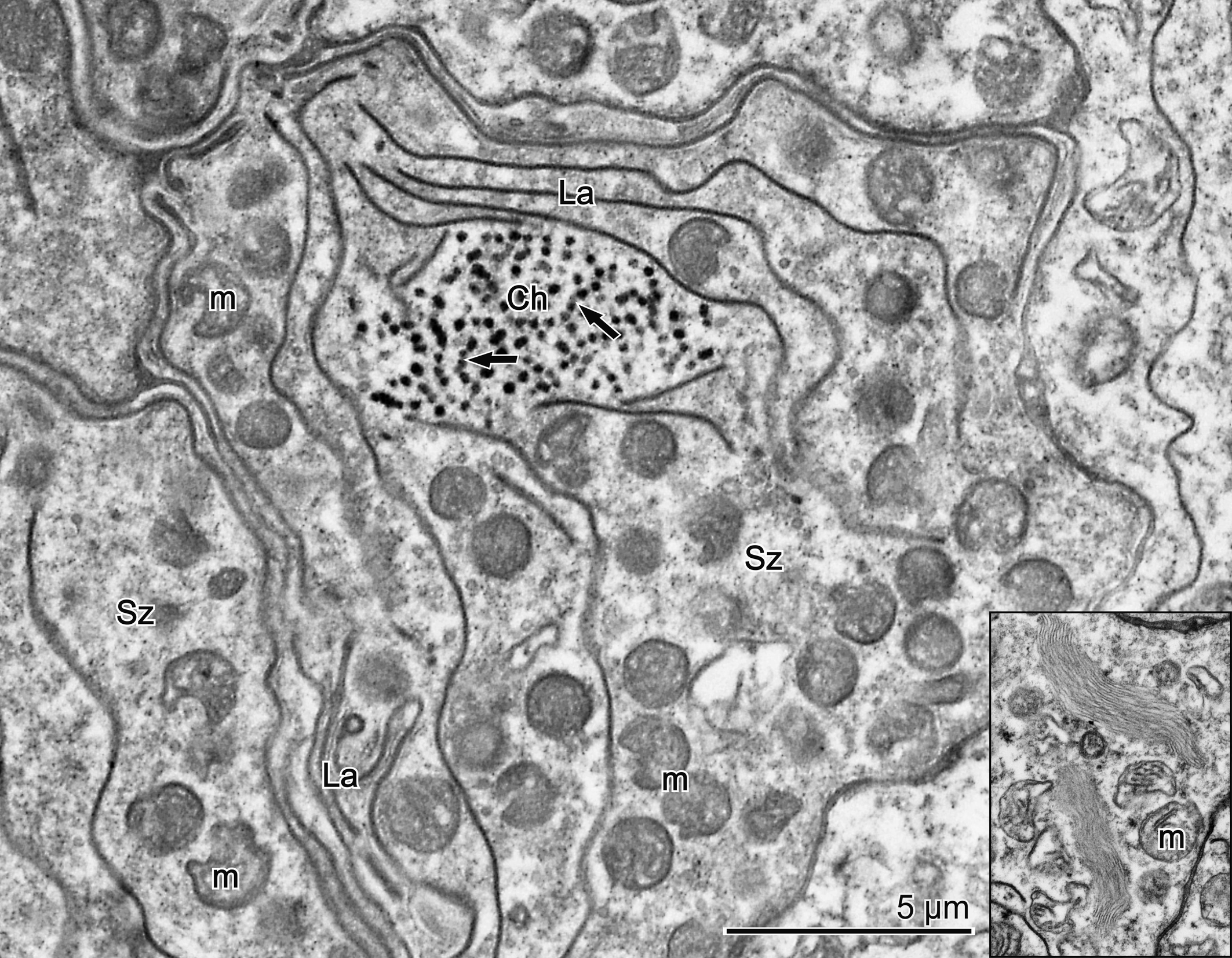
Spermatozoa are gathered in the lower part of the testis, filling the entrance to the deferent duct (Figs 2A′, 5B). These are the pleomorphic cells, ca. 8–12 x 9–18 in size; n=10 (Figs 5B, 6A). Chromatin (Figs 5B, 6A) is assembled sub-centrally as the granules (80–90 nm in diameter; n=10), surrounded by several electron-dense lamellae. Chromatin granules frequently form the strings of beads (Fig. 6). Numerous mitochondria are spread out within the cytoplasm and between the electron-dense lamellae. They are roundish and contain irregular cristae, frequently exhibiting a concavity on one side (Fig. 6A, C). Typical spongy bodies (OSER) are no longer encountered, even though their irregular remnants, located in the marginal parts of spermatozoa, may still be discerned. The sperm cytoplasm may also occasionally contain bundles of some fibrillar material (Fig. 6 inset).
Reproductive system and structure of the testis in Diplaegidia
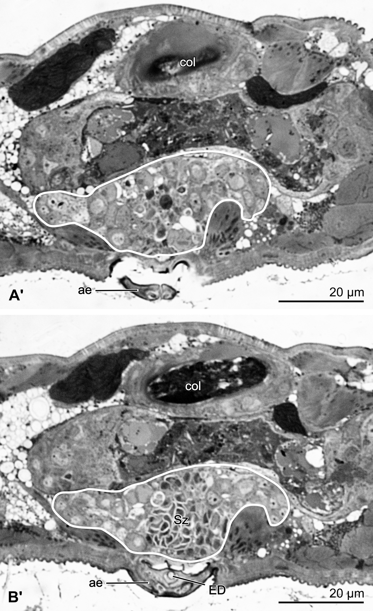
The male reproductive system of Diplaegidia columbae is relatively small and compact. It is made up of an unpaired testis located ventrally in the posterior part of the mite's body, just dorsally to the aedeagus (Figs 7, 8), and a short, thick unpaired deferent duct, running posteriorly from the testis. The deferent duct passes into the ejaculatory duct, cuticle-lined and circular in the cross section, which turns anteriad to enter the aedeagus (Fig. 8B′). It finally terminates at the aedeagus tip. No accessory gland is discernible.
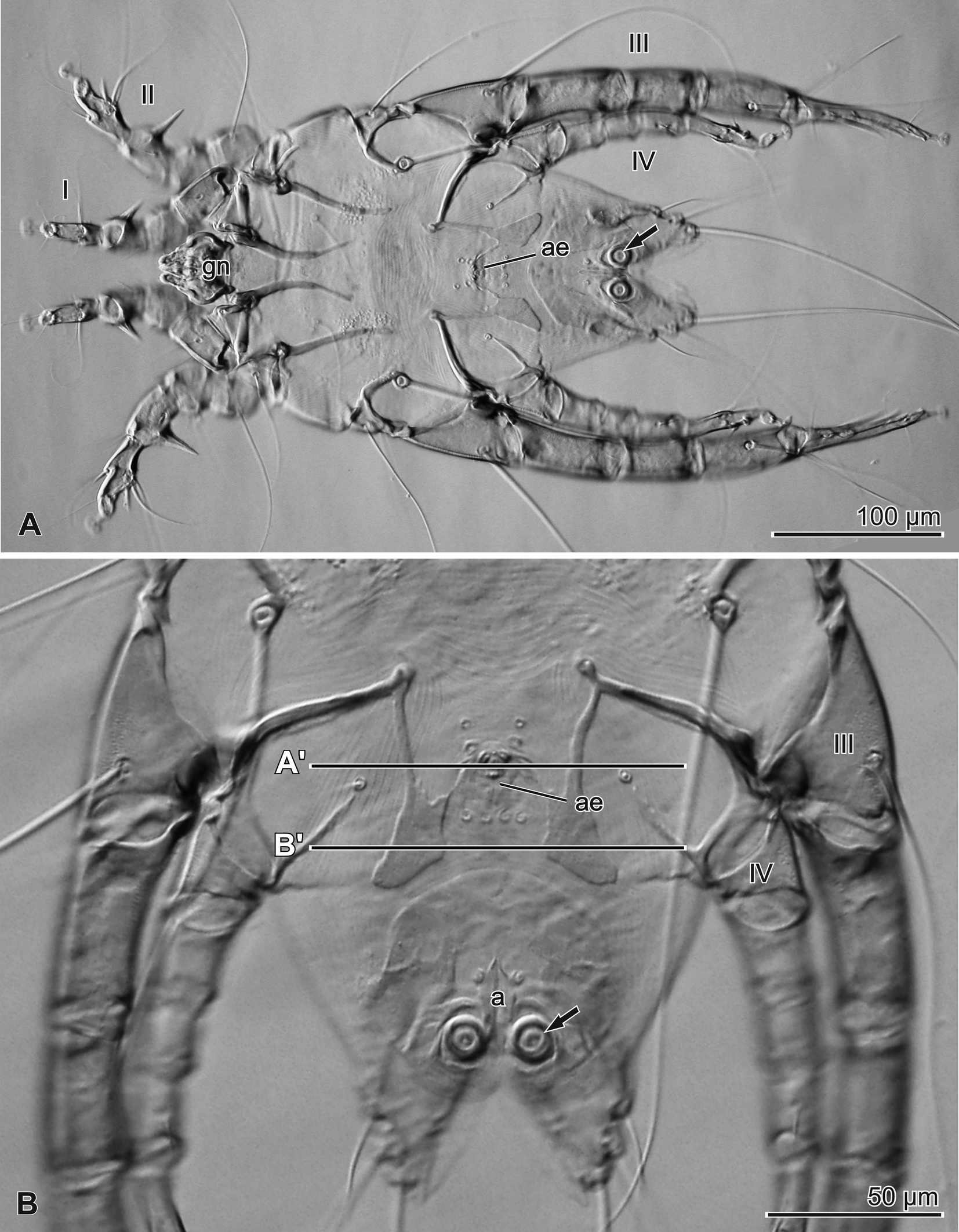
Testis in D. columbae (Fig. 8) contains germinal cells spread out randomly. Spermatogonia and primary spermatocytes are located separately, whereas the secondary spermatocytes and spermatids form groups with two or four isogenic cells, respectively, resultant from meiosis. All spermiogenesis stages, except for spermatozoa, may be found in the anterior part of the testis (Fig. 8A′), whereas spermatozoa form a central pellet in the posterior part of the testis (Figs 8B′, 9).
The spaces between the germinal cells, except the spermatozoa, are filled with scarce somatic stroma cells (Figs 9, 12).
In our material, the germarium has not been encountered in the testis, thus prompting a conclusion that this structure is to be found in the younger males only.
Spermatocytogenesis in Diplaegidia
Since the germarium has not yet been observed, neither did we encounter the early spermatogonia. More advanced spermatogonia or early spermatocytes (Figs 9, 10A) are the roundish or slightly elongated cells with smooth surface, roundish nucleus, and a cytoplasm containing a moderate number of ribosomes, as well as Golgi bodies.
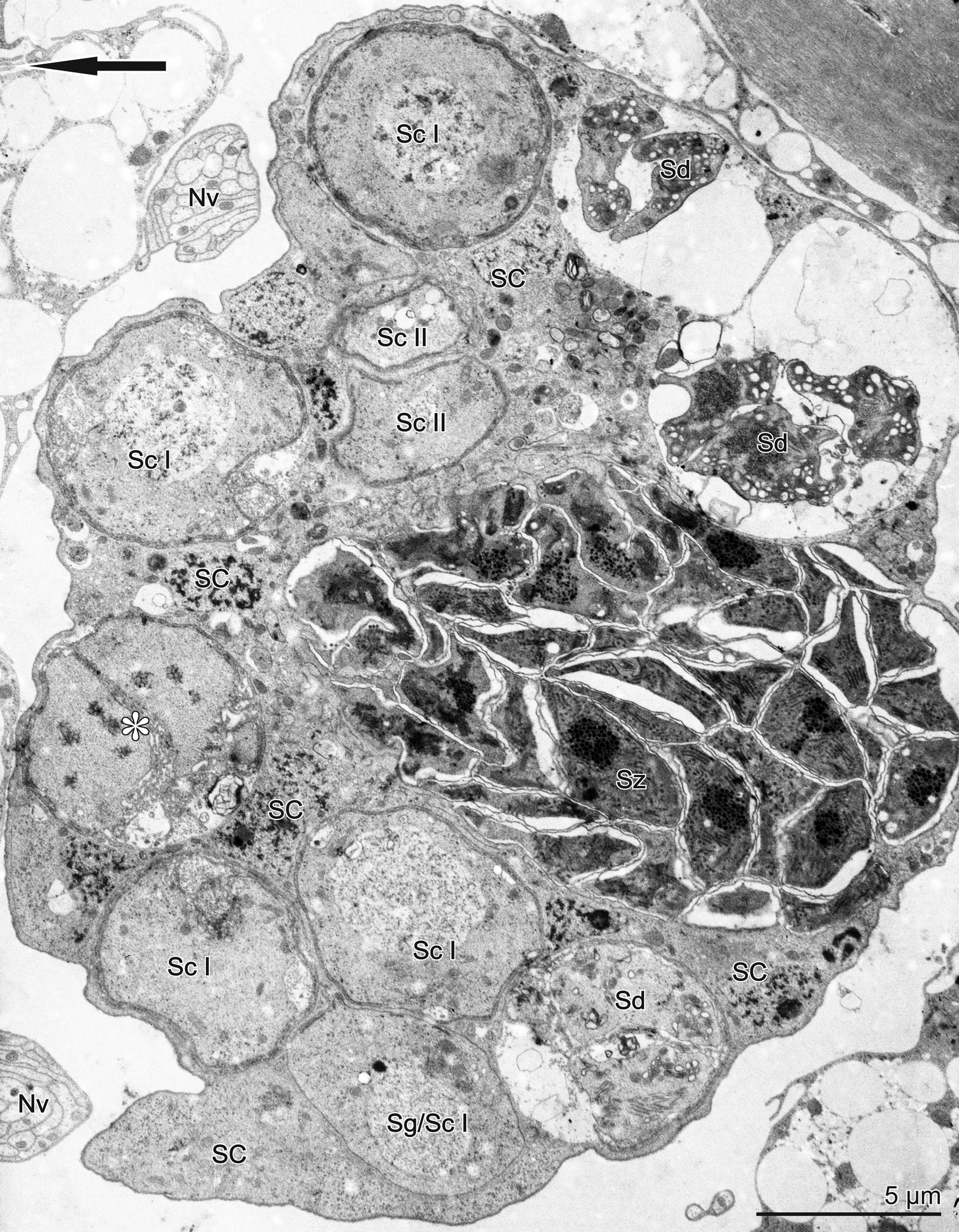
As spermatocytogenesis progresses, the first primary spermatocyte division is completed and two-cellular isogenic groups of secondary spermatocytes are formed (Figs 9, 10C), whereas following the second division, isogenic groups of spermatids may be observed (Figs 9, 10D, 11).
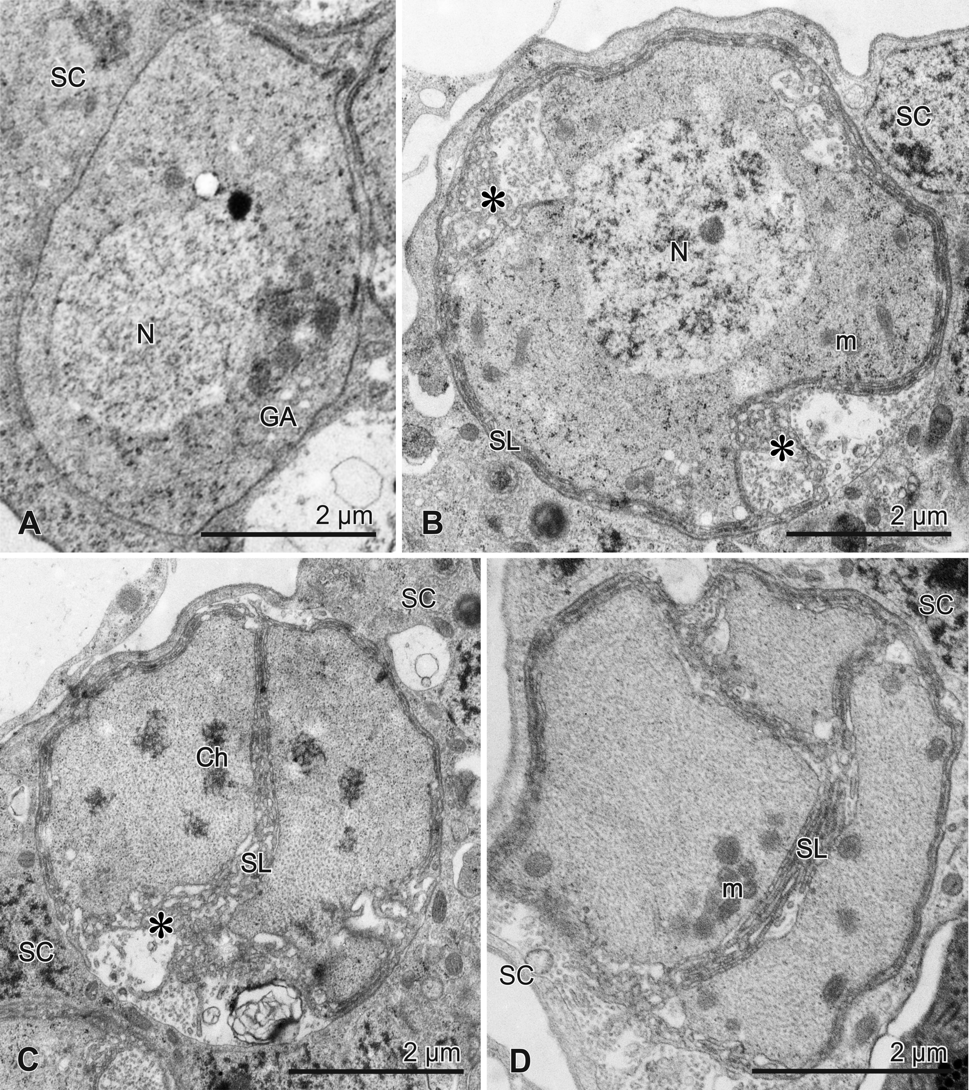
Primary spermatocytes (6.2–6.6 μm in diameter; n=5) become spherical (Figs 9, 10B). Their surface is covered with a layer of anastomosing membranes of Golgi body origin, the so-called spongy layer. Spongy layer is typical for spermatocytes, even though it persists up to the early spermatids (Fig. 10D). Spermatocyte surface may form shallow concavities, filled with irregularly distributed spongy layer membranes, forming the externally located spongy bodies (Fig. 10B). During the first meiotic division (Fig. 10C), secondary spermatocytes appear, whilst the nuclear envelope disintegrates in such cells. The chromatin is visible as an electron-dense foci, apparently representing the chromosomes. Meiotic figures have not been observed, though.
Spermiogenesis and sperm structure in Diplaegidia
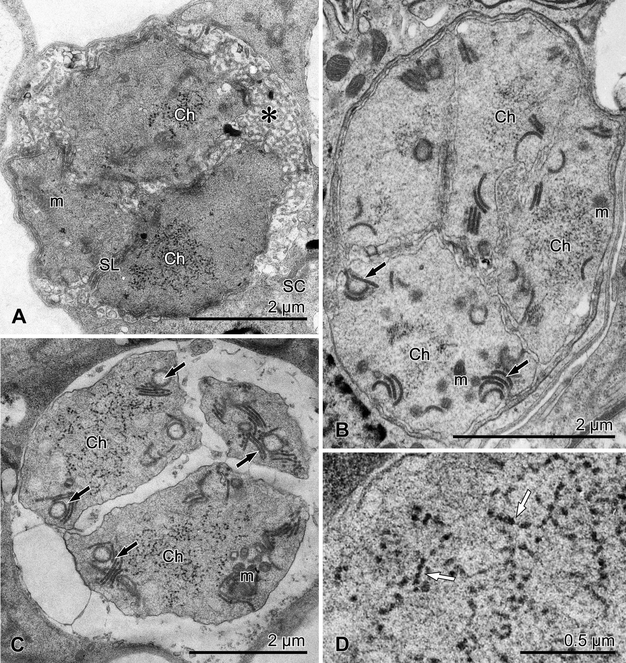
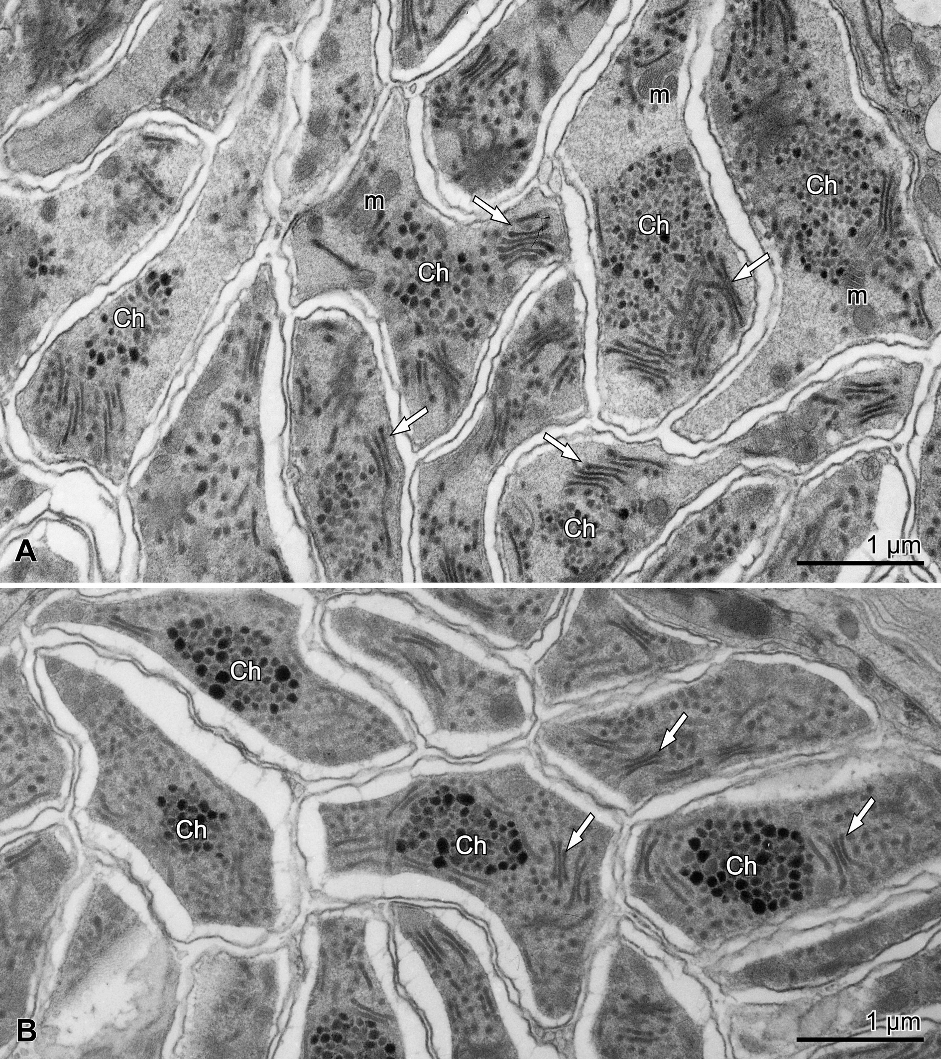
Early spermatids form four-cellular isogenic groups and are characterized by scarce peripherally located mitochondria (Fig. 10D). The condensed chromatin is no longer visible. The spongy layer is initially well-developed, but in the more advanced spermatids (Fig. 11A), it becomes loose, to eventually disappears altogether (Fig. 11A–C). In such spermatids, fine granular chromatin embedded in cytoplasm may be recognized in the cell centre (Fig. 11 A–C). The chromatin granules (30–40 nm in diameter; n=10) are frequently arranged in linear aggregates, resembling a string of beads (Fig. 11D). This is when the new structures, made up of short, arcuate dense lamellae and vesicles, distributed rather peripherally in the cells, may well be encountered (Fig. 11B, C). Both arcuate lamellae and vesicles show a double-membranous structure and may represent the same structure, which shows a different shape, depending on the plane of the section itself (see Discussion).
Spermatozoa are grouped in the central part of the testis, forming a pellet at the entrance of the deferent duct (Figs 8B′, 9). In the section, they are variable in shape, usually appearing elongated (ca. 1.4–2.0 x 2.7–3.5 μm; n=10) (Figs 12, 13), containing more or less centrally located granules of chromatin, surrounded with stacks of short and straight profiles of electron-dense lamellae, and scarce mitochondria with lamellar cristae. The vesicles are no longer encountered. The flagellum or axoneme are not encountered.
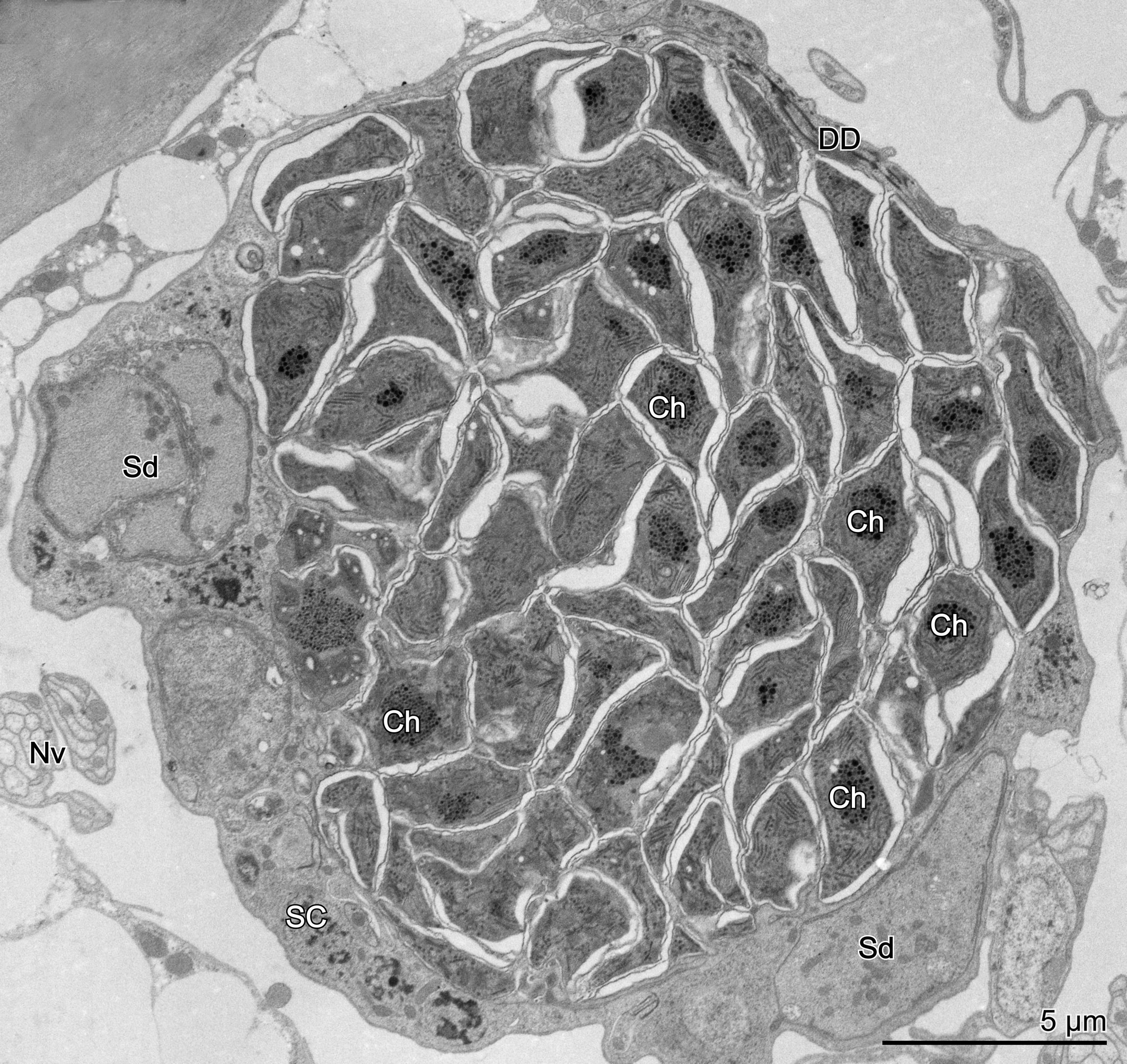
The chromatin granules in the less developed sperm cells (Fig. 13A) are larger than in the advanced spermatids (ca. 80–90 nm in diameter; n=10), although subsequently, in the more mature sperm cells (Fig. 13B) their diameter may reach ca. 100–120 nm; n=10. Furthermore, in such cells, the chromatin granules are more electron-dense and much more focused around the central part of the spermatozoon.
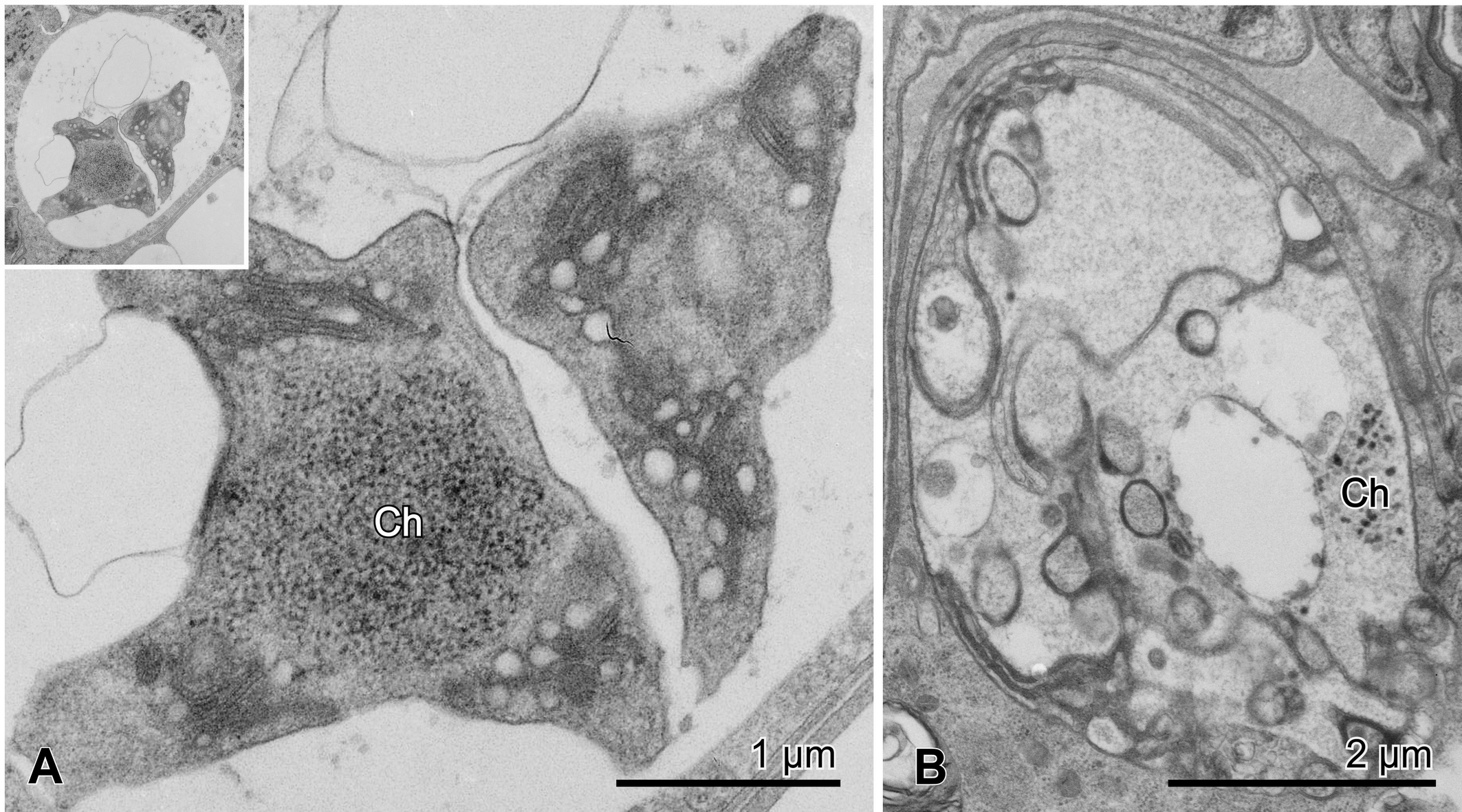
Despite the fact that the cells pass through regular spermatogenesis stages, some groups of spermatids may occasionally exhibit degenerative symptoms (Fig. 14).
Discussion
In Glycyphagus domesticus male, the reproductive system (Witaliński and Walzl 1995) is similar to those of many other astigmatic mites that have been studied to date (for review see: Witaliński 2014). It contains paired, symmetrically positioned testes that both contain functional germaria, whereas the left testis is in the anterior part replaced by a male accessory gland. Each germarium comprises the closely adhering germinal cells, whereas further stages of spermatogenesis are spread out in the central and lower parts of the testis; the entrances of the deferent ducts, filled with spermatozoa, are visible in the lower part of each testis.
The germarium in several astigmatan species contains the so-called testicular central cell (TCC) surrounded with spermatogonia. The actual origin of TCC is not clear, but some studies on Histiostoma feroniarum (Witaliński et al. 2014) indicate that TCC belongs to the germinal cell line, and it may be instrumental in regulating the actual proliferation of the adjacent germ cells (Florek and Witaliński 2010). In G. domesticus, the testicular central cell has not yet been encountered (Witaliński and Walzl 1995; the present study), nevertheless, it was proposed that during the development of the testes, such cells, connected via cellular bridges with the surrounding spermatogonia, may indeed appear (Witaliński 2014).
As opposed to G. domesticus and many Astigmata, numerous other Astigmata (e.g. Psoroptidae – Lekimme et al. 2005, Sarcoptidae – Witaliński 1988, and Pyroglyphidae – Walzl 1992), as well as D. columbae, exhibit unpaired testis, the situation resulting apparently from the secondary fusing of the paired testis anlages. In D. columbae, the germarium has not been encountered. This said, it may well be found in the younger males, in which functioning germarium can be a source of the spermatogonia. Subsequently, the spermatogonia undergo spermatogenesis, whereas the germarium may disappear altogether. Such a structure and function of the testis might imply a time-limited spermatogonia production, and a smaller number of spermatozoa in result. On the other hand, an allegedly larger number of spermatozoa, produced thanks to the still functioning germarium in G. domesticus, can be diminished, as this species is haplo-diploidal. Arrhenotoky, corroborated by no meiotic division, in which a primary spermatocyte is leading to two secondary spermatocytes and then to four spermatids/spermatozoa, ultimately results in four-fold smaller number of spermatids and spermatozoa.
A distinctive feature of spermatocytes and early spermatids in both species under study appears to be a spongy layer covering the surface of the cells, a structure of Golgi origin. Interestingly, such structure may be frequently observed in other Astigmata (Witaliński et al. 1986; Witaliński and Afzelius 1987; Florek and Witaliński 2010; Witaliński et al. 2014; Rożej-Pabijan and Witaliński 2018), but also during spermatogenesis in other mites like Gamasida (Witaliński 1976, 1988; Witaliński and Dallai 1991). However, its role is still unclear. It was postulated that the formation of spongy layer may represent a form of elimination of superfluous cytoplasm and/or formation of new plasmalemma (Witaliński 1988). During the spermatocyte stage, a spongy layer thickness increases, but later on, in spermatid stage, most of spongy layer membranes are transferred into the spongy bodies, which subsequently disappear. Thus, the thickness of a spongy layer diminishes in the course of spermiogenesis to eventually form a singular plasmalemma on the surface of late spermatids and spermatozoa.
Spermatids exhibit two other new structures, i.e. electron dense lamellae and spongy bodies. The electron dense lamellae, supposedly originating from ER cisterns (Witaliński et al. 1986; Witaliński 2014), which remains in line with a well-documented multifunctional plasticity of the ER (Baumann and Walz 2001; Woeltz et al. 2002; Schwarz and Blower 2016; Obara et al. 2023). The lamellae are a permanent structure in the spermiogenesis of Astigmata, including the species presently under study. In G. domesticus, they are relatively long and persist up to the spermatozoon stage, located around the chromatin. This in turn might imply their protective role for the chromatin in the case of mechanical stress to the spermatozoa, during and after insemination (Witaliński 2014; Rożej-Pabijan and Witaliński 2018). In D. columbae spermatids, electron-dense lamellae are short and curved, and frequently form the stacks adhering to the vesicles; such vesicles have a double-membranous wall of similar appearance as the electron-dense lamellae. This may imply that the vesicles are not new, separate structures, but represent cross sections rather than axial sections through the arcuate lamellae. Stacks of the lamellae may persist up to the spermatozoa stage, even though they are straight rather than curved. This may actually underpin the observation that the vesicles are no longer encountered. Furthermore, an association of electro-dense lamellae and chromatin in spermatozoa is not evident.
Interestingly, similar electron-dense lamellae have also been observed in other cells, in the so-called mitochondria-rich cells (MRCs) (Witaliński and Liana 2010). Several dozen of MRCs were encountered in the anterior part of the bodies in feather mites D. columbae, but also in Falculifer rostratus (Pterolichoidea). Such cells are filled with a number of large mitochondria, placed within an elaborate system of electron-dense lamellae. Owing to the mitochondria, a certain role of MRCs was proposed in heat generation, even though the actual contribution of such electron-dense lamellae to that process remains unknown as yet.
An unusual structure in G. domesticus spermatids appears as a spongy body, a large and very regular aggregation of membranes called OSER (Organized Smooth Endoplasmic Reticulum). Such structures have been reported in a variety of cells, tissues and organisms like plants, fungi and animals (for further references on the subject see Snapp et al. 2003), including Astigmata (Florek and Witaliński 2010; Witaliński 2014). The OSER is represented at least in two variants, lamellar OSER and sinusoidal OSER. However, the actual basis for their formation and their exact role in the cell still remains unclear. In OSER formation, the key role is postulated for tight binding interactions between the cytoplasmic domains of ER resident proteins, but motor proteins interacting between ER membranes and cytoskeletal elements may also be involved. As far as OSER function is concerned, there seems to be a likelihood for some sequestering processes within the cell, much like in the case of other membranous organelles (e.g. mitochondria, nucleus, or chloroplasts). On the other hand, the actual role of a large, well-developed OSER in the spermatids of Astigmata is still awaiting persuasive clarification.
Less regular structures similar to OSER may be also observed in spermatids in the other two astigmatan species, called a spongy body in Carpoglyphus lactis (Florek and Witaliński 2010), and an area of anastomosing membranes in Chaetodactylus osmiae (Witaliński 2014).
Spermatogenesis in Astigmata leads to the production of spermatozoa, characterized by several unusual features, i.e. 1) they are multiform cells, 2) there is no acrosome/acrosomal complex, 3) chromatin is embedded within the cytoplasm and is not delimited by a nuclear envelope, 4) there is no axoneme/flagellum as in all mites and ticks (e.g., Breucker and Horstmann 1968; Reger 1974; Alberti 1980a, b).
The multiform spermatozoa in mites (Parasitiformes and Acariformes) may be encountered at least in several groups of Astigmata (Liana and Witaliński 2005). Their sizes vary within a large range, from ca. 2 µm in Histiostoma feroniarum (Histiostomatoidea) and 3–4 µm in Myocoptes musculinus, Notoedres cati and Sarcoptes scabiei (Sarcoptoidea), up to 18 µm in Canestrinia sellnicki (Canestrinioidea), and 22–27 µm in Scutulanyssus obscurus (Analgoidea). Multiform spermatozoa represent an autapomorphic character in Astigmata, but small and large sperm cells may also be observed in the closely related superfamilies (e.g. Histiostomatoidea and Canestrinioidea) (Liana and Witaliński 2005). The functional reason for multiform sperm cells can be combined with other unusual feature of Astigmata spermatozoa, i.e., a chromatin embedded in cytoplasm instead of being closed in the nucleus (see later in the Discussion). Both modifications seem to be a response to a strong stress of sperm, occurring during and after insemination, when the spermatozoa are forced to crawl along a narrow tube which connects the inseminatory orifice and spermatheca.
The other autapomorphy is related to the fact that there is no acrosome/acrosomal complex (Witaliński et al. 1986; Witaliński and Afzelius 1987; Witaliński 1988), which is allegedly related to early spermatozoa penetration into the oocytes, i.e. before they manage to form a vitelline envelope or chorion at the oocyte surface.
The third autapomorphy of Astigmata spermatozoa is related to the specific organization of chromatin. Chromatin threads are embedded in the sperm cytoplasm, rather than sequestered in a compact cell nucleus, delimited with a nuclear envelope. During spermatogenesis, the nuclear envelope of primary spermatocyte disappears, and the spermatid chromatin is discernible as the thin threads embedded in the cytoplasm. Subsequently, when chromatin condensation progresses, in most Astigmata the threads of chromatin become thicker (Liana and Witaliński 2005). However, in the recently studied species G. domesticus and D. columbae, chromatin condenses in the form of granules rather than threads, and such chromatin granules persist up to the spermatozoon stage. During the chromatin condensation in the spermatids, the chromatin granules appear to be connected with the thin threads (DNA?), which promote their linear aggregation much like the strings of beads (Fig. 11D). It must be noted, however, that in the previous study (Liana and Witaliński 2005), the chromatin organization in G. domesticus sperm cells has been erroneously described as made up of threads instead of granules.
The last feature of astigmatan spermatozoa consists in a lack of flagellum or axoneme, a common feature for mites and ticks sperm cells, resultant from centriole absence in the cells of those animals. It follows, that sperm motility is secured through other, actin-based mechanisms, e.g., in ticks (Witaliński and Dallai 1994) and Astigmata. In astigmatan sperm, however, the ameboid movements have been proposed, even though they are neither persuasively evidenced, nor indeed clarified in sufficient detail (Witaliński 1988). Interestingly, in other animal group showing an amoeboid sperm motility, the nematodes, sperm motility is based on the unique Major Sperm Protein (MSP) rather than on any actin-based mechanisms (Roberts and King 1991; Italiano et al. 2001; Ma et al. 2012)
Acknowledgements
We are most grateful to Dr. Roberto Nannelli, Istituto Sperimentale per la Zoologia Agraria, Florence, Italy, who kindly provided us with Glycyphagus domesticus (De Geer), and Prof. Jacek Dabert, A. Mickiewicz University, Poznań, Poland, who identified Diplaegidia columbae (Buchholz) males, as well as to the academic staff of the Department of Cell Biology and Imaging, Institute of Zoology and Biomedical Research, Jagiellonian University, Kraków, Poland, for making the TEM available for our research effort.
References
- Alberti G. 1980a. Zur Feinstruktur der Spermien und Spermiocytogenese der Milben (Acari). I. Anactinotrichida. Zool. Jb. Anat. 104: 77-138.
- Alberti G. 1980b. Zur Feinstruktur der Spermien und Spermiocytogenese der Milben (Acari). II. Actinotrichida. Zool. Jb. Anat. 104: 144-203.
- Anderson R.G., Orci L., Brown M.S., Garcia-Segura L.M., Goldstein J.L. 1983. Ultrastructural analysis of crystalloid endoplasmic reticulum in UT-1 cells and its disappearance in response to cholesterol. J. Cell Sci., 63:1-20. https://doi.org/10.1242/jcs.63.1.1
- Baumann O., Walz B. 2001. Endoplasmic reticulum of animal cells and its organization into structural and functional domains. Int. Rev. Cytol., 205: 149-214. https://doi.org/10.1016/S0074-7696(01)05004-5
- Breucker H., Horstmann E. 1968. Die Spermatozoen der Zecke Ornithodorus moubata (Murr.). Z. Zellforsch. Mikrosk. Anat., 88: 1-22. https://doi.org/10.1007/BF00492230
- Florek M., Witaliński W. 2010. Spermatogenesis and sperm structure in Carpoglyphus lactis (L.) (Acari: Astigmata). Arthropod Struct. Develop., 39: 41-51. https://doi.org/10.1016/j.asd.2009.09.001
- Italiano J.E., Jr., Stewart M., Roberts T.M. 2001. How the assembly dynamics of the nematode major sperm protein generate amoeboid cell motility. Int. Rev. Cytol., 202: 1-34. https://doi.org/10.1016/S0074-7696(01)02002-2
- Karnowsky M.J. 1965. A formaldehyde-glutaraldehyde fixative of high osmolality for use in electron microscopy. J. Cell Biol., 27: 137A-138A.
- Lekimme M., Leclercq-Smekens M., Devignon C., Leclipteux T., Poumay Y., Losson B. 2005. Ultrastructural morphology of the male and female genital tracts of Psoroptes spp. (Acari: Astigmata: Psoroptidae). Exp. Appl. Acarol., 36: 305-324. https://doi.org/10.1007/s10493-005-6076-8
- Liana M., Witaliński W. 2005. Sperm structure and phylogeny of Astigmata. J. Morphol., 265: 318-324. https://doi.org/10.1002/jmor.10361
- Ma X., Zhao Y., Sun W., Shimabukuro K., Miao L. 2012. Transformation: how do nematode sperm become activated and crawl′ Protein & Cell., 3: 755-761. https://doi.org/10.1007/s13238-012-2936-2
- Obara C.J., Moore A.S., Lippincott-Schwartz J. 2023. Structural diversity within the endoplasmic reticulum - from the microscale to the nanoscale. Cold Spring Harb Perspect Biol., 15(6):a041259. https://doi.org/10.1101/cshperspect.a041259
- Reger J.F. 1974. The origin and fine structure of cellular processes in spermatozoa of the tick Dermacentor andersoni. J. Ultrastruct. Res., 48: 420-434. https://doi.org/10.1016/S0022-5320(74)80012-2
- Roberts T.M., King K.L. 1991. Centripetal flow and directed reassembly of the Major Sperm Protein (MSP). Cytoskeleton in the ameboid sperm of the nematode, Ascaris suum. Cell Motil. Cytoskel., 20: 228-241. https://doi.org/10.1002/cm.970200306
- Rożej-Pabijan E., Witaliński W. 2018. Gonads and gametogenesis in Chaetodactylus osmiae (Acariformes: Astigmata: Chaetodactylidae) a parasite of solitary bees. Acarologia, 58: 801-824. https://doi.org/10.24349/acarologia/20184269
- Snapp E.L., Hegde R.S., Francolini M., Lombardo F., Colombo S., Pedrazzini E., Borgese N., Lippincott-Schwartz J. 2003. Formation of stacked ER cisternae by low affinity protein interactions. J. Cell Biol., 163: 257-269. https://doi.org/10.1083/jcb.200306020
- Schwarz D.S., Blower M.D. 2016. The endoplasmic reticulum: structure, function and response to cellular signaling. Cell. Mol. Life Sci., 73: 79-94. https://doi.org/10.1007/s00018-015-2052-6
- Walzl M.G. 1992. Ultrastructure of the reproductive system of the house dust mites Dermatophagoides farinae and D. pteronyssinus (Acari, Pyroglyphidae) with remarks on spermatogenesis and oogenesis. Exp. Appl. Acarol., 16: 85-116. https://doi.org/10.1007/BF01201494
- Witaliński W. 1976. Spermatogenesis in free living mite, Pergamasus viator Halaš. (Parasitidae, Mesostigmata). II. Spermiogenesis. Z. mikrosk.-anat. Forsch., 90: 657-680.
- Witaliński W. 1988. Spermatogenesis and postinseminational alterations of sperm structure in a sarcoptid mite, Notoedres cati (Hering) (Acari, Acaridida, Sarcoptidae). Acarologia, 29: 411-421.
- Witaliński W. 2014. Gonads and gametogenesis in astigmatic mites (Acariformes: Astigmata). Arthropod Struct. Develop., 43: 323-340. https://doi.org/10.1016/j.asd.2014.04.003
- Witaliński W., Afzelius B.A. 1987. Spermatogenesis in an itch mite, Sarcoptes scabiei (L.) (Acari, Sarcoptidae). J. Submicrosc. Cytol., 19: 615-625.
- Witaliński W., Dallai R. 1991. Ultrastructural study of spermatogenesis in gamasid mites (Acari, Gamasida). Acta Zool., Stockholm, 72: 49-54. https://doi.org/10.1111/j.1463-6395.1991.tb00315.x
- Witaliński W., Dallai R. 1994. Actin in spermatozoon of a soft tick, Argas (A.) polonicus (Ixodida, Acari). Folia Histochem. Cytobiol., 32: 257-264.
- Witaliński W., Jończy J., Godula J. 1986. Spermatogenesis and sperm structure before and after insemination in two acarid mites, Acarus siro L. and Tyrophagus putrescentiae (Schrank) (Acari, Astigmata). Acarologia, 27: 41-51.
- Witaliński W., Liana M. 2010. Mitochondria-rich cells in the astigmatic mites, Diplaegidia columbae (Buchholz) (Analgidae) and Falculifer rostratus (Buchholz) (Falculiferidae) (Acari: Astigmata). Arthropod Struct. Develop., 39: 334-339. https://doi.org/10.1016/j.asd.2010.04.002
- Witaliński W., Rożej-Pabijan E., Podkowa D. 2014. Gonads in Histiostoma mites (Acariformes: Astigmata): structure and development. Arthropod Struct. Develop., 43: 385-401. https://doi.org/10.1016/j.asd.2014.04.006
- Witaliński W., Szlendak E., Boczek J. 1990. Anatomy and ultrastructure of the reproductive systems of Acarus siro (Acari: Acaridae). Exp. Appl. Acarol., 10: 1-31. https://doi.org/10.1007/BF01193970
- Witaliński W., Walzl M.G. 1995. Reproductive systems in selected acaridid mites (Acaridida). In: Kropczyńska D., Boczek J., Tomczyk A. (Eds), The Acari. Physiological and Ecological Aspects of Acari-Host Relationships. Dabor, Warszawa, p. 109-116.
- Venable J.H., Coggeshall R. 1965. A simplified lead citrate stain for use in electron microscopy. J. Cell Biol., 25: 407-408. https://doi.org/10.1083/jcb.25.2.407
- Voeltz G.K., Rolls M.M., Rapoport T.A. 2002. Structural organization of the endoplasmic reticulum. EMBO reports, 3: 944-950. https://doi.org/10.1093/embo-reports/kvf202
 to post any comment or correction.


2026-05-13
Date accepted:
2026-09-20
Date published:
2026-10-01
Edited by:
Mąkol, Joanna

This work is licensed under a Creative Commons Attribution 4.0 International License
2026 Witaliński, Wojciech and Podkowa, Dagmara
 Download article Download low definition
Download the citation
RIS with abstract
(Zotero, Endnote, Reference Manager, ProCite, RefWorks, Mendeley)
RIS without abstract
BIB
(Zotero, BibTeX)
TXT
(PubMed, Txt)