


The flat mite genera Capedulia Smith Meyer and Phytoptipalpus Trägårdh (Trombidiformes: Tenuipalpidae) from the eastern Canary Islands
Ferragut, Francisco  1
and Baumann, Julia
2
1
and Baumann, Julia
2
1✉ Instituto Agroforestal Mediterráneo. Universitat Politècnica de València. Valencia, Spain.
2Institute of Biology, University of Graz. Graz, Austria.
2026 - Volume: 66 Issue: 3 pages: 841-854
https://doi.org/10.24349/oy45-jcgzZooBank LSID: 16D1CDB1-94DE-4C61-8356-A2757F6EF9CB

Original research
Keywords
Abstract
Introduction
The Canary Islands constitute an archipelago of seven main islands located in the North Atlantic Ocean, off the northwest coast of Africa. Lanzarote and Fuerteventura are the easternmost islands, separated from the African mainland by about 100 km. These two islands differ markedly from the rest of the archipelago by their geography, environmental conditions, and resident biotas. All the Canary Islands are of volcanic origin and have never been connected to the continent. They emerged from the ocean floor between 1 and 23 million years ago as a result of a westward-propagating geological process, and consequently, the eastern islands are the oldest (Carracedo and Troll 2021; Whittaker et al. 2023). This geological history has subjected Lanzarote and Fuerteventura to prolonged erosional processes, resulting in arid, highly eroded landscapes under extreme climatic conditions, with sparse vegetation. Nevertheless, evolutionary processes have led to the emergence of a remarkable number of endemic plant and animal species.
The flat mite fauna (Tenuipalpidae) of the Canary Islands remains unknown. This study presents the first results of surveys conducted on native plants in Lanzarote and Fuerteventura. Two new species belonging to the genera Capedulia Smith Meyer and Phytoptipalpus Trägårdh are described. Capedulia canariensis sp. nov. represents the fifth species to be placed in this small genus, which exhibits a notable disjunct distribution between the African continent, the eastern Mediterranean region, and Macaronesia. Phytoptipalpus majorensis sp. nov. is recorded from host plants belonging to the family Fabaceae.
Material and methods
The mites were collected by the first author in Lanzarote and Fuerteventura Islands between 22 January and 4 February 2024. They were extracted from native plants by beating twigs and leaves on a fine sieve, and preserved in ethanol 70%. Specimens were later preclarified in Nesbitt fluid, mounted in Heinze-PVA medium, and stored in an oven at 50°C until totally dried and clarified. Mounted specimens were examined using differential interference contrast (DIC) and Phase contrast techniques with a Nikon Eclipse Ni-U compound microscope (Nikon Corporation, Tokyo, Japan). Illustrations were performed on a graphic tablet using Concepts software (https://concepts.app ). Measurements of morphological traits were obtained with the software NIS-Elements D 3.1 and are given in the text in micrometres (μm). The data are presented as the female holotype in bold followed in parenthesis by the range of all the females measured, or as the range of all the measurements in the case of males. Distances between setae were measured from the centre of setal insertions. Setal length was taken from its base to the tip of the seta. The length of legs was obtained from the base of the trochanter to the end of tarsus, excluding the ambulacrum.
The generic concept of Capedulia was proposed originally by Smith Meyer (1979) based on the first described species; as four further species have been described since then, we propose an updated diagnosis of the genus. We follow the original definition of Phytoptipalpus later extended and updated by Smith Meyer and Van Dis (1993) and Ueckermann et al. (2019). Terminology of the idiosomal and leg setation follows Lindquist (1985). Nomenclature for idiosomal cupules is based on Grandjean (1939).
Results
Family Tenuipalpidae Berlese, 1913
Genus Capedulia Smith Meyer, 1979
Type species — Capedulia calendulae Smith Meyer, 1979, by original designation.
Diagnosis
Female. Idiosoma soft, lacking dorsal and ventral sclerotized plates. Palps one-segmented; palp tarsus bearing two terminal phaneres and one solenidion. Subcapitular seta m absent. Anterior margin of prodorsum rounded, without median projections or a notch. Dorsal opisthosoma with 10 pairs of setae (c1, c2, c3, d1, d3, e1, e3, f3, h1, and h2 present); dorsal setae setiform, smooth or barbed. Genital, ventral, and anal regions membranous; anal plate with two pairs of setae, ps2 situated posterolaterally to ps3, both setae nearly aligned transversely. Leg chaetotaxy for legs I–IV: coxae 2-2-1-1 (except in C. maritima, with one ventral seta on coxa II); trochanters 1-1-2-1 (except in C. maritima, with trochanter IV nude); femora 3-3-2-1/2 or 4-4-2-1; genua 1-1-0-0, 2-2-0-0, 2-2-1-0, or 3-2/3-1-0; tibiae 4-4-3-3; tarsi 7/8(+1)-7/8(+1)-3/5-3/5. Tarsi I–II each with one solenidion ω″. Dorsal setae on femora and tibiae I–III setiform. Tarsal claws and empodia pad-like, with tenent hairs; claw pads with 2 inner tenent hairs shorter than the single outer tenent hair.
Male. Tarsi I–II with two solenidia, ω′, ω″.
Capedulia canariensis Ferragut sp. nov.
ZOOBANK: BACC5C7C-5AEB-421B-A73D-9A48F79FF623
(Figures 1–3)
Type material and depository
Female holotype, 20 female paratypes, and two male paratypes on Traganum moquinii (Amaranthaceae); Lanzarote, Caleta de Famara beach, 29°07′30″N, 13°34′31″W, 5 m a.s.l. (above sea level); 4 February 2024. Two females on Bassia tomentosa (Amaranthaceae); Lanzarote, near Famara resort, 29°06′45″N, 13°32′54″W, 30 m a.s.l.; 4 February 2024. Four females on Traganum moquinii (Amaranthaceae); Fuerteventura, Dunas de Corralejo, 28°41′14″N, 13°50′39″W, 2 m a.s.l.; 22 January 2024. Female holotype, 13 female and one male paratypes were deposited in the Museo de Ciencias Naturales de Tenerife, Santa Cruz de Tenerife, Canary Islands. The remaining specimens are in the Acari collection, Instituto Agroforestal Mediterráneo, Universidad Politécnica de Valencia, Spain.
Diagnosis
As per genus definition with the additional specific characters. Female: dorsal setae h2 longer than 100 μm; ratio setae h2/h1 3.3–4.1, ratio f3/h2 0.5–0.7. Setal formulae of genua 2-2-1-0; dorsal seta on genua I–II absent. Femur IV with two setae (d, v′). Tarsus I with one solenidion and two eupathidia, tarsus II with one solenidion and one eupathidium. Male: tarsus I with two solenidia and two eupathidia, tarsus II with two solenidia and one eupathidium.
Description
Female (27 examined, 10 measured. Figures 1–2)
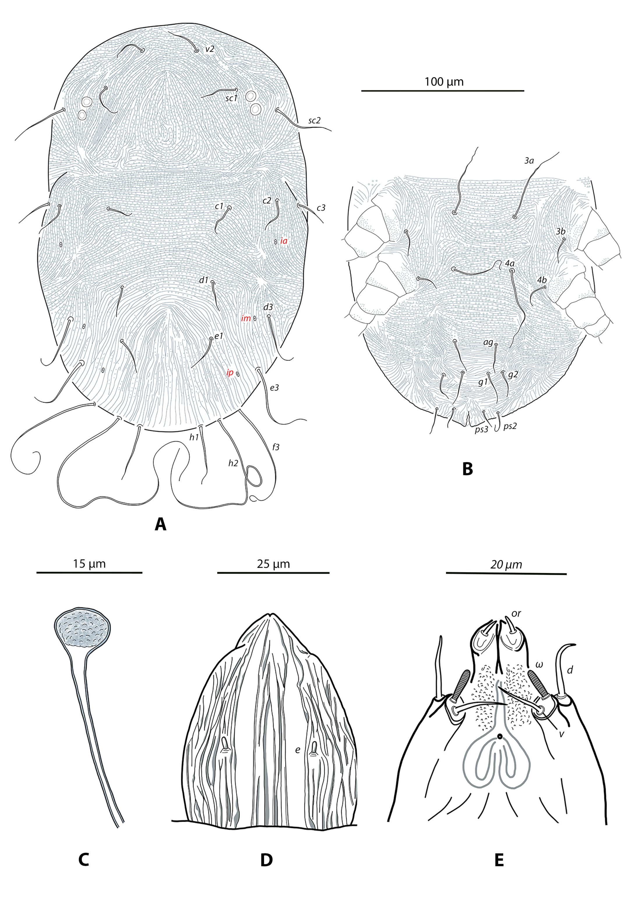
Dorsum. (Figure 1A). Absence of anterior prodorsal lobe. Prodorsal and opisthosomal surfaces soft, without developed shields. Dorsum thoroughly striated, striae forming characteristic ornamentation patterns. On the prodorsum, transversal striae between setae v2 and posteromedially; median inverted V-shaped pattern. Dorsum of opisthosoma with transverse striae between c1–d1; longitudinal or oblique striae on the laterals; striae forming an inverted V-shaped or U-shaped pattern which anterior angle is situated between setal pairs d1–e1. The separating spaces between consecutive striae covered by abundant microtubercles giving a granulate appearance, only absent on the cuticle posterior to setal row D. Opisthonotal cupules difficult to see, placed as follows: ia posterior and slightly paraxial to the insertion of c2, im mesad sockets of d3, ip mesad insertion of e3. Body measurements: v2–h1 231 (191–233), sc2–sc2 126 (122–137). Distances v2–v2 32 (32–37), sc1–sc1 78 (76–85), c1–c1 78 (73–90), c2–c2 131 (126–164), c3–c3 155 (143–188), d1–d1 56 (55–62), d3–d3 117 (117–133), e1–e1 52 (50–60), e3–e3 107 (105–111), f3–f3 83 (80–89), h1–h1 33 (32–37), h2–h2 57 (55–61). Dorsal setae fine, simple, acuminate, smooth or very finely barbed; barbs only visible under higher magnifications. Setal measurements: v2 21 (21–25), sc1 21 (21 –30), sc2 43 (33–49), c1 25 (19–27), c2 20 (20–26), c3 20 (19–27), d1 20 (20–27), d3 35 (33–41), e1 23 (22–29), e3 48 (36–60), f3 69 (69–78), h1 34 (32–38), h2 105 (105–151).
Venter. (Figure 1B) Ventral cuticle thoroughly striate and microtuberculate (granulate); striae are nude on coxa II and genital and anal plates with microtubercles absent. Longitudinal striae between 1a–1b and on the coxal areas of all legs; the remaining striae mostly transversal. Ventral setae smooth and setiform. Setal measurements: 1a 63 (60–81), 1b 17 (13–19), 1c 11 (11–14), 2b 12 (12–20), 2c 23 (20–25), 3a 59 (55–73), 3b 13 (11–20), 4a 50 (40–65), 4b 13 (13–19), ag 26 (19–27), g1 27 (20–30), g2 22 (18–23), ps2 18 (13–18), ps3 15 (11–16).
Spermathecal apparatus (Figure 1C). Insemination duct relatively short and broad, 48 (48–51) long, slightly flaring near the spherical vesicle. Seminal receptacle spherical, often slightly flattened around distal margin, 6–7 long, 7–9 wide. Lumen of the seminal vesicle appears granulated.
Gnathosoma (Figures 1D–E). Dorsal gnathosoma suboval, 62 (59–64) long, 38 (38–43) wide at level of palpal supracoxal setae e; setae e rod-like, about 3 long. Dorsal integument laterally folded leaving two smooth and narrow bands (Figure 1D). Ventral cuticle finely granulated near the buccal cone; infracapitular setae m absent. One pair of oral setae visible. Palp one-segmented, with a smooth blunt dorsal seta 9 (9–12) long, and two ventral phaneres, one smooth 8 (7–9) and one solenidion 6 (6–8).
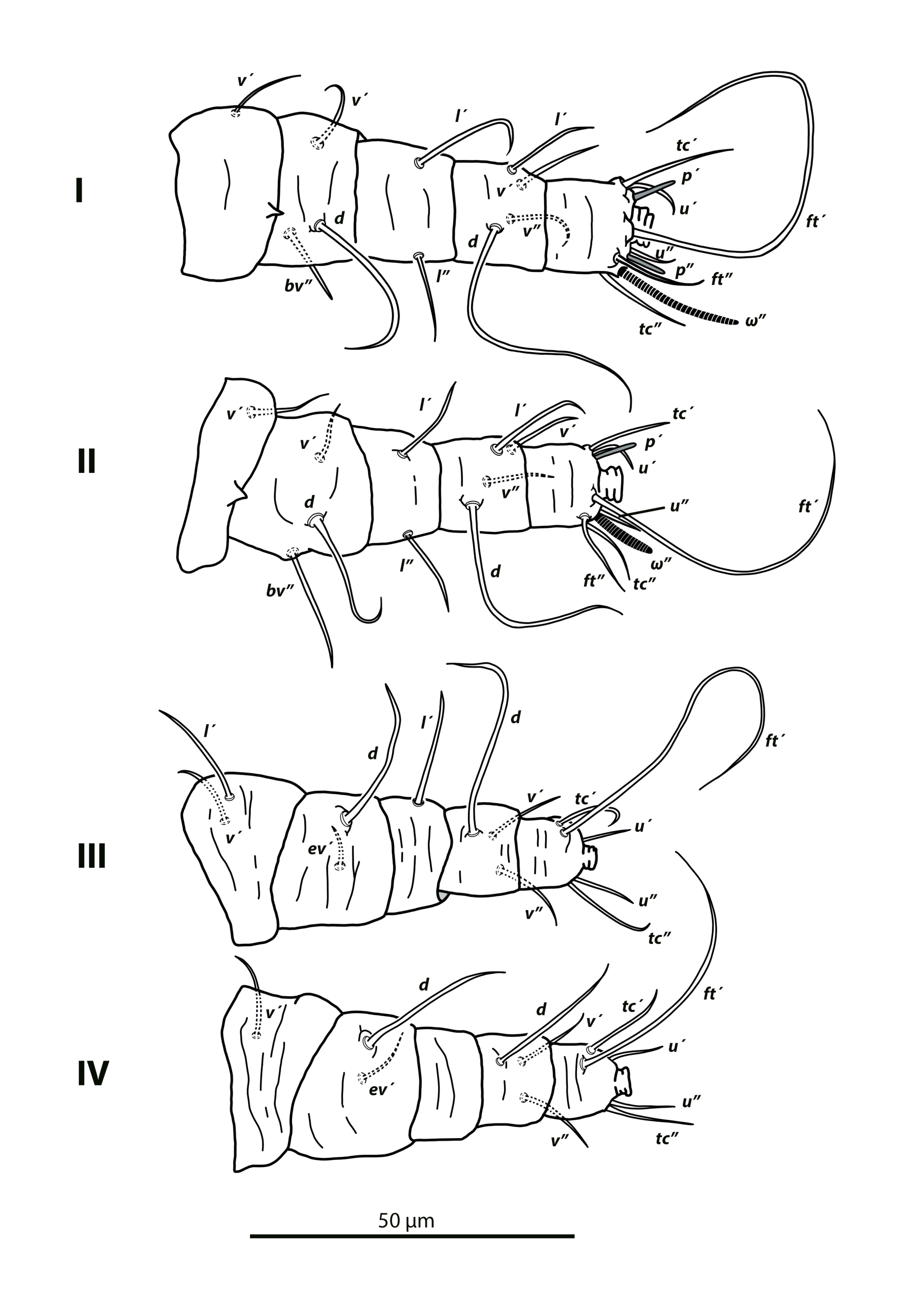
Legs (Figure 2). Length of leg I 74 (70–76), leg II 55 (51–54), leg III 57 (55–59), leg IV 59 (58–63). Setae smooth and pointed or blunt, only exceptionally a minute barb can be discerned. Setal formulae for legs I–IV (coxae to tarsi): 2-1-3-2-4-8(1), 2-1-3-2-4-7(1), 1-2-2-1-3-5, 1-1-2-0-3-5, respectively. Leg chaetotaxy as follows: trochanters I–II, IV v′; trochanter III l′, v′; femora I–II d, v′, bv″; femora III–IV d, ev′; genua I–II l′, l″; genu III l′; genu IV nude; tibiae I–II d, l′, v′, v″; tibiae III–IV d, v′, v″; ta I–II u′, u″, pζ′, pζ″, tc′, tc″, ft′, ft″, ω″; ta III–IV u′, u″, tc′, tc″, ft′. Dorsal setae on femora I–III and genua I–II setiform and smooth. Tarsi I and II each with one solenidion ωʺ, narrow, curved on tarsus I, and twice as long 16 (14–17) as on tarsus II 8 (8–9), which is broader.
Male (2 examined and measured. Figure 3).
Dorsum (Figure 3A). Prodorsum and opisthonotum entirely striate. Prodorsal ornamentation as in female, striae microtuberculate giving a granulate appearance. Opisthonotal surface with longitudinal striae on the transverse band between setae c1–c3 and d1–d3 and; transversal striae between d1–d3 and e1–e3; a median inverted V-shaped pattern on the cuticle posterior to e1. Absence of microtubercles on most of the opisthonotum. Body measurements: v2–h1 152–164, sc2–sc2 113–116. Distances v2–v2 28–32; sc1–sc1 71–72, c1–c1 39–57, c2–c2 113–121, c3–c3 130–141, d1–d1 40–46, d3–d3 94–99, e1–e1 50–59, e3–e3 74–82, f3–f3 60–63, h1–h1 30, h2–h2 45–49. Dorsal setae as in female, the longer (h2) with a few minute barbs difficult to see. Setal measurements: v2 14–16, sc1 12–17, sc2 22, c1 9–11, c2 10, c3 12–14, d1 10, d3 16–21, e1 11–13, e3 26–27, f3 34–39, h1 19–27, h2 80–101.
Venter (Figure 3B). Ventral cuticle striate and microtuberculate (granulate). Striae between coxae I–II oriented as in female; striae forming a bird nest-shaped pattern between 3a–4a; transversal striae posterior to 4a, including the anogenital lobes. Setae g1 and g2 smooth and blunt, placed on the lobes, g2 posteroantiaxial to g1. Anal setae ps2 on the posterior edge of the lobes and a bit longer than ps3. Setal measurements: 1a 49–57, 1b 10–11, 1c 9–11, 2b 9–10, 2c 12–14, 3a 45–49, 3b 10–12, 4a 20–43, 4b 14, ag 12–16, g1 19, g2 14–15, ps2 15–18, ps3 11–16.
Aedeagus (Figure 3C). Penis sheath sclerotised, elongate, (64–67 long from the apodemal plate to the distal end), containing the thin ejaculatory duct.
Gnathosoma. Ornamentation of dorsal and ventral cuticle similar to that found in the female. Palp dorsal seta smooth and blunt, 7–9 long; solenidion 5–7, companion seta 6. One visible pair of oral setae.
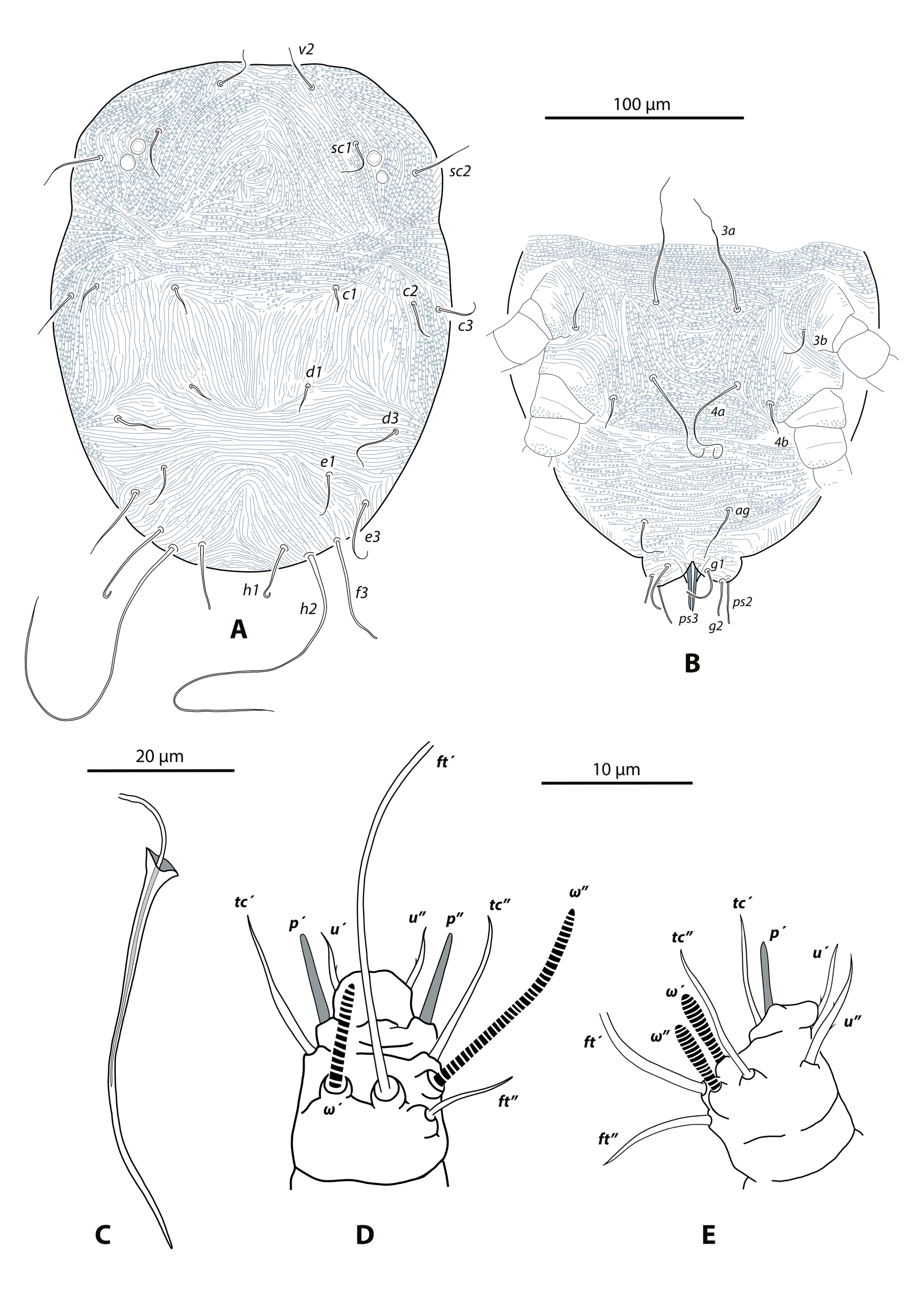
Legs (Figures 3D–E). Same leg chaetotaxy as in female, except for the tarsi I–II. Tarsus I with 8 (+2) setae: ft′, ft″, tc′, tc″, pζ′, pζʺ, u′, u″, ω′, ω″. Tarsus II with 7 (+2) setae, eupathidium pζʺ absent. On tarsus I solenidion ω′ 8–9, ω″ 14–16; on tarsus II both solenidia subequal, 8–9 long.
Immature stages. Unknown.
Etymology
The specific name canariensis refers to the Canary Islands, home of the specimens collected. This name also aims to highlight the geographical origin of the species, belonging to a genus with an evident disjunct distribution.
Remarks
Capedulia is a small genus consisting of five species of small, soft-bodied mites characterized by: i) the extreme reduction in palp segmentation, ii) the simple, setiform, and finely barbed dorsal setae, and iii) by having a single antiaxial, longer tenent hair, and a pair of paraxial, shorter tenent hairs on the pad like claws (Smith Meyer, 1979). Members in the genus are rather homogeneous in their general habitus, and they can be differentiated using the leg chaetotaxy (Table 1), the length of dorsal setae and the ornamentation of dorsal cuticle. All the species have a striate integument on the dorsal and ventral surfaces, and in all species except C. prasadi Ueckermann & Tiedt, the space between the striae is mostly occupied by small and rounded tubercles (microtubercles, sensu Ueckermann et al. 2018), referred to as ''lobes» in early publications. According to the illustrations for the previously described species and the examination of the new species, the dorsal cuticle in the gnathosoma is laterally folded, forming one central and two lateral bands wrinkled separated by smooth integument (see Figure 1D and Smith Meyer, 1979; Ueckermann and Tiedt, 1999). Moreover, the pair of subcapitular setae m is apparently absent in the known species.
Download as
Leg segments
C. calendulae 1,2,5
C. canariensis sp. nov.
C. maritima 3,4,5
C. prasadi 2,5
C. xeroclei 2
Coxae
2-2-1-1
2-2-1-1
2-1-1-1
2-2-1-1
2-2-1-1
Trochanters
–
1-1-2-1
1-1-2-0 *, **
1-1-2-1
1-1-2-1
Femora
3-3-2-1
3-3-2-2
3-3-2-2
4-4-2-1
4-4-2-1
Genua
2-2-1-0
2-2-1-0
1-1-0-0
2-2-0-0
3-(2–3)-1-0
Tibiae
4-4-3-3
4-4-3-3
4-4-3-3
4-4-3-3
4-4-3-3
Tarsi
8(+1)-8(+1)-5-5 *
8(+1)-7(+1)-5-5
7(+1)-7(+1)-3-3 *
8(+1)-7(+1)-5-5 *, **
8(+1)-8(+1)-5-5
Until now, details of the leg chaetotaxy in Capedulia species has not been recorded, with only simple setal counts being reported in the literature. This is the first study to identify all leg setae for a Capedulia species. We also emphasize the importance of including these data in species descriptions, as the known species can be distinguished primarily based on differences in leg setal patterns. Examination of the buccal cone of the new species revealed only a single pair of oral setae. The detection of additional, minute oral setae, if present, lies beyond the resolving power of light microscopy and should be verified using scanning electron microscopy.
Capedulia canariensis sp. nov. can be separated from C. calendulae Smith Meyer by having an additional seta (d) on femur IV and seta pζ″ on tarsus II absent. It differs from C. prasadi for lacking seta l′ on femora I–II, for having seta d on femur IV, and seta l′ on genu III. Additionally, C. prasadi does not have microtuberculate dorsal and ventral surfaces (Ueckermann and Tiedt, 1999). The new species differs from C. xeroclei Ueckermann & Tiedt by the absence of seta l′ on femora I–II, seta d on genu I, and pζ″ on tarsus II (all present in C. xeroclei); and by the presence of d on femur IV (absent in C. xeroclei). Capedulia maritima Gerson & Smith Meyer is the most disparate species in the genus, with a reduced leg setation and long dorsal setae (mainly sc2, c3, d3, e3). Capedulia maritima differs from the Canarian species by having genua I–II with just one seta present (l′). Gerson and Smith-Meyer (1980) also indicate that the species has greatly reduced setation on tarsi I and II - tarsus I with 5(+1) and tarsus II with 4(+1); however, the actual setal count on tarsi I and II is 7(+1) (pers. com. J.J. Beard). Capedulia canariensis sp. nov. and C. maritima both share an elongate dorsal opisthosomal setae h2, exceeding 100 μm; however, setae h2 is significantly longer than h1 in the former, but similar in length in the latter.
Interestingly, the genus shows a disjunct distribution, with three South African members (C. calendulae, C. prasadi, C. xeroclei), one species from the Middle East (C. maritima) and one from the Canary Islands. This pattern is consistent with the biogeographic concept of Rand Flora which attempts to explain the disjunct distribution in many plant lineages across Macaronesia–Northern Africa, Southwest Asia, and East–South Africa (Sanmartín et al. 2010; Pokorny et al. 2015). One of the hypotheses on the evolutionary origin of this flora indicates that the extant species are the result of long-distance dispersal events followed by in situ diversification, and the routes taken by the plants would follow a northern direction, from Southern Africa to Northern Africa via the east of the continent. Once in northern Africa, plants and the mites associated with them may have been dispersed across the area now occupied by the Sahara Desert, an area that has experienced repeated episodes of dry and wet climate in relatively recent times. All of the C. canariensis sp. nov. individuals collected were found on two species of Amaranthaceae (formerly Chenopodiaceae), with most collected on Traganum moquinii, a succulent, multibranched shrub that lives on the sand and dunes of the coastal regions in Southwestern Morocco, western Sahara, Cape Verde, and all the Canary Islands with the exception of the westernmost islands in the archipelago, La Palma and El Hierro (Sauerbier et al. 2023).
The adults of the new species are red in life, and live on the leaves and twigs of the aerial part of the plant, without any indication of association with the roots, as can be seen with C. calendulae and C. maritima (Smith Meyer, 1979; Gerson and Smith Meyer, 1980). The new species was found in association with two species of phytoseiid mites, Neoseiulella extraseta Ferragut and Typhlodromus (Anthoseius) rhenanoides Athias-Henriot, and an unidentified eupalopsellid mite. All these predators had red gut contents, suggesting that they use the new flat mite as prey.
Genus Phytoptipalpus Trägårdh, 1904
Type species — Phytoptipalpus paradoxus Trägårdh, 1904, by original designation.
Phytoptipalpus majorensis Ferragut sp. nov.
ZOOBANK: 8BCA0F97-9437-4D52-B2EF-70DCCD356337
(Figures 4–5)
Type material and depository
Female holotype and 15 female paratypes on Ononis hesperia (Fabaceae); Fuerteventura, La Pared, Pueblo del Mar, 28°12′28″N, 14°13′27″W, 69 m a.s.l.; 30 January, 2024. Female holotype and seven female paratypes were deposited in the Museo de Ciencias Naturales de Tenerife, Santa Cruz de Tenerife, Canary Islands. The remaining specimens are in the Acari collection, Instituto Agroforestal Mediterráneo, Universidad Politécnica de Valencia.
Diagnosis
Female. As per the genus definition with the following additional attributes. Thirteen pairs of opisthonotal setae, with f2 present. Four pairs of legs. Anterior margin of prodorsum bilobed (with a broad shallow notch). Surface of prodorsum and opisthonotum with fine irregular fissures and grooves. Dorsal setae obovate to lanceolate and serrated; prodorsal setae lanceolate or narrowly lanceolate, opisthonotal obovate to broadly lanceolate. Infracapitulum extending to distal part of genu I. Palp setation (trochanter to tarsus) 0-1-0-2-3. Setal formula on leg segments: trochanters 1-1-2-1; femora 4-4-2-1; genua 3-3-0-0. Dorsal setae on femora I–III, genua I–II, and tibiae III–IV broadly lanceolate-serrate; dorsal setae on tibiae I–II simple and barbed. Sperm access system with a very long and convoluted insemination duct and a tubular sclerotized structure terminating in a small and spherical vesicle.
Description
Female (16 examined, 10 measured. Figures 4–5).
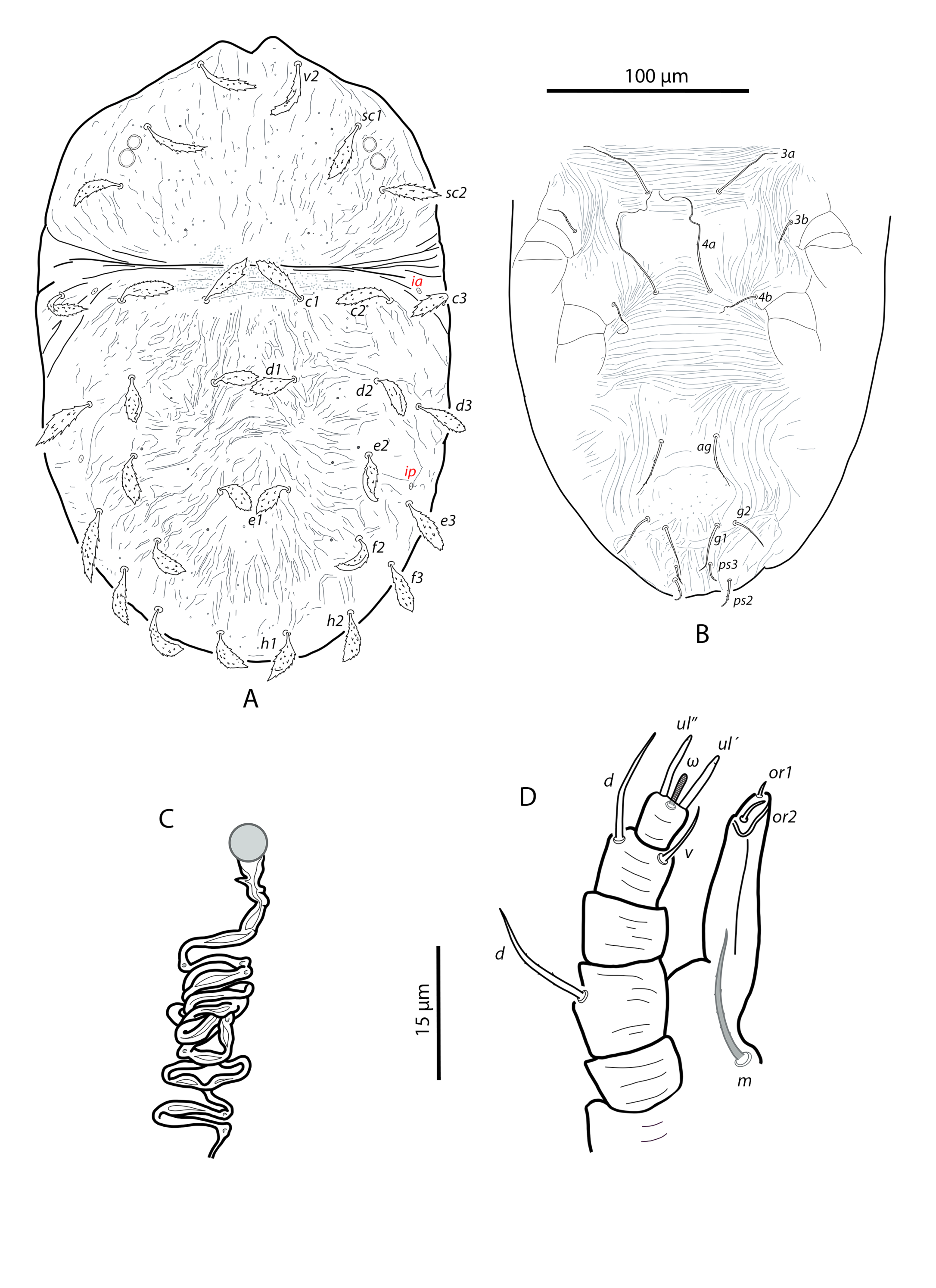
Dorsum (Figure 4A). Dorsum well sclerotized; anterior margin of prodorsum with broad median shallow notch 8–10 μm deep, lateral lobes of notch dome-shaped and often asymmetrical. Surface of prodorsum with fine irregular fissures and grooves, becoming longitudinally oriented medially, and obliquely oriented laterally. Numerous minute pore-like perforations visible in the dorsal cuticle, prodorsum with three pairs and opisthonotum with at least five pairs distinctly larger and longitudinally aligned. Opisthonotum with deep and irregular furrows and fissures, more strongly developed than on the prodorsum. Furrows aligned longitudinally between setae c1–d1; irregular to transverse between d1–e1. Opisthonotal shield sparsely finely punctate across the surface. Body measurements: v2–h1 284 (266–299), sc2–sc2 134 (123–134). Distances v2–v2 51 (45–52), sc1–sc1 112 (98–112), c1–c1 56 (45–56), c2–c2 139 (126–139), c3–c3 191 (170–191), d1–d1 37 (29–37), d2–d2 128 (114–133), d3–d3 172 (153–172), e1–e1 31 (29–32), e2–e2 118 (110–124), e3–e3 155 (141–155), f2–f2 96 (93–97), f3–f3 129 (113–129), h1–h1 33 (29–35), h2–h2 88 (74–90). Setae on prodorsum of similar length, lanceolate to narrowly lanceolate, serrate, usually ending in a fine tip; barbs more densely distributed near the base. Most dorsal setae on the opisthosoma similar in shape to those on the prodorsum but broader; setae d1, e1, h1 obovate. Setal measurements: v2 34 (28–34), sc1 29 (27–32), sc2 28 (27–32), c1 26 (25–30), c2 28 (26–32), c3 26 (25–28), d1 21 (21–24), d2 21 (20–24), d3 30 (29–32), e1 19 (19–21), e2 23 (19–24), e3 33 (29–34), f2 22 (20–24), f3 29 (27–30), h1 23 (21–26), h2 31 (26–31). Cupules difficult to discern, only ia and ip clearly distinguishable; ia in membranous region between the insertions of c2–c3; ip just anterior to e3.
Venter (Figure 4B). Intercoxal area between setae 1a and 3a with fine transverse striae, and almost smooth between 3a and 4a. Cuticle posterior to 4a with broadly spaced transverse striae, becoming longitudinal and sinuous laterad the genital region. Genital shield almost smooth, with a few punctures posteriorly. Anal plates with a few striae and minute punctures. Ventral seta 1a smooth, the remaining ventral and coxal setae finely or moderately barbed. Aggenital (ag), genitals (g1, g2) and pseudanal (ps2, ps3) setae barbed and slightly tapered. Setal measurements: 1a 100 (76–103), 1b 30 (30–39), 1c 19 (19–24), 2b 41 (36–61), 2c 36 (34–40), 3a 93 (80–96), 3b 30 (26–34), 4a 84 (75–85), 4b 23 (19–28), ag 27 (23–28), g1 24 (22–25), g2 20 (19–22), ps2 15 (12–15), ps3 11 (8–12).
Spermathecal apparatus (Figure 4C). Very long and convoluted insemination duct of varying diameter, alternating from narrow to expanded sections along its length. The duct terminates in a more sclerotized and tubular section with irregular walls (with a minute lateral digit observed in some females), which leads to a small, spherical, membranous seminal receptacle, about 4 μm in diameter.
Gnathosoma (Figure 4D). Anterior tip of infracapitulum extending to the distal part of genu I. Ventral surface of infracapitulum finely granulate, dorsal surface plicate. Supracoxal setae e about 3 long; subcapitular seta m minutely barbed, 18 (16–19). Two pairs of oral setae; or1 minute, about one-third length of or2. Palps 5-segmented; setal formula 0-1-0-2-3 (1 solenidion+2 eupathidia). Tibial seta d 14 (13–14), seta v 10 (8-10); tarsal solenidion ω 5 (5–6); tarsal eupathidia ul′–ul″ 9 (8–9), 8 (7–8), respectively.
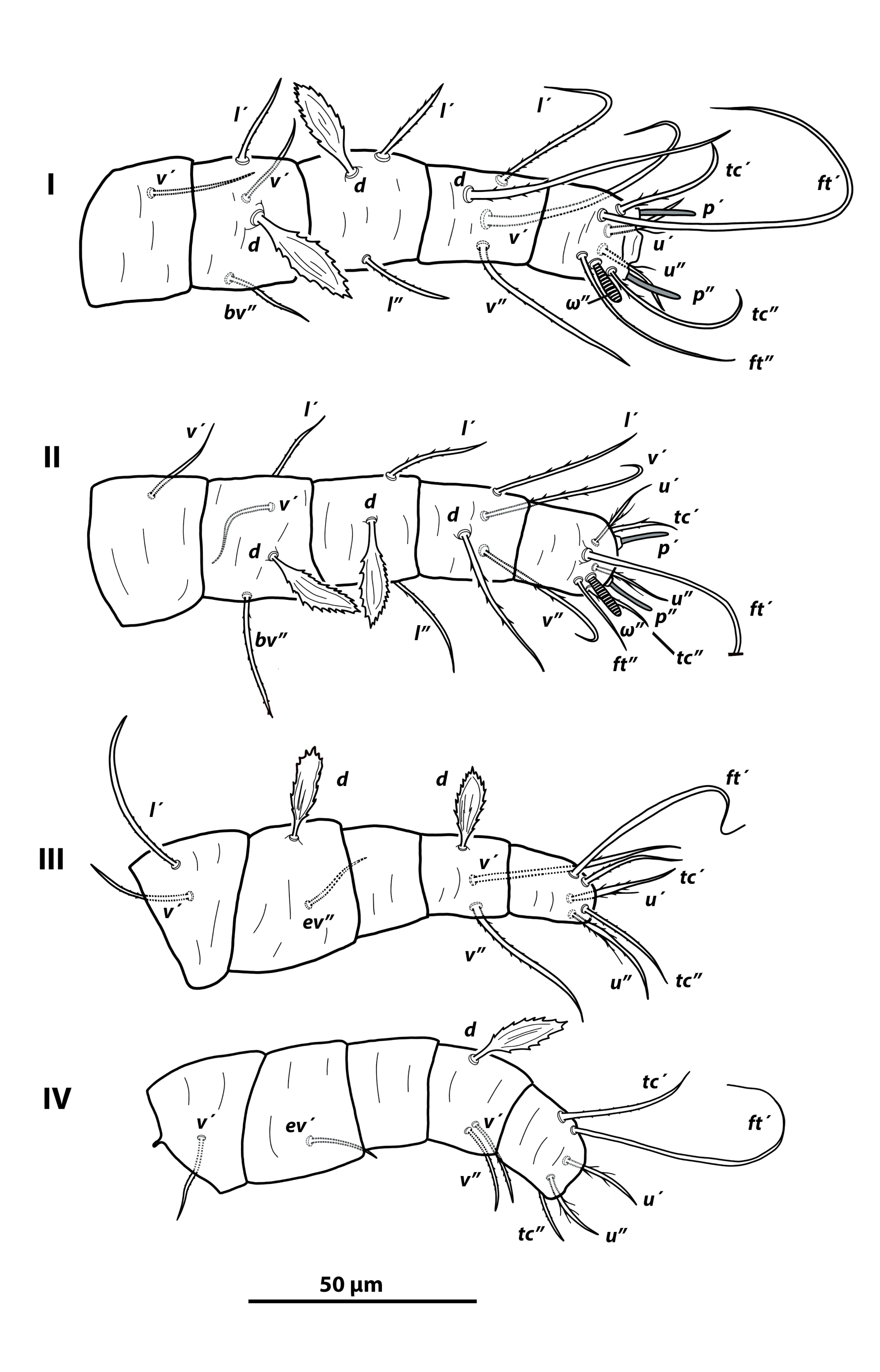
Legs (Figure 5). Length of leg I 103 (88–107), leg II 89 (77–93), leg III 81 (75–86), leg IV 83 (7589). Setal formula for legs I–IV (coxae to tarsi): legs I–II 2-1-4-3-4-9(1 ω), leg III 1-2-2-0-3-5, leg IV 1-1-1-0-3-5, respectively. Tarsi I and II each with one cigar-shaped solenidion ω'' (tarsus I 9 (8–9), tarsus II 8 (7–9)). Leg chaetotaxy as follows: trochanters I–II, IV v′; trochanter III l′, v′; femora I–II d, l′, v′, bv″; fe III d, ev′; fe IV ev′; genua I–II d, l′, l″; genua III–IV nude; tibiae I–II d, l′, v′, v″; tibiae III–IV d, v′, v″; ta I–II u′, u″, pζ′, pζ″, tc′, tc″, ft′, ft″, ω″; ta III–IV u′, u″, tc′, tc″, ft′. Dorsal setae on femora I–III, genua I–II, and tibiae III–IV broadly lanceolate and serrate around margin.
Other stages. Unknown.
Etymology
The specific name majorensis derives from the local demonym of ''majoreros'', as the people of Fuerteventura Island are known.
Remarks
Seven species of Phytoptipalpus are characterized by the presence of dorsal seta f2, four pairs of legs, a dorsum without reticulation, and lanceolate to spatulate dorsal setae: P. ceibae (De Leon), P. euphratica (Al-Gboory), P. harveyi Smith Meyer & Van Dis, P. lepsis Hassan, Ashfaq & Li, P. lithos Hassan, Ashfaq & Li, P. multistriatum (Meyer), and P. phoenicis Alatawi & Kamran (De Leon, 1962; Smith Meyer, 1979; Al-Gboory, 1987; Baker and Tuttle, 1987; Smith Meyer and Van Dis, 1993; Hassan et al. 2003; Alatawi et al. 2015). With the exception of P. ceibae, which is native to Mexico and the Caribbean, the remaining taxa have a disjunct distribution from Southern Africa to countries in the Middle East, occurring on a range of unrelated plant families. These species differ mainly in the setation of the leg segments, particularly the genua, which exhibit five distinct setal patterns (Table 2). The new species Phytoptipalpus majorensis sp. nov. has a unique setal formula on genua I–IV, 3-3-0-0, due to the presence of seta l″. These differences in leg setal patterns among species, together with other morphological traits, are summarized in Table 2.
Download as
Trochanters
Femora
Genua
Tibiae
Setae on palptarsus
Anterior prodorsum
P. ceibae (De Leon) 1,2
0-0-2-0
3-3-2-1
2-2-0-0
4-4-3-3
3
without notch
P. euphratica (Al-Gboory) 3
1-1-1-1
3-3-2-1
1-1-1-0
3-3-3-3
3
without notch
P. harveyi Smith Meyer & Van Dis 4
1-1-2-1
4-4-2-1
2-2-0-0
4-4-3-3
3
bilobed
P. lepsis Hassan, Ashfaq & Li 5
1-1-2-1
4-4-2-1
2-2-1-0
4-4-3-3
3
bilobed
P. lithos Hassan, Ashfaq & Li 5
1-1-2-1
4-4-2-1
2-2-0-0
4-4-3-3
2
bilobed
P. majorensis sp nov.
1-1-2-1
4-4-2-1
3-3-0-0
4-4-3-3
3
bilobed
P. multistriatum (Smith Meyer) 6,7
1-1-2-1
4-4-1-1
1-1-0-0
4-4-3-3
3
without notch
P. phoenicis Alatawi & Kamran 8
1-1-1-1
4-4-2-1
2-2-1-1
4-4-3-3
3
bilobed
The spermathecal apparatus has been described in few species of Phytoptipalpus, precluding the definition of a general form, but the structure shows some variation. In P. alexandriae Smith Meyer & Van Dis and P. occultuae Ueckermann, Ochoa & Bauchan, the insemination duct is short and terminates in an oval, vacuolated receptacle (Smith Meyer & Van Dis, 1993; Ueckermann et al. 2019). In contrast, in P. salicicola (Al-Gboory) and P. calligonus Rad & Asadi, the insemination duct is very long, variously folded, and composed of alternating narrow and expanded segments along its length (Çobanoḡlu et al. 2016; Rad and Asadi, 2021). This latter morphology agrees with that observed in the species described herein and also with that reported for some Aegyptobia species.
Specimens collected were red in life. As only adult females were found, it may indicate that this is how the species overwinters. The geographic range of the host plant, Ononis hesperia, includes the central and eastern Canary Islands, Southwestern Morocco, western Sahara, and Mauritania (Sauerbier et al. 2023). A notable proportion of Phytoptipalpus species (eight from twenty–five species, or 32%) have been collected from the plant family Fabaceae (see Fabaceae, Caesalpiniaceae, Leguminosae and Mimosaceae in Castro et al. 2026). This is the first record of Phytoptipalpus on the plant genus Ononis.
Acknowledgements
The first author was provided with a permit to collect plants by the Cabildo de Fuerteventura (Resolución 1087/2024). The authors wish to thank Jennifer Beard for pointing out some inaccuracies in the leg setal counts in the original descriptions of several Capedulia species (unpublished data based on the examination of type specimens). We would also like to thank Ronald Ochoa and Elizeu de Castro for sharing some publications.
References
- Alatawi F.J., Kamran M., Negm M.W. 2015. False spider mites (Acari: Tenuipalpidae) associated with date palm orchards in Saudi Arabia: description of two new species and new records. Syst. Appl. Acarol., 20: 809-830. https://doi.org/10.11158/saa.20.7.9
- Al-Gboory I. 1987. Taxonomic studies of false spider mites (Acari: Tenuipalpidae) in central Iraq. PhD dissertation, Institut für angewandte Zoologie der Rheinischen Friedrich-Wilhelms-Universität Bonn, Bonn, Germany, 205 pp.
- Baker E.W., Tuttle D.M. 1987. The false spider mites of Mexico (Tenuipalpidae: Acari). USDA, Agricultural Research Service, Tech. Bull., 1706: 1-236.
- Berlese A. 1913. Acarotheca Italica. Tipografia di M. Ricci, Firenze, Italy, 221 pp.
- Carracedo J.C., Troll V.R. 2021. North-East Atlantic Islands: The Macaronesian Archipelagos. In: D. Alderton, A. E. Scott (Eds), Encyclopedia of Geology (2nd edition), Academic Press, Cambridge, Massachusetts, USA, pp. 674-699. https://doi.org/10.1016/B978-0-08-102908-4.00027-8
- Castro E.B., Mesa N.C., Feres R.J.F., Moraes G.J.de, Ochoa R., Beard J.J., Demite P.R. 2026. Tenuipalpidae Database.
- Available from: http://www.tenuipalpidae.ibilce.unesp.br (accessed on 21/04/2026).
- Çobanoḡlu S., Ueckermann E.A., Saḡlam H.D. 2016. The Tenuipalpidae of Turkey, with a key to species (Acari: Trombidiformes). Zootaxa, 4097: 151-186. https://doi.org/10.11646/zootaxa.4097.2.1
- De Leon D. 1962. Two new false spider mites from Mexico and a new distribution record. Proceedings of the Entomological Society of Washington, 64 (3): 203-205.
- Gerson U., Smith Meyer M.K.P. 1980. A new species of Capedulia (Acari: Tenuipalpidae) from a marine habitat in Israel. Isr. J. Entomol., 14: 9-12.
- Grandjean F. 1939. Les segments post-larvaires de l'hysterosoma chez les Oribates (Acariens). Bull. Soc. Sci. Nat. France, 64: 273-284.
- Hassan M., Ashfaq M., Li Y. 2003. Two new species of the genus Phytoptipalpus (Acari: Tenuipalpidae) recorded in Pakistan. J. Southwest Agric. Univ. Chongqing, 25 (6): 525-529.
- Lindquist E.E. 1985. External anatomy. In: Helle, W & Sabelis, M.W. (Eds.), Spider Mites: Their Biology, Natural Enemies and Control. Vol. 1a. Elsevier, Amsterdam, pp. 3-28.
- Pokorny L., Riina R., Mairal M., Meseguer A., Culshaw V., Cendoya J., Serrano M., Carbajal R., Ortiz S., Heuertz M., Sanmartín I. 2015. Living on the edge: timing of Rand Flora disjunctions congruent with ongoing aridification in Africa. Front. Genet., 6-2015. https://doi.org/10.3389/fgene.2015.00154
- Rad D.B., Asadi M. 2021. A new species of the genus Phytoptipalpus (Acari: Tenuipalpidae) from Iran. Int. J. Acarol, 47 (1): 1-7. https://doi.org/10.1080/01647954.2020.1866665
- Sanmartin I., Anderson L., Alarcón M., Ronquist, F., Aldasoro J.J. 2010. Bayesian island biogeography in a continental setting: the Rand Flora case. Biol. Letters, 6: 703-707. https://doi.org/10.1098/rsbl.2010.0095
- Sauerbier H., Cabrera F., Muer T. 2023. Flora Vascular de Canarias. Publ. Turquesa, La Laguna, Tenerife, 1514 pp.
- Smith Meyer M.K.P. 1979. The Tenuipalpidae (Acari) of Africa with keys to the world fauna. Entomology Memoir of the Department of Agricultural Technical Services, Pretoria, Republic South Africa, 50: 1-135.
- Smith Meyer M.K.P., Van Dis J.C.S. 1993. Notes on some species of the related genera Aegyptobia Sayed, Phytoptipalpus Trägärdh and Pentamerismus McGregor, with descriptions of eight new species (Acari: Tenuipalpidae). Genus, 4: 309-357.
- Trägärdh I. 1904. Acariden aus Ägypten und dem Sudan. Zugleich ein Beitrag zur Kenntnis der Entwicklungsgeschichte der Gattungen Phytoptipalpus, Pimeliaphilus, Pterygosoma und Podapolipus. In: Jägerskiöld, L.A. (Ed.), Results of the Swedish Zoological Expedition to Egypt and the White Nile 1901, 2. Uppsala, Library of the Royal University of Uppsala. 138 pp.
- Ueckermann E.A., Tiedt L.R. 1999. Two new species of the genus Capedulia Meyer (Acari: Tenuipalpidae). Int. J. Acarol, 25 (3): 201-209. https://doi.org/10.1080/01647959908684154
- Ueckermann E.A., Palevsky E., Gerson U., Recht E., Theron P.D. 2018. The Tenuipalpidae (Acari: Trombidiformes) of Israel. Acarologia, 58 (2): 483-525. https://doi.org/10.24349/acarologia/20184255
- Ueckermann E.A., Ochoa R., Bauchan G.R., Neser S. 2019. An amazing sub-cambium flat mite from South Africa (Acari: Trombidiformes: Tenuipalpidae). Acarologia, 59 (4): 507-530. https://doi.org/10.24349/acarologia/20194351
- Whittaker R.J., Fernández-Palacios J.M., Matthews T.J. 2023. Island Biogeography. Oxford University Press-Academic, UK, 496 pp. https://doi.org/10.1093/oso/9780198868569.001.0001
to post any comment or correction.


2026-04-29
Date accepted:
2026-09-14
Date published:
2026-09-21
Edited by:
Castro, Elizeu B.

This work is licensed under a Creative Commons Attribution 4.0 International License
2026 Ferragut, Francisco and Baumann, Julia
 Download article Download low definition
Download the citation
RIS with abstract
(Zotero, Endnote, Reference Manager, ProCite, RefWorks, Mendeley)
RIS without abstract
BIB
(Zotero, BibTeX)
TXT
(PubMed, Txt)