


First species of Novophytoptus Roivainen (Eriophyoidea, Phytoptidae) from flower buds of archaic angiosperm Drimys winteri J. R. Forst & G. Forst from Chile: Ancestral association or host shift from monocots?
Chetverikov, Philipp E.  1
; Kudryavtzev, Andrey T.2
; Peralta-Alba, Lourdes3
and Rojas-Espinoza, Francisco
4
1
; Kudryavtzev, Andrey T.2
; Peralta-Alba, Lourdes3
and Rojas-Espinoza, Francisco
4
1✉ X-BIO Institute, University of Tyumen, 6 Volodarskovo Street, Tyumen, 625003, Russia & Zoological Institute of Russian Academy of Sciences, Universitetskaya Nab. 1, Saint-Petersburg, 199034, Russia.
2Zoological Institute of Russian Academy of Sciences, Universitetskaya Nab. 1, Saint-Petersburg, 199034, Russia.
3Laboratory of Entomology and Acarology, Curico Office, Agricultural and Livestock Service (SAG), Ministry of Agriculture, Carmen N° 560, 3rd Floor, 3340001, Curico, Maule Region, Chile.
4Laboratory of Entomology and Acarology, Valparaiso Office, Agricultural and Livestock Service (SAG), Ministry of Agriculture, Camino La Polvora Km 12.7, ZEAL Complex, 2340000, Valparaiso, Valparaiso Region, Chile.
2026 - Volume: 66 Issue: 3 pages: 805-817
https://doi.org/10.24349/pdbm-9lsrZooBank LSID: 81AB16C5-C8BA-41D1-88CA-FAC47EE1712E

Original research
Keywords
Abstract
Introduction
The magnoliid plant family Winteraceae (Canellales) represents a classic relict element of the Gondwanan Antarctic flora, currently restricted to disjunct humid temperate and tropical regions of the Southern and Northern Hemispheres (Thomas et al. 2014). In the temperate rainforests of southern Chile, Drimys winteri J.R. Forst. & G. Forst. stands as a foundational evergreen tree species (Ladio et al. 2026), representing an archaic mesangiosperm lineage, magnoliids (Fig. 1), that evolved independently from the major radiations of monocots and eudicots (Zuntini et al. (2024)). Investigating the specialized acarofauna associated with such evolutionarily ancient hosts contributes to a better understanding of the historical biogeography of ecosystems, helping to reconstruct ancient colonization patterns and delineate the boundaries of host-specificity in phytophagous acariform lineages (Lindquist and Oldfield 1996).
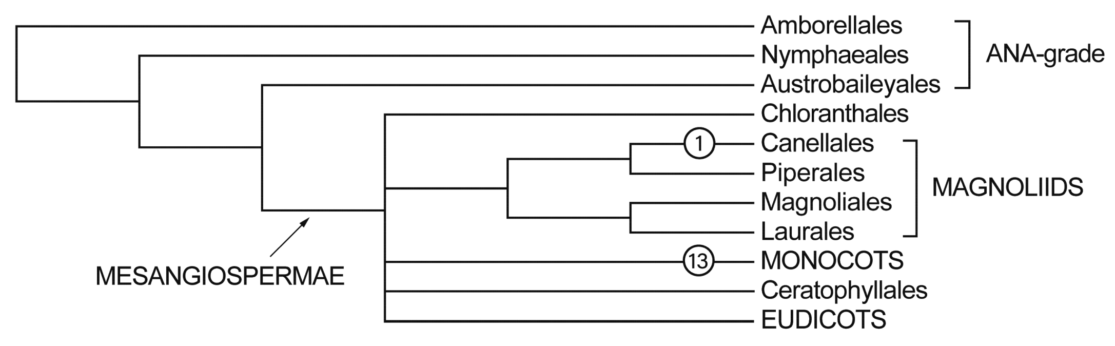
Morphologically simplified and greatly miniaturized, obligate phytophagous mites of the superfamily Eriophyoidea are renowned for their extreme host specificity, with the vast majority of species exhibiting strict mono- or oligophagy (Skoracka et al. 2010). While host-plant associations usually appear conservative at the species level, a completely different pattern emerges at higher taxonomic ranks, where broad host ranges and significant host shifts become apparent. This is clearly reflected in many species-rich morphogenera, as seen in Epitrimerus Nalepa 1898 and Eriophyes von Siebold 1851 recorded on both gymnosperms and angiosperms, or Phytoptus Dujardin 1851 and Aceria Keifer 1944 associated both with monocots and dicots. Crucially, among such supraspecific groups, a few well-supported monophyletic lineages (e.g., Calacarus Keifer 1940, Diptilomiopus Nalepa 1916, Oziella Amrine et al. 2003, and Retracrus Keifer 1965) confirm that such host shifts do occur, as their members utilize hosts across different plant families and orders.
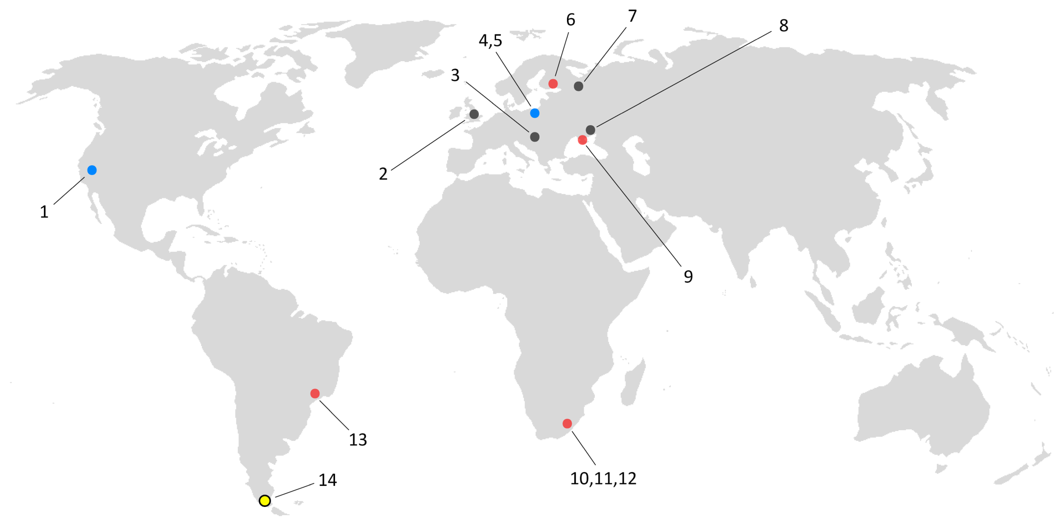
The genus Novophytoptus Roivainen, 1947 is a morphologically well-defined and advanced taxon within the archaic family Phytoptidae. It is easily distinguishable by greatly elongated body, the absence of several leg and opisthosomal setae (including bv I and II, solenidion φ I, and c1), exceptionally long prodorsal shield setae sc, and external genitalia situated in the area delimited by tubercles of the opisthosomal setae c2 and d (Amrine et al. 2003). All previously described members of this genus have been considered strict monocot associates (Fig. 2), exclusively colonizing herbaceous plants of the families Poaceae, Cyperaceae, and Juncaceae (Chetverikov et al. 2023). On these hosts, Novophytoptus species demonstrate a highly uniform ecology: they inhabit the subepidermal air cavities of leaves and stems, penetrating them through presumably self-made circular holes in the epidermis (Flechtmann, 2004; Chetverikov and Petanović 2016). Their feeding activity results in relatively mild, localized parenchymal necrosis, which typically manifests externally as ''brown stripes» running between adjacent longitudinal veins, causing no major alterations to the host's organs.
In this paper, we aim to describe a new Novophytoptus species from southern Chile and discuss its unique destructive association with Drimys winteri, a Gondwanan mesangiosperm host outside of the monocots. We provide a detailed morphological description of the new species, investigate its exoskeleton using confocal laser scanning microscopy (CLSM), document the damage caused by the mites, provide the sequence of two gene markers (Cox1 and 28S rRNA) for this taxon, and discuss future perspectives for investigating the phylogeny of Novophytoptus.
Material and methods
Collection and morphological measurements
As part of the activities of Surveillance of pests in native species, carried out by the Agricultural and Livestock Service of Chile (SAG), branches of Drimys winteri were collected in southern Chile (Timaukel, Magallanes Region) and transferred to the laboratories of Servicio Agricola y Ganadero (SAG) of the Ministry of Agriculture of Chile in Valparaíso and Curicó for processing. Eriophyoid mites were collected from flower buds using a fine minuten pin and a stereomicroscope. They were mounted in Hoyer's medium and cleared on a heating block at 94 °C for 5 hours (Upton 1993, Amrine & Manson 1996). Some material was stored in vials with 96% ethanol in a freezer for subsequent DNA extraction, PCR and sequencing of two marker genes (D1D2 28S rDNA and mitochondrial Cox1) using previously described methodology (Klimov et al. 2018). The slide-mounted specimens were examined with differential interference contrast light microscopy (DIC LM) and phase-contrast light microscopy (PC LM). In the species descriptions, measurements of the holotype (female) are presented along with ranges based on paratypes; for males, only ranges are given. All measurements are given in micrometers (μm) as lengths unless stated otherwise. Classification and terminology of eriophyoid genital anatomy and external morphology follow that of Amrine et al. (2003), Chetverikov (2014), and Lindquist (1996) respectively. Host plant morphology follows Gottsberger et al. (1980), Svoma (1998) and Doust (2001). Slide-mounted specimens of Novophytoptus spp. from the Acarological collection of Zoological Institute of Russian Academy of Sciences (ZIN RAS) as well as unpublished microphotographs of previously described novophytoptines from North America, Europe and South Africa were used as comparative materials.
Confocal microscopy
Exoskeletons of Novophytoptus specimens mounted in Hoyer's medium were investigated using confocal laser scanning microscopy (CLSM) with a spectral confocal and multiphoton system Leica TCS SP2 (Leica Microsystems GmbH, Wetzlar, Germany) with the same settings of the confocal microscope as described by Chetverikov (2014). Image stacks were rendered three-dimensionally using the reconstruction software Amira®5.3.2 (FEI Visualization Science Group, Hillsboro, OR, USA).
Results
Superfamily Eriophyoidea Nalepa 1898
Family Phytoptidae Murray 1877
Subfamily Novophytoptinae Roivainen 1953
Genus Novophytoptus Roivainen 1947
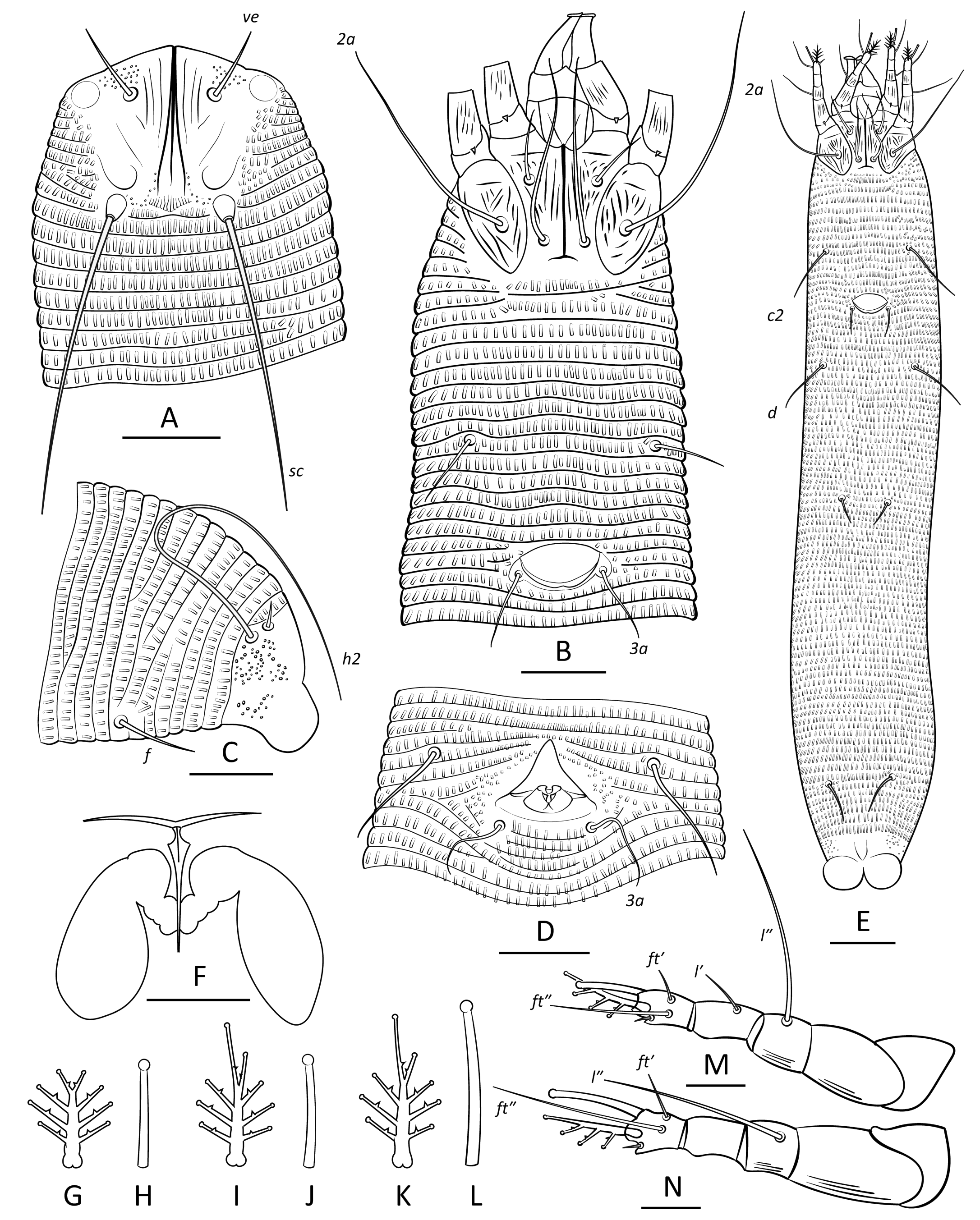
Novophytoptus floris n. sp. – Figure 3
ZOOBANK: AC76FE41-D6F9-4560-A928-357B10D71479
Female holotype (n = 11)
Body vermiform, 371 (309–422), 64 (54–67) wide at the level of setae c2.
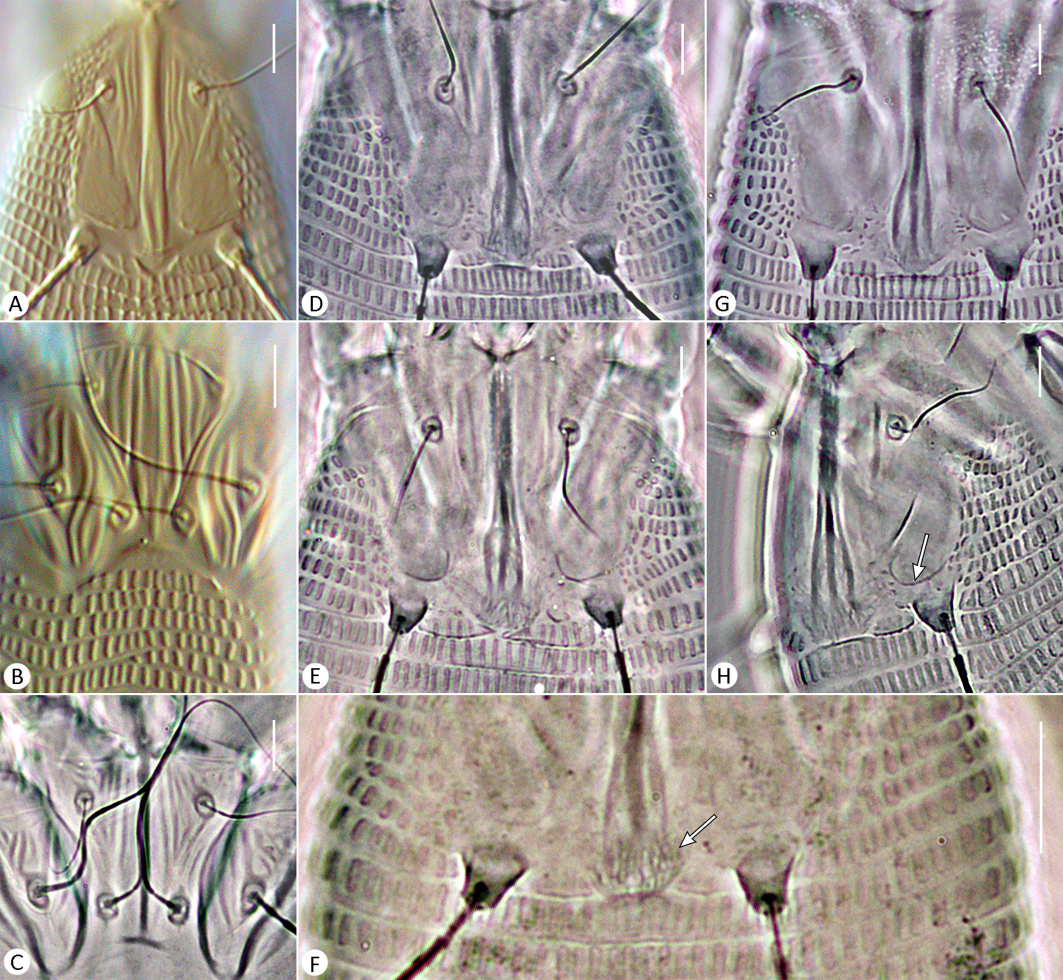
Prodorsal shield subpentagonal, 30 (28–32) long, 31 (27–31) wide, with two ocellus-like spots lateral to setae ve, and clusters of sparse microtubercles anterior to the ve tubercles. Median line of prodorsal shield distinct, complete, and entire. Admedian lines complete, converging in the anterior third to form a single thick ridge. Two to three short, curved submedian lines (I, II, and III) present between the admedian line and the ve tubercle in the anterior half of the shield. Lateral areas between the ve and sc tubercles on each side with two faint, short, oblique lines and a more distinct, curved line resembling a fishhook and preceding the sc tubercle. A subtriangular area, delimited by two converging rows of sparse microtubercles, situated between the sc tubercles, the posterior ends of the admedian lines, and the posterior margin of the shield. A thin, obtusely angled, V-shaped line enclosing 12–15 very faint, subparallel longitudinal ridges located between the posterior ends of the admedian lines and the first dorsal opisthosomal annulus. Setae ve 14 (13–15), directed anterolaterad, tubercles 13 (11–14) apart; sc 43 (41–48) long, directed posterolaterad, tubercles 19 (18–24) apart. Distance between tubercles ve–sc 20 (18–21).
Gnathosoma elongate, directed straight forward at slightly ventral angle, 25 (24–27); pedipalp coxal seta ep 4 (3–4), pedipalp genual seta d absent, subapical pedipalp tarsal seta ν 1 (0.5–1). Ventrally, basal gnathosoma covered by smooth subtriangular suboral plate with two faint diverging ridges forming V-shaped figure.
Leg I 29 (27–31), tarsus 5 (6–7), u′ 2 (2–3), ft′ 2 (2–4), ft″ 14 (13–16), ω 6 (6–7) with tiny spherical knob, empodium I 4/4-rayed, 7 (6–8), each ray with one short secondary branch, the medial ray of the terminal pair twice as long as the lateral ray; tibia 5 (5–6), ventrally striated, l′ 2 (2–4), genu 5 (4–6), with 3–5 longitudinal striae ventrally, l″ 17 (16–19), femur 11 (9–12), with 6–10 longitudinal striae ventrally, bv absent. Leg II 29 (27–31), tarsus 5 (4–6), u′ 2 (1–3), ft′ 3 (2–4), ft″ 16 (15–18), ω 9 (7–9) with tiny spherical knob, empodium II 4/4-rayed, 7 (6–8), similarly shaped as empodium I; tibia 5 (4–6), smooth ventrally, genu 5 (3–6), with 3–4 longitudinal striae ventrally, l″ 16 (15–18), femur 11 (9–12), with 4–9 longitudinal striae ventrally, bv absent.
Coxal plates I and II ornamented with short, irregularly distributed ridges, some of which are subparallel near the lateral and medial margins of the coxae. Prosternal apodeme inversely T-shaped, 19 (18–20). Setae 1b 13 (11–16), 12 (11–13) apart; 1a 27 (25–32), 8 (7–8) apart; 2a 45 (42–55), 22 (19–24) apart; 14 (13–16) coxigenital annuli ahead of epigynum. Genital coverflap semi-circular, smooth, 10 (8–12), 14 (14–16) wide; setae 3a 13 (11–15), 16 (14–18) apart. Internal genitalia (n = 5). Spermatheca sac-shaped, large, 8–11, 4–7 wide; spermathecal tube with narrower medial and swollen lateral segments, 3–4, longitudinal bridge 6–9, anterior genital apodeme 10–14 wide.
Opisthosoma vermiform, slightly expanded caudally, with 91 (88–96) dorsal and 97 (92–104) ventral annuli with elongated microtubercles; last 4–5 dorsal annuli with less numerous and smaller microtubercles. Lateral surface of caudal lobe with a group of small round microtubercles. Setal lengths: c2 15 (14–18), d 24 (21–26), e 12 (11–14), f 16 (15–17), h1 5 (4–6), h2 61 (58–71); 9 (9–10) annuli from rear shield margin to c2; 16 (14–18) annuli between c2–d; 21 (20–24) annuli between d and e; 38 (35–39) annuli between e and f; 7 (6–8) annuli between f and h1.
Male (n = 4)
Body vermiform, 347–403, 70–78 wide at the level of setae c2.
Prodorsal shield subpentagonal, 29–34 long, 29–31 wide, ornamented similarly to females. Setae ve 13–14, directed anterolaterad, tubercles 16–17 apart; sc 31–34 long, directed posterolaterad, tubercles 21–22 apart. Distance between tubercles ve–sc 21–22.
Gnathosoma elongate, directed straight forward at slightly ventral angle, 28–31; pedipalp coxal seta ep 3–5, pedipalp genual seta d absent, subapical pedipalp tarsal seta ν 1–1.5. Suboral plate subtriangular, with two ridges forming V-shaped figure.
Leg I 31–32, tarsus 5–6, u′ 1–2, ft′ 2–3, ft″ 17–18, ω 6–7 with tiny spherical knob, empodium I 4/4-rayed, 5–6, each ray with one short secondary branch, terminal paired rays short and equally long; tibia 7–8, l′ 4–6, genu 4–6, l″ 19–21, femur 11–12, bv absent. Leg II 31–32, tarsus 5–6, u′ 2–3, ft′ 3–5, ft″ 18–21, ω 8–9 with tiny spherical knob, empodium II 4/4-rayed, 4–6, similarly shaped to empodium I; tibia 5–6, genu 5–6, l″ 18–21, femur 11–12, bv absent. Coxal plates ornamented similarly to those of females. Prosternal apodeme inversely T-shaped, 16–17. Setae 1b 12–15, 13–14 apart; 1a 28–31, 7–9 apart; 2a 44–53, 22–24 apart; 12–13 coxigenital annuli before epiandrium.
Genital area 14–15, 15–17 wide, with a small, papilla-shaped genital cone in the center, preceded by a narrow transverse genital slit and a thin cuticular subtriangular flap; setae 3a 14–16, 16–18 apart, setae eu non apparent. Small papilla-shaped genital cone preceded by narrow transverse genital slit and thin cuticular subtriangular flap situated in the centre of genital area.
Opisthosoma with 91–100 dorsal and 85–97 ventral annuli with elongated microtubercles. Lateral surface of caudal lobe with a group of small, round microtubercules. Setal lengths: c2 12–15, d 22–24, e 12–15, f 15–17, h1 4–6, h2 45–53; 11–12 annuli from rear shield margin to c2; 12–16 annuli between c2–d; 20–23 annuli between d and e; 38–40 annuli between e and f; 6–8 annuli between f and h1.
GenBank data
PZ786220 (D1D2 28S rRNA, 1026 bp), PZ785228 (Cox1, 443 bp).
BLAST search results
The rDNA sequence of N. floris n. sp. showed the highest similarity to GenBank sequences of Novophytoptus rostratae (KY921989) and N. cf. rostratae (KT070284, KT070285), with 99–100% query coverage and 92.6–94.8% sequence identity. For the Cox1 gene, the nucleotide/amino acid sequences of N. floris n. sp. were most similar to GenBank sequences of Novophytoptus cf. rostratae (KT070241/AKP20854) and N. luzulis (MT712730/AKP20855), with 99/100% query coverage and 82.5/88.4% sequence identity.
Host plant
Drimys winteri J.R. Forst. & G. Forst. (Canellales: Winteraceae).
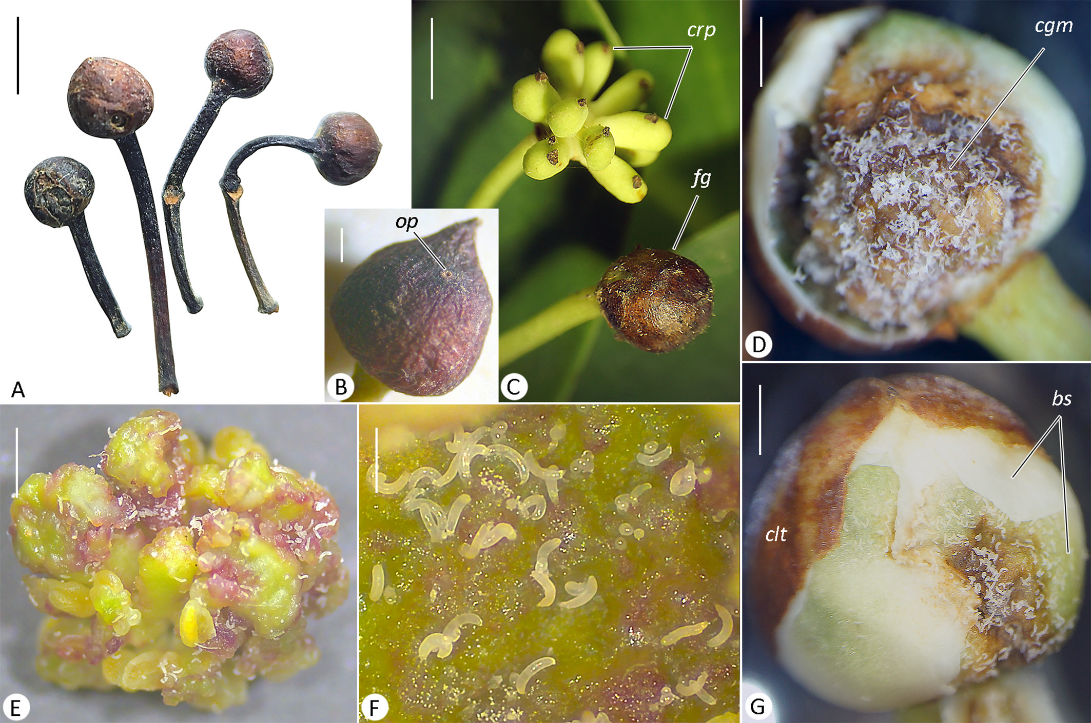
Relation to the host. Mites form large colonies consisting of thousands of individuals inside flower buds, blocking anthesis and inducing the formation of subspherical flower galls that are often situated on curved pedicels (Fig. 4 A, B, C). In D. winteri, the young flower bud prior to anthesis is enclosed by a completely sealed calyptra (fused sepals), which tears during blooming. Flower buds infested by N. floris n. sp. retain the calyptra, suggesting that the mites penetrate the bud at a very early stage of its development. Mites feed on the green bud scales (cataphylls) beneath the calyptra (Fig. 4 F, G), as well as on the tissues of internal juvenile carpels, reprogramming their normal ontogenesis to form irregular cell masses (Fig. 4 D, E). Disruption of flower development prevents its opening, pollination, and the formation of fruits and seeds (Fig. 4 E). The mites emerge from the galls via presumably self-made circular openings usually located at the apical pole of the gall (Fig. 4 B).
Remarks
Males were rarely encountered and remained confined to the interior of galls, whereas females were abundant both within and on the surface of galls. The latter displayed rapid movement, consistent with active dispersal.
Type locality
CHILE: Magallanes Region, Timaukel, 53°44′59.70″ S, 70°05′51.39″ W
Type material
Female holotype on slide v2107А-5, 10 paratype females and 4 males in slide series v2107А. All specimens from type locality, collected on 22 April 2025, coll. C. Arriagada. Type material is deposited in Acarological collection of ZIN RAS. Some paratypes have been also deposited in SAG in the Colección Acarológica SAG Laboratorio Valparaíso, Valparaíso and Curico (Chile).
Additional material. Numerous adults and immatures in slide series v2093, v2094, and v2108 collected on 22 April 2025 and 3 February 2026 from flower galls of Drimys winteri (type locality, the same collector).
Etymology
The specific epithet floris is a substantive derived from the Latin flos (''flower''), indicating the association of N. floris n. sp. with the flower buds of its host plant.
Differential diagnosis
Among Novophytoptus spp. the new species is closest to N. zuluensis Chetverikov et al. 2023 described from a sedge Carex zuluensis C.B.Clarke (Poales: Cyperaceae) from South Africa. The main differences are in the ornamentation of the prodorsal shield and coxae, and lengths of prodorsal shield and setae sc and f (Fig. 5, Table 1).
Download as
Characters
N. zuluensis
N. floris n. sp.
Prodorsal shield design
Median and admedian lines
Admedian lines are very close to each other and slightly converge at their posterior ends; the basal part of the median line is indistinct
Admedian lines are basally widely diverging; the basal part of the median line is clearly visible
Fish-hook shaped line in front of tubercles of sc
Distinct, well-separated, and not marked with microtubercles
Less distinct, medially joining admedian line, marked with sparse microtubercles
Thin subparallel lines associated with transverse V-shaped ridge adjacent to basal part of medians
Present
Absent
Microtubercules in the area between sc and posterior part of admedians
Absent
Present
Ornamentation of coxae I and II
Composed of long, stout, subparallel ridges
Composed of shorter and thinner ridges that are more irregularly distributed
Length of prodorsal shield
21–24
28–32
Length of sc
60–79
41–48
Length of f
9–13
15–17
Reference
Chetverikov et al. (2023)
this paper
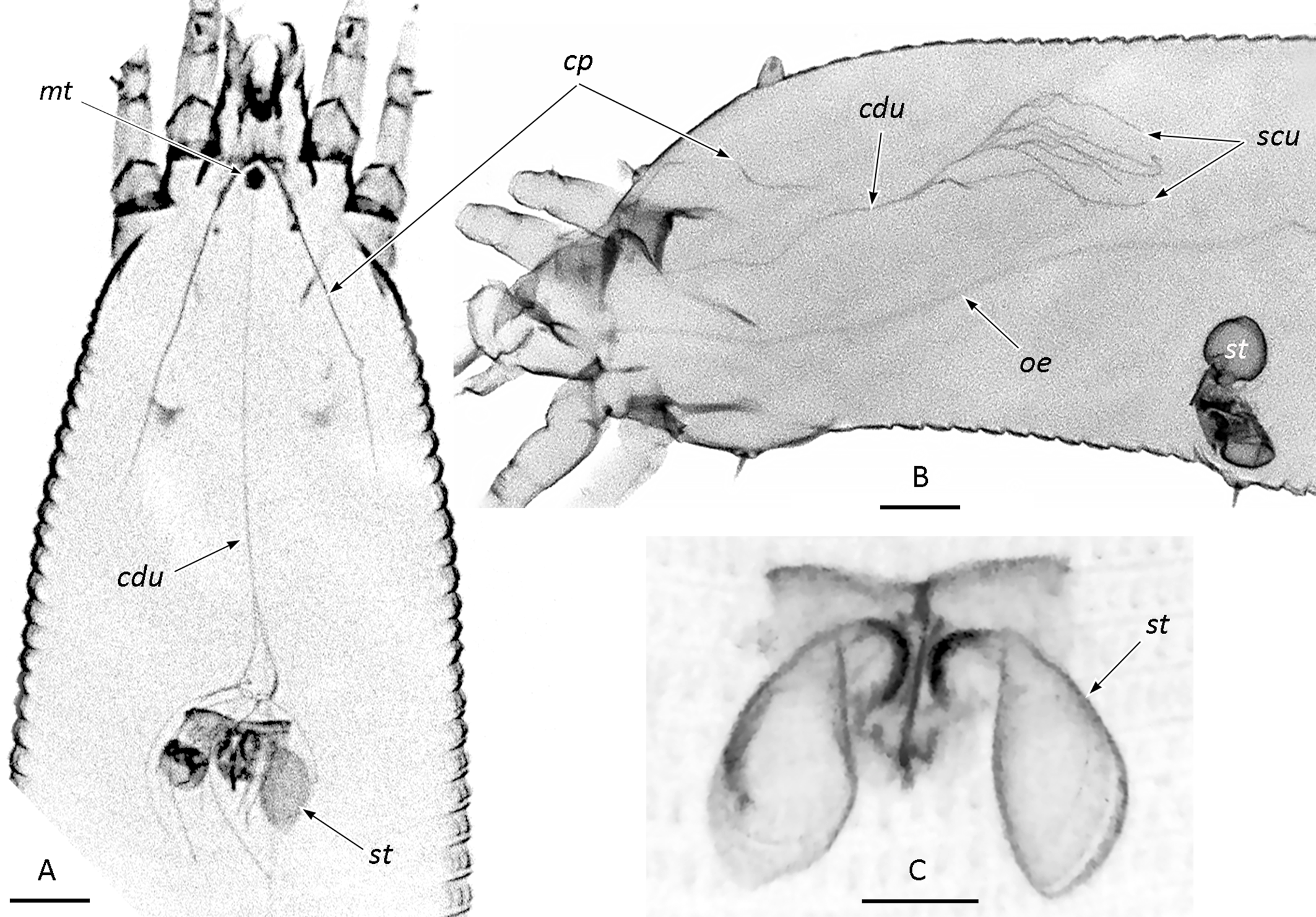
CLSM observations (Fig. 6)
Exoskeletons of the investigated females of N. floris n. sp. (n = 7) produced strong autofluorescence under a blue laser (405 nm), allowing visualization of internal cuticle-lined structures. In addition to distinct elements of the spermathecal apparatus, tiny paired cuticular canals of the anal glands (not shown), a subspherical motivator, a thin, capillary-like oesophagus (~1 µm in diameter) transitioning into the midgut in the middle of the opisthosoma, and the canals of the prosomal glands were observed. Their topography and relative positions are consistent with previous histological data reported by Nuzzaci and Alberti (1996) and Propistsova et al. (2023). The observable structures of the unpaired gland include a series of narrow secretory canaliculi joining into a common duct that runs medially, subparallel to the esophagus, and terminally penetrates the motivator. The canals of the paired podocephalic glands run convergently, very close to the dorsal cuticle of the anterior opisthosoma and prosoma, opening slightly anterior to and below the motivator under the cheliceral bases.
Discussion
In this paper, we describe the first non-monocot-associated species of the genus Novophytoptus. This is the only member of the genus known to infest reproductive plant organs and the second species recorded from South America (Fig. 2), following the Brazilian species N. silvai Flechtmann, 2004, associated with Cyperus giganteus Vahl (Cyperaceae). Most Novophytoptus species have been described from Europe (Roivainen 1947, Chetverikov and Sukhareva 2007, Skoracka et Boczek 2000, Pye 2012, Chetverikov 2015, Chetverikov and Petanović 2016), with fewer species and records reported from North America (Keifer 1962, Oldfield 1973, Chetverikov et al. 2017), South Africa (Chetverikov et al. 2021, 2023) and Asia (Chetverikov 2016, Klimov et al. 2018). Australasia remains the only large biogeographic region where Novophytoptus has not yet been registered. Following our finding of N. floris n. sp. on a Gondwanan host plant genus Drimys in Chile, targeted surveys in Australasia have become of particular evolutionary interest, given that members of the family Winteraceae are well represented in this region (Thomas et al. 2014). Remarkably, while the small genus Drimys is strictly confined to the New World, most other winteracean lineages (excluding the monotypic Takhtajania from Madagascar) are widely distributed across Australia, Tasmania, and New Guinea (Thomas et al. 2014). If N. floris n. sp. is a true Gondwanan relict that co-evolved with its host, then a search for eriophyoid mites on Australasian Winteraceae (Bubbia, Pseudowintera, Tasmannia, and Zygogynum) may lead to the discovery of new non-monocot-associated Novophytoptus species.
Like its congeners, N. floris n. sp. exhibits a concealed lifestyle, colonizing target plant organs at an early developmental stage to feed and reproduce within enclosed microhabitats. Our long-term observations across various Palearctic Novophytoptus species from monocots have revealed a consistent pattern in the spatial distribution of the sexes, which is fully shared by N. floris n. sp.: males occur exclusively within infested plant organs, whereas dispersing individuals collected outside host tissues are invariably females. This pattern may be linked to a pronounced sexual dimorphism in tarsal morphology (Chetverikov and Petanović 2016). Compared to females, males possess shorter tarsal solenidia and shorter empodia, the latter characterized by a reduced length of the terminal pair of empodial rays. Such morphological constraints likely limit male locomotion outside feeding sites, restricting their biological role to spermatophore deposition.
Compared to Novophytoptus spp. from monocots, N. floris n. sp. exhibits unique biological features, distinguished by: (1) infesting young flower buds rather than vegetative organs; (2) feeding on the cells of juvenile carpels and cataphylls—which are technically external, albeit enclosed within the unexpanded bud—instead of consuming the juice of parenchymal cells within subepidermal cavities; and (3) inducing complex gall formation via reprogramming the normal morphogenesis of the flower bud, rather than causing simple parenchymal necrosis. To emerge from the gall, N. floris n. sp. forms exit holes in the calyptra, mirroring the behavior of other Novophytoptus species that penetrate the host epidermis in sedges, rushes, and grasses. Previously, it was hypothesized that these mites create exit holes purely mechanically; this was supported by observations of rotational body movements during the presumed drilling of the plant cuticle (Chetverikov and Petanović 2016). In this study, we visualized the ducts of the prosomal glands in Novophytoptus and found that at least the unpaired prosomal gland (and possibly the podocephalic glands) is remarkably well-developed. Although the chemical composition of the prosomal gland secretion remains unknown, an intriguing hypothesis is that it facilitates the enzymatic degradation of plant tissues during exit-hole formation. Supplementing mechanical drilling with chemical treatment would significantly ease the penetration of tough plant barriers—a process otherwise difficult to explain via purely mechanical means, given that the eriophyoid gnathosoma possesses piercing, needle-like stylets and lacks chewing mouthparts (Lindquist 1996).
While our findings shed light on the basic biology of N. floris n. sp., its full life cycle remains to be described with reference to the host's phenological stages and climate. Moreover, evaluating how the mite affects seed production and population regeneration in Drimys winteri is essential to understand its overall pest status and ecological significance. The morphological similarity of reproductive organs among Drimys species also implies that associations with Novophytoptus mites may extend beyond D. winteri—a hypothesis that warrants testing through expanded surveys across South America. Notably, data on flower visitors to D. brasiliensis in Brazil reveal abundant Coleoptera (mainly Curculionidae), Diptera, and Thysanoptera (Gottsberger et al. 1980). Given the documented phoresy of eriophyoid mites on psyllids (Liu et al. 2016), insects that regularly visit D. winteri flowers could act as phoretic carriers for N. floris n. sp., promoting host-specific dispersal. In this context, the terminally forked, elongated, and asymmetrical empodia of females might be better suited for attachment to such carriers than those of males.
Drimys winteri, the host species of N. floris n. sp., exhibits a striking biogeographic pattern in Chile. Its main range extends across the temperate rainforests of southern Chile and adjacent Argentina (approximately 36–56°S), whereas a disjunct population occurs in the unique coastal relict forest of Fray Jorge (~31°S) nearly 1000 km north of the nearest part of the main distribution area (Ladio et al. 2026, Squeo et al. 2016). This isolated site, sustained by coastal fog (camanchaca), is regarded as a relict of a formerly continuous temperate forest belt, thus providing a powerful system to study how fragmentation and subsequent isolation have affected associated biota (Cornelius et al. 2000, Pérez et al. 2017, Cadiz et al. 2018). Determining the presence of N. floris n. sp. on D. winteri in Fray Jorge and comparing genetic variation between the isolated and southern populations could reveal how historical disjunction has shaped the mite's phylogeography.
To date, the phylogeny of the genus Novophytoptus and the evolution of its host-plant associations have not been investigated. However, our discovery of N. floris n. sp. on Drimys in Chile, combined with the unresolved taxonomic position of Novophytoptus within the family Phytoptidae (Chetverikov et al. 2021), highlights the need for a phylogenetic approach. Currently, GenBank contains only 24 sequences for this genus, representing five species from monocots and comprising five genes previously shown to be highly suitable for phylogenetic studies in Acariformes (Klimov et al. 2018): Cox1 (10 sequences), 28S (5), 18S (5), Hsp70 (3), and EF-1α (1). Although the 28S and Cox1 sequences of N. floris n. sp. showed the highest BLAST similarity to N. rostratae from Cyperaceae, this similarity is uninformative due to the limited reference data in GenBank. Two hypotheses can explain the host association of N. floris n. sp. with Drimys: (1) it represents a host shift from monocots, assuming that monocots were the ancestral hosts of Novophytoptus; or (2) Drimys and potentially other non-monocot taxa represent the true complex of primary host lineages, whereas the group of Novophytoptus species on Poales results from a recent divergence following a transition to herbaceous monocots. To test these hypotheses, additional sampling across different continents, targeted searches for Novophytoptus on non-monocot hosts, and a comprehensive multi-gene analysis (e.g. using the five markers available in GenBank) are needed.
Acknowledgements
We thank Mr C. Arriagada (SAG) for collecting plant samples in southern Chile. This study was supported by the Russian Science Foundation, grant № 25-14-00118 to the first author.
References
- Amrine J.W.Jr. & Manson D.C.M. 1996. Chapter 1.6.3 Preparation, mounting and descriptive study of eriophyoid mites. In: Lindquist E.E., Sabelis M.W. & Bruin J. (Eds). Eriophyoid Mites: Their Biology, Natural Enemies and Control. World Crop Pests, vol. 6: 383-396. Amsterdam: Elsevier Science Publishing. https://doi.org/10.1016/S1572-4379(96)80023-6
- Amrine J.W.Jr., Stasny T.A.H. & Flechtmann C.H.W. 2003. Revised keys to the world genera of the Eriophyoidea (Acari: Prostigmata). Michigan: Indira Publishing House. p. 244
- Cadiz F.J., Esquerre Gheur D., Cadiz V.H. & de Mello Martins F. 2018. Phylogeography of Plectostylus Beck, 1837 (Gastropoda: Stylommatophora: Orthalicoidea): Origin and isolation of the Fray Jorge Forest relicts in northern Chile. J. Zool. Syst. Evol. Res. https://doi.org/10.1111/jzs.12242
- Chetverikov P.E. 2014. Comparative confocal microscopy of internal genitalia of phytoptine mites (Eriophyoidea, Phytoptidae): new generic diagnoses reflecting host-plant associations. Exp. Appl. Acarol., 62(2): 129-160. https://doi.org/10.1007/s10493-013-9734-2
- Chetverikov P.E. 2015. Hidden diversity of endoparasitic eriophyoid mites: two new Novophytoptus Roivainen, 1947 (Acari: Eriophyoidea: Phytoptidae) species from the parenchymatous tissues of rushes (Juncaceae). Zootaxa, 4006(3): 481-505.
- Chetverikov P.E. 2016. New species and records of phytoptid mites (Acari: Eriophyoidea: Phytoptidae) on sedges (Cyperaceae) from the Russian Far East. Zootaxa, 4061(4): 367-380. https://doi.org/10.11646/zootaxa.4061.4.3
- Chetverikov P.E. & Sukhareva S.I. 2007. Supplementary descriptions and biological notes on eriophyid mites (Acari: Eriophyidae) of the genus Novophytoptus Roivainen, 1947. Acarina, 15(1): 261-268.
- Chetverikov P.E., Amrine J., Bauchan G., Ochoa R., Sukhareva S.I. & Vishnyakov A.E. 2017. Supplementary description of Novophytoptus stipae Keifer 1962 (Acariformes, Eriophyoidea) with LT-SEM observation on mites from putatively conspecific populations: cryptic speciation or polyphagy of novophytoptines on phylogenetically remote hosts? Syst. Appl. Acarol., 22(2): 253-270. https://doi.org/10.11158/saa.22.2.9
- Chetverikov P.E. & Petanović R.U. 2016. Longest endoparasitic eriophyoid mite (Acari, Eriophyoidea): description of Novophytoptus longissimus n. sp. and remarks on size limits in eriophyoids. Syst. Appl. Acarol., 21(11): 1547-1563. https://doi.org/10.11158/saa.21.11.10
- Chetverikov P.E., Craemer C., Cvrković T., Klimov P.B., Petanović R.U., Romanovich A.E., Sukhareva S.I., Zukoff S., Bolton S. & Amrine J. 2021. Molecular phylogeny of the phytoparasitic mite family Phytoptidae (Acariformes: Eriophyoidea) identified the female genitalic anatomy as a major macroevolutionary factor and revealed multiple origins of gall induction. Exp. Appl. Acarol., 83: 31-68. https://doi.org/10.1007/s10493-020-00571-6
- Chetverikov P.E., Craemer C., Gankevich V.D., Kremenetskaya M.V., Kuzmin I.V. & Zhuk A.S. 2023. Endoparasitic Gall Mites: Two New Novophytoptus Species (Eriophyoidea, Phytoptidae) from Southern African Sedges (Cyperaceae, Carex) and New Hypotheses on the Phylogeny of Novophytoptines. Diversity, 15(3): 416. https://doi.org/10.3390/d15030416
- Cornelius C., Cofré H. & Marquet P.A. 2000. Effects of habitat fragmentation on bird species in a relict temperate forest in semiarid Chile. Conserv. Biol., 14(2): 534-543. https://doi.org/10.1046/j.1523-1739.2000.98409.x
- Doust A.N. 2001. The developmental basis of floral variation in Drimys winteri (Winteraceae). Int. J. Plant Sci., 162(4): 697-717. https://doi.org/10.1086/320790
- Flechtmann C.H.W. 2004. Eriophyid mites (Acari: Eriophyidae) from Brazilian sedges (Cyperaceae). Int. J. Acarol., 30(2): 157-164. https://doi.org/10.1080/01647950408684385
- Gottsberger G., Silberbauer-Gottsberger I. & Ehrendorfer F. 1980. Reproductive biology in the primitive relic angiosperm Drimys brasiliensis (Winteraceae). Plant Syst. Evol., 135(1): 11-39. https://doi.org/10.1007/BF00983005
- Keifer H.H. 1962. Eriophyid studies B-8. Bureau of Entomology of California Department of Agriculture. 20 pp.
- Klimov P.B., O'Connor B.M., Chetverikov P.E., Bolton S.J., Pepato A.R., Mortazavi A.L., Tolstikov A.V., Bauchan G.R. & Ochoa R. 2018. Comprehensive phylogeny of acariform mites (Acariformes) provides insights on the origin of the four-legged mites (Eriophyoidea), a long branch. Mol. Phylogenet. Evol., 119: 105-117. https://doi.org/10.1016/j.ympev.2017.10.017
- Ladio A.H., Ezcurra C., Ochoa J. & Castillo L.D. 2026. Drimys winteri JR Forst. & G. Forst. Winteraceae. In: Ladio A.H., Ezcurra C., Ochoa J.J. & Castillo L.D. (Eds). Ethnobotany of the Mountain Regions of Patagonia: 1-6. Cham, Switzerland: Springer. https://doi.org/10.1007/978-3-031-89165-6
- Lindquist E.E. 1996. Chapter 1.1 External anatomy and systematics 1.1.1. External anatomy and notation of structures. In: Lindquist E.E., Sabelis M.W. & Bruin J. (Eds). Eriophyoid Mites: Their Biology, Natural Enemies and Control. World Crop Pests, vol. 6: 3-31. Amsterdam: Elsevier. https://doi.org/10.1016/S1572-4379(96)80003-0
- Lindquist E.E. & Oldfield G.N. 1996. Chapter 1.5.1 Evolution and phylogeny 1.5.1 Evolution of eriophyoid mites in relation to their host plants. In: Lindquist E.E., Sabelis M.W. & Bruin J. (Eds). Eriophyoid Mites: Their Biology, Natural Enemies and Control. World Crop Pests, vol. 6: 277-300. Amsterdam: Elsevier Science Publishing. https://doi.org/10.1016/S1572-4379(96)80018-2
- Liu S., Li J., Guo K., Qiao H., Xu R., Chen J., Xu C. & Chen J. 2016. Seasonal phoresy as an overwintering strategy of a phytophagous mite. Sci. Rep., 6: 25483. https://doi.org/10.1038/srep25483
- Nuzzaci G. & Alberti G. 1996. Internal anatomy and physiology. In: Lindquist E.E., Sabelis M.W. & Bruin J. (Eds). Eriophyoid Mites: Their Biology, Natural Enemies and Control. World Crop Pests, vol. 6: 101-150. Amsterdam: Elsevier Science Publishing. https://doi.org/10.1016/S1572-4379(96)80006-6
- Oldfield G.N. 1973. Sperm storage in female Eriophyoidea (Acarina). Ann. Entomol. Soc. Am., 66(5): 1089-1092. https://doi.org/10.1093/aesa/66.5.1089
- Pérez F., Hinojosa L.F., Peralta G., Montenegro P., Irarrázabal C. & Cossio M. 2017. Genetic patterns of Myrceugenia correifolia, a rare species of fog-dependent forests of Mediterranean Chile: is it a climatic relict? Front. Plant Sci., 8: 1097.
- Propistsova E.A., Makarova A.A., Chetverikov P.E. & Polilov A.A. 2023. Anatomy of the miniature four-legged mite Achaetocoptes quercifolii (Arachnida: Acariformes: Eriophyoidea). Arthropod Struct. Dev., 72: 101235. https://doi.org/10.1016/j.asd.2023.101235
- Pye D.R. 2012. New eriophyoid mites (Acari: Prostigmata: Eriophyoidea) in Britain: one new genus, four new species, 19 new records and two incursions. Zootaxa, 3578: 43-68. https://doi.org/10.11646/zootaxa.3578.1.3
- Roivainen H. 1947. Eriophyid news from Finland. Acta Entomol. Fenn., 3: 1-51.
- Skoracka A. & Boczek J. 2000. Novophytoptus glyceriae sp. n. and N. ammophilae sp. n., the new species of eriophyoid mites (Acari: Eriophyoidea) from grasses. Acarina, 8(2): 95-101.
- Skoracka A., Smith L., Oldfield G., Cristofaro M. & Amrine J.W. 2010. Host-plant specificity and specialization in eriophyoid mites and their importance for the use of eriophyoid mites as biocontrol agents of weeds. Exp. Appl. Acarol., 51(1): 93-113. https://doi.org/10.1007/s10493-009-9323-6
- Squeo F.A., Loayza A.P., López R.P. & Gutiérrez J.R. 2016. Vegetation of Bosque Fray Jorge National Park and its surrounding matrix in the Coastal Desert of north-central Chile. J. Arid Environ., 126: 12-22. https://doi.org/10.1016/j.jaridenv.2015.10.013
- Svoma E. 1998. Studies on the embryology and gynoecium structures in Drimys winteri (Winteraceae) and some Annonaceae. Plant Syst. Evol., 209(3): 205-229. https://doi.org/10.1007/BF00985229
- Thomas N., Bruhl J.J., Ford A. & Weston P.H. 2014. Molecular dating of Winteraceae reveals a complex biogeographical history involving both ancient Gondwanan vicariance and long‐distance dispersal. J. Biogeogr., 41(5): 894-904. https://doi.org/10.1111/jbi.12265
- Upton M.S. 1993. Aqueous gum-chloral slide mounting media: an historical review. Bull. Entomol. Res., 83(2): 267-274. https://doi.org/10.1017/S0007485300034763
- Zuntini A.R., Carruthers T., Maurin O., Bailey P.C., Leempoel K., Brewer G.E. & Knapp S. 2024. Phylogenomics and the rise of the angiosperms. Nature, 629(8013): 843-850. https://doi.org/10.1038/s41586-024-07324-0
to post any comment or correction.


2026-07-15
Date accepted:
2026-09-07
Date published:
2026-09-17
Edited by:
Kreiter, Serge

This work is licensed under a Creative Commons Attribution 4.0 International License
2026 Chetverikov, Philipp E.; Kudryavtzev, Andrey T.; Peralta-Alba, Lourdes and Rojas-Espinoza, Francisco
 Download article Download low definition
Download the citation
RIS with abstract
(Zotero, Endnote, Reference Manager, ProCite, RefWorks, Mendeley)
RIS without abstract
BIB
(Zotero, BibTeX)
TXT
(PubMed, Txt)