


Redescription of Steneotarsonemus (Mahunkacarus) abruptus (Acari: Heterostigmata: Tarsonemidae) and its first record from Western Siberia, Russia
Khaustov, Alexander A.  1
1
1✉ X-BIO Institute, University of Tyumen, Tyumen, 6 Volodarskogo Str., 625003, Russia.
2026 - Volume: 66 Issue: 3 pages: 705-713
https://doi.org/10.24349/8eyc-zarzZooBank LSID: 44CCF190-9CBE-4D20-A4B7-D193D7727240

Original research
Keywords
Abstract
Introduction
The family Tarsonemidae is one of the largest groups in the mite cohort Heterostigmata (Acari: Trombidiformes) and currently includes 50 genera and more than 600 species (Lin & Zhang 2002; Khaustov & Tolstikov 2025). Family members exhibit a wide range of feeding habits, including phytophagy, mycophagy, predation on mite eggs, and parasitism or parasitoidism of insects (Lindquist 1986). Tarsonemid mites inhabit soils and litter, various plants, bracket fungi, subcortical galleries of insects, etc. and many species utilize insects for phoretic dispersal (Lindquist 1986).
The genus Steneotarsonemus comprises 88 species of phytophagous mites grouped into six subgenera (Seeman et al. 2016; Ganguly et al. 2021; Mondal et al. 2021; Ganguly & Karmakar 2022; Khaustov & Mashkov 2026). Some species are considered as pests of poaceous and other crops. For example, S. spinki is a serious pest of rice (Kayal et al. 2021), S. panshini and S. spirifex are pests of wheat (Livshits et al. 2011), and S. ananas is a pest of pineapple (Seeman et al. 2016). Steneotarsonemus spinki may also transmit viral diseases (Shikata et al. 1984).
The subgenus Steneotarsonemus (Mahunkacarus) Wainstein comprises five species, namely: S. (M.) abruptus Livshits et al. 1982, S. (M.) apertimerus Mahunka, 1971, S. (M.) badulini Wainstein, 1979, S. (M.) gibber Suski, 1970, and S. (M.) mayae Lin & Zhang, 2005 (Seeman et al. 2016). The main diagnostic character of the subgenus is vestigial setae sc1 and reduced prodorsal bothridia in female (Lindquist 1986).
The female of Steneotarsonemus abruptus Livshits, Mitrofanov & Sharonov, 1982 was described from European Russia from Smooth meadow-grass Poa pratensis (Livshits et al. 1982). Since that time it has never been found in other localities. During the study of phytophagous mites in Western Siberia, Russia, we found females of S. abruptus on Bush grass Calamagrostis arundinacea (Poaceae). The original description of S. abruptus is incomplete. Therefore, I provide a redescription of female based on the type material and additional specimens from Western Siberia. This article is a continuation of a series of publications dedicated to the redescription of species described by Livshits, Mitrofanov, and Sharonov from Russia (Khaustov & Mashkov 2025, 2026; Khaustov 2026).
Materials and methods
The collection of the type materials of Tarsonemidae described by Livshits, Mitrofanov and Sharonov was studied. The collection is currently deposited in the University of Tyumen, Tyumen, Russia. Siberian specimens were collected from samples of plants using Berlese funnels and mounted on slides in Hoyer's medium. The terminology follows that of Lindquist (1986), except the ventral subcapitular seta is labeled as m (Grandjean 1944). All measurements are given in micrometers (μm) for the holotype and non-type specimens (in parentheses). For leg chaetotaxy the number of solenidia is given in parentheses. Mite morphology was studied using Carl Zeiss AxioImager A2 compound microscope with phase contrast and differential-interference contrast (DIC) optical systems. Photomicrographs were taken with an AxioCam ICc5 digital camera.
Abbreviations: ap1-ap4—apodemes 1-4, appr—prosternal apodeme, appo—poststernal apodeme, apsej—sejugal apodeme, Tr—trochanter, Fe—femur, Ge—genu, Ti—tibia, Ta—tarsus, TiTa—tibiotarsus.
Results
Family Tarsonemidae Canestrini and Fanzago, 1877
Genus Steneotarsonemus Beer, 1954
Subgenus Mahunkacarus Wainstein, 1979
Type species: Steneotarsonemus gibber Suski, 1970, by original designation.
Diagnosis
As of Lindquist (1986) with modifications of Mitrofanov & Sharonov (1988).
Steneotarsonemus (Mahunkacarus) abruptus Livshits, Mitrofanov & Sharonov, 1982
Steneotarsonemus abruptus Livshits, Mitrofanov & Sharonov, 1982, 25, Fig. 1
Steneotarsonemus (Mahunkacarus) abruptus: Mitrofanov & Sharonov, 1988, 47
Figures 1–5
Diagnosis
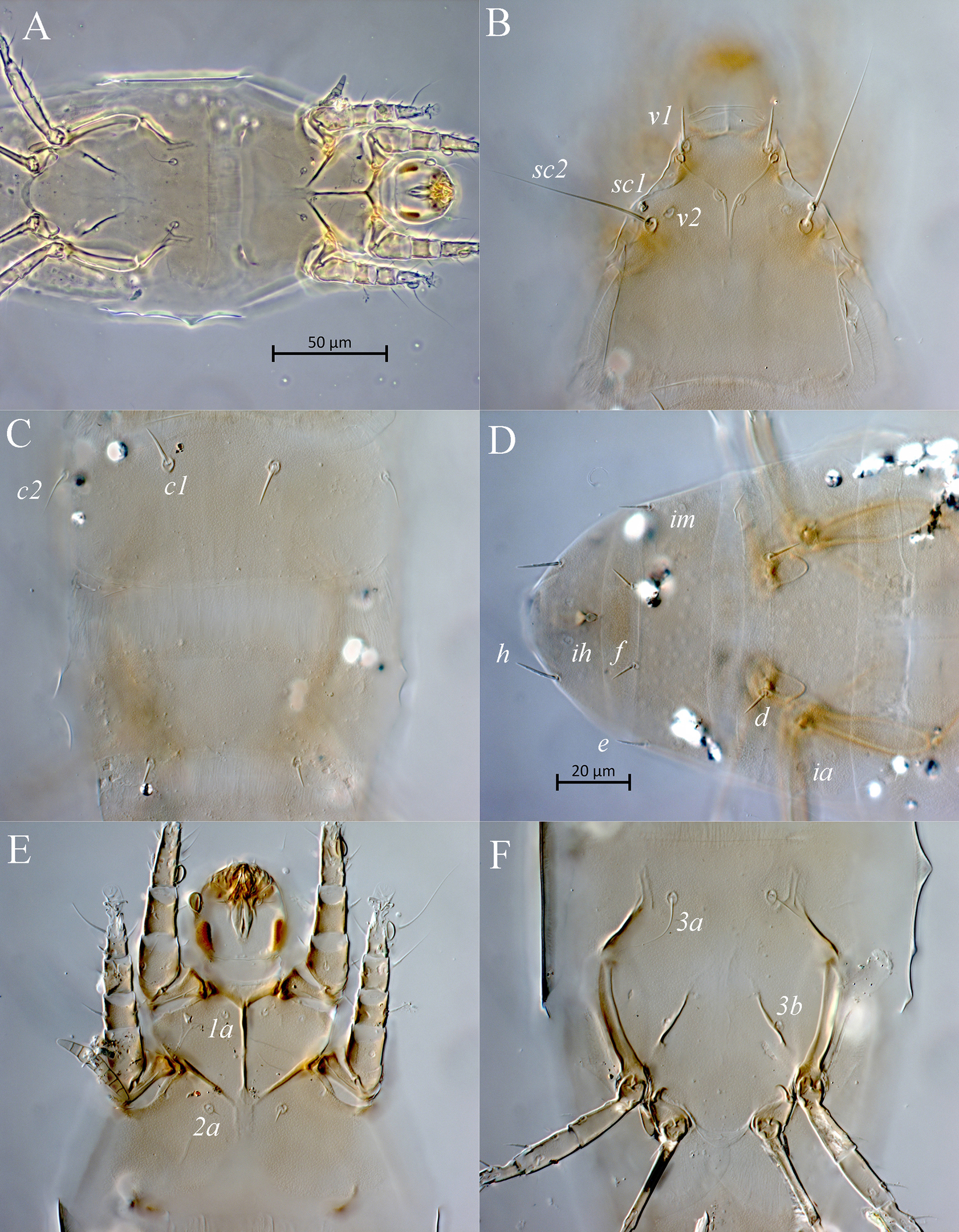
Female — Setae v1 situated anteriad stigmata; tracheal trunks without postatrial sclerotized sacs; setae 1a and 2a situated distinctly posteriad apodemes 1 and 2, respectively; prosternal apodeme indistinct posteriad median ends of apodemes 2; apodemes 3 with anterior projections; gnathosoma with ventrolateral pigmented spots.
Description
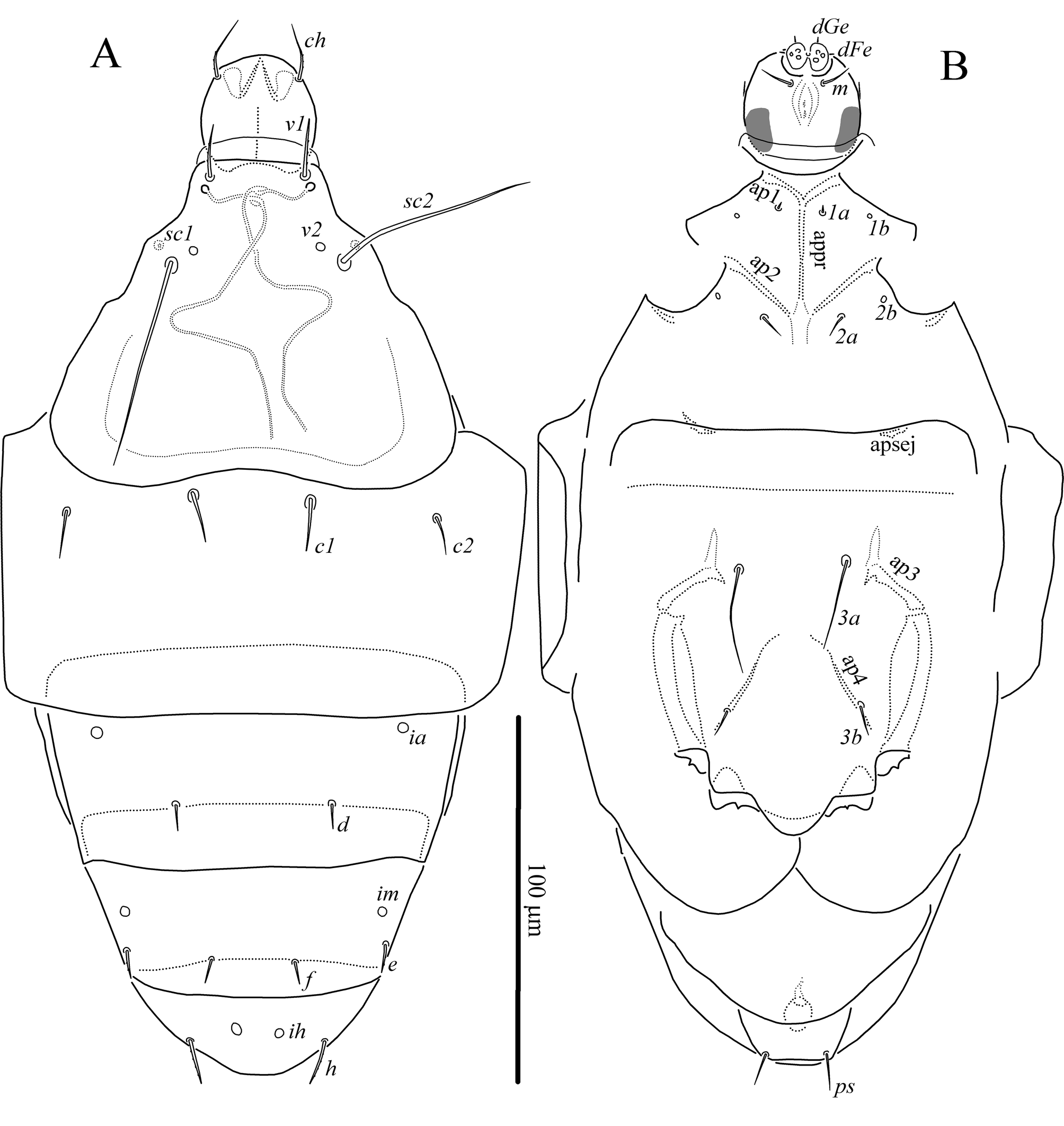
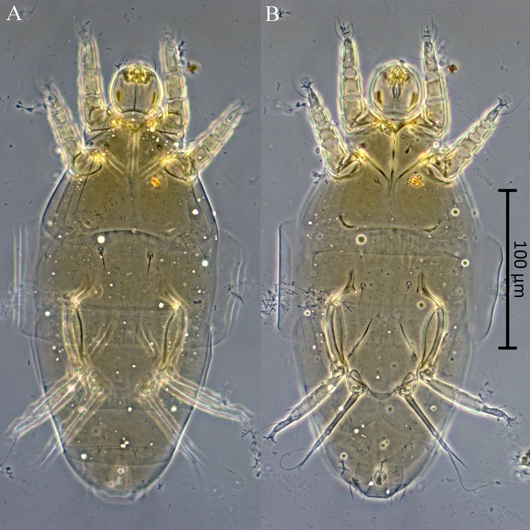
Female — (Figs 1–5). Idiosomal dorsum (Figs 1A, 4A, 5B–D). Length of idiosoma 245 (230), width 110 (100–105). All dorsal shields with uniform very small and hardly visible round puncta; posterior parts of prodorsal shield and tergites C, D, and EF with delicate striae (Figs 5B–D). Anterior margin of prodorsal shield straight. Stigmata located posteriad setae v1. Tracheal trunks without postatrial sclerotized sacs. Setae v1, e, and h weakly barbed, other dorsal setae smooth; setae sc2 and c2 pointed, other dorsal setae blunt-tipped; trichobothria sc1 vestigial (Fig. 5B). Setae sc2 situated in anterior half of prodorsal shield; pits v2 located anteromesad setae sc2; setae c1 situated slightly anteriad imaginary line connecting bases of setae c2. Lengths of dorsal setae: v1 16 (14–15), sc2 55 (53–56), c1 14 (13), c2 14 (13–15), d 8 (9–10), e 9 (9–10), f 7 (8), h 12 (12). Distances between setae: v1–v1 25 (23), sc2–sc2 45 (42), c1–c1 31 (26–29), c2–c2 96 (88–90), c1–c2 33 (30–32), d–d 41 (37–40), e–e 67 (60–64), e–f 22 (20–22), f–f 22 (20–22), h–h 35 (31–32).
Idiosomal venter (Figs 1B, 4B, 5A, E, F). Ventral plates with very small and hardly visible puncta (Figs 5E, F). All ventral setae smooth; setae 1a needle-like, other ventral setae pointed. Pits 1b and 2b small, round. Setae 1a and 2a located posteriad apodemes 1 and 2, respectively. Apodemes 1 fused with prosternal apodeme; apodemes 2 not fused with prosternal apodeme (Fig. 5A); prosternal apodeme indistinct posteriad median ends of apodemes 2; sejugal apodeme represented by small crescent-shaped structures; apodemes 3 with anterior projections; apodemes 4 thin and long; poststernal apodeme indistinct (Fig. 5A). Tegula length 7 (6–7), maximum width 16 (15). Anterior margin of posterior sternal plate with delicate striae (Fig. 5A). Lengths of ventral setae: 1a 3 (3), 2a 8 (7), 3a 28 (36–43), 3b 9 (9–10), ps 13 (11–13).
Gnathosoma (Figs 1, 5A, E). Gnathosomal capsule subcircular in outline. Length of gnathosomal capsule 32 (28–31), width 30 (29–32). Dorsomedian apodeme well developed. Gnathosoma with dorsal pair of setae ch 25 (18–20) distinctly longer than subcapitular setae m 8 (7–8); both setae pointed; setae ch weakly barbed, postpalpal setae situated laterally. Cheliceral levers well developed, nearly 2/3 the length of cheliceral stylets. Palpi very short, with two pairs of blunt-tipped setiform structures (dFe and dGe) distally and three pairs of tiny projections ventrally. Pharynx as on Figure 5E. Gnathosoma with ventrolateral pigmented spots (Fig. 5E).
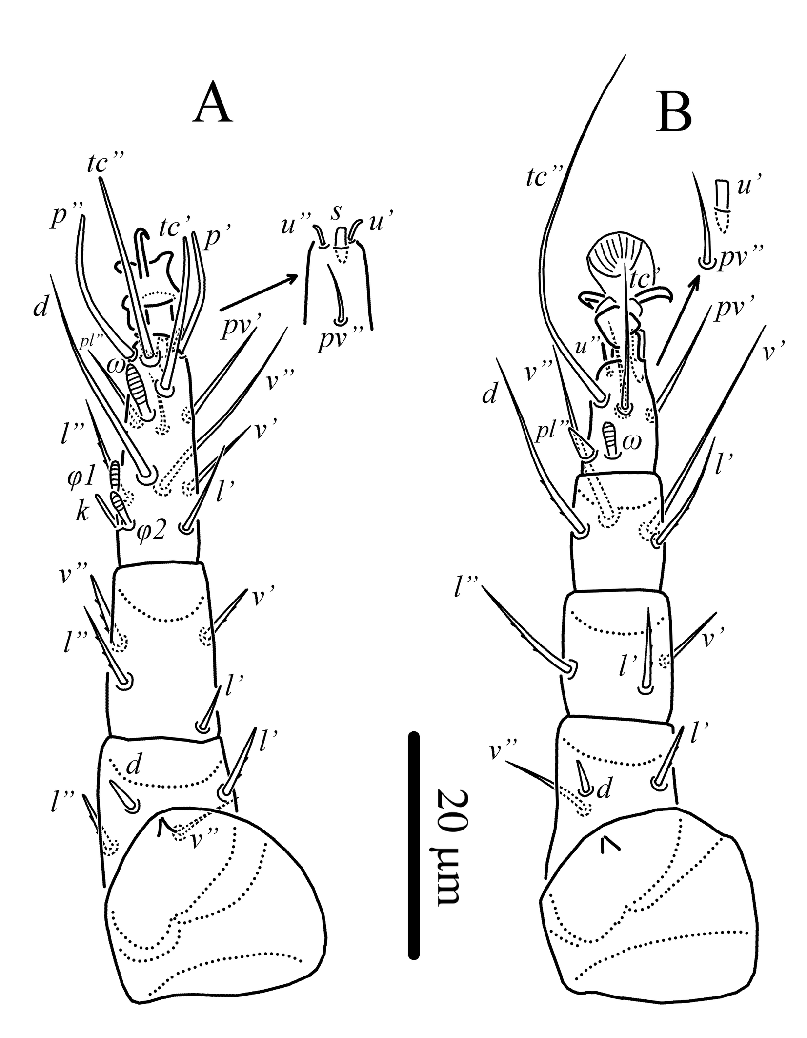
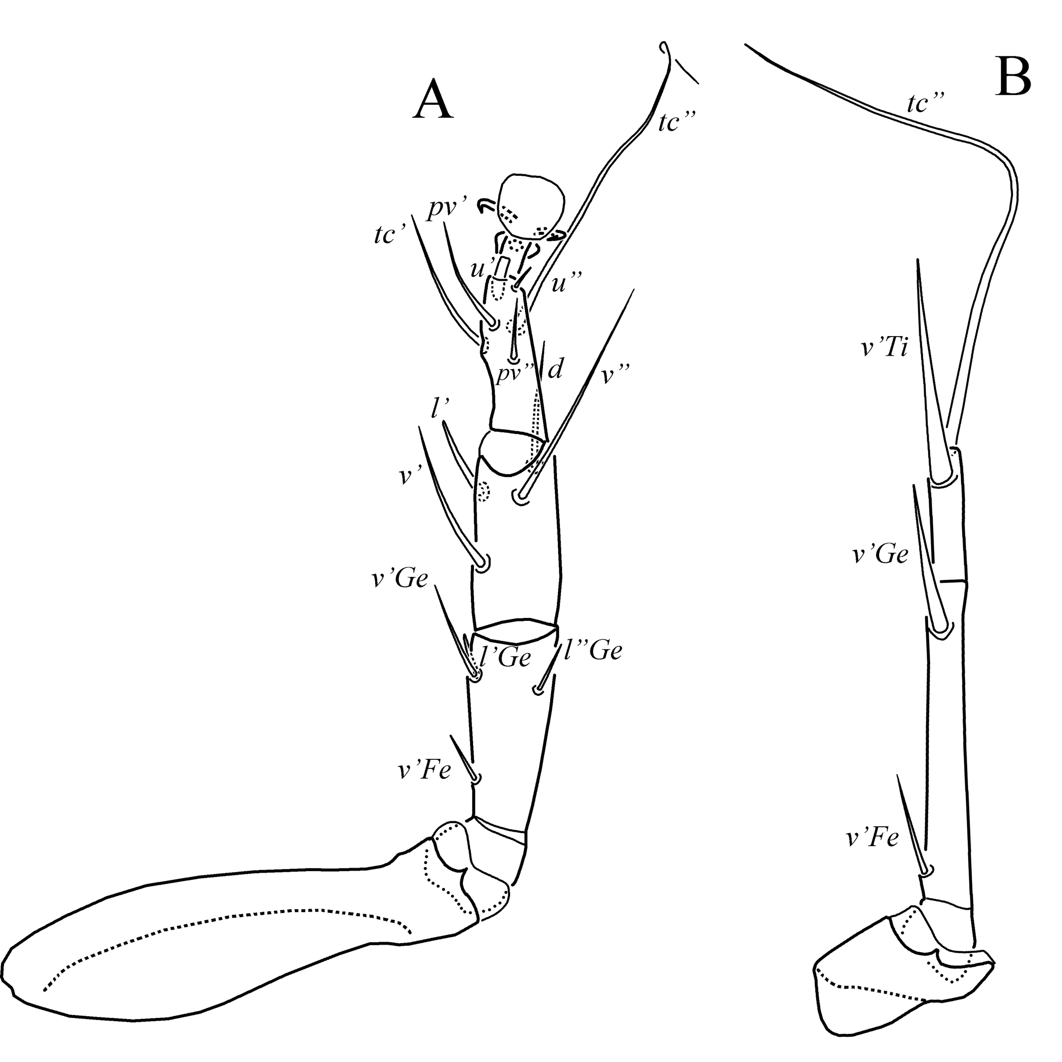
Legs (Figs 2, 3). Setae (u) of tarsus I, u″ of tarsi II and III short, blunt-tipped and well visible, included in setal count. Number of setae and solenidia on femur, genu, tibia, and tarsus, respectively: leg I: 4 (d, l′, l″, v″)-4 (l′, l″, v′, v″)-6(2)(d, l′, l″, v′, v″, k, φ1, φ2) + 10(l) (tc′, tc″, p′, p″, pl″, s, pv′, pv″, u′, u″, ω); leg II: 3 (d, l′, v″)-3 (l′, l″, v′)-4 (d, l′, v′, v″)-7(l) (pl″, tc′, tc″, u′, u″, pv′, pv″, ω); leg III: 1 (v′)+3 (l′, l″, v′)-4 (d, l′, v′, v″)-6 (tc′, tc″, u′, u″, pv′, pv″); leg IV: 1+1 (v′Fe, v′Ge)-1+1 (v′Ti, tc″). Leg I (Fig. 2A). Tarsal claw small, narrow, hooked distally. Solenidion ω slightly clavate, φ1 and φ2 slightly capitate; seta k 5 (4) rod-shaped. Lengths of solenidia: ω 6 (5–6), φ1 6 (5), φ2 4 (3). Setae (tc) and (p) of tibiotarsus smooth, blunt-tipped, eupathid-like; tc″ situated between p′ and p″; setae (tc) longer than (p). Seta s of tibiotarsus truncated distally; seta d of femur spiniform; setae l′, l″ of femur, l′, v′, v″ of genu blunt-tipped, other leg setae pointed; at least setae l′, l″ of femur, l″, v′, v″ of genu, and l″ of tibiotarsus weakly barbed, other setae smooth. Leg II (Fig 2B). Solenidion ω 4 (4–5) weakly clavate. Seta u′ truncated distally; setae d of femur and pl″ of tarsus spiniform; seta l′ of femur blunt-tipped, other setae pointed; at least setae l′ of femur, l′, l″ of genu, d, l′ of tibia weakly barbed. Empodium striated dorsally. Leg III (Fig. 3A). Empodium and seta u′ as on tarsus II. All setae smooth; setae l′ of genu and tibia blunt-tipped, other setae pointed. Leg IV (Fig. 3B). All setae smooth; seta v′ of tibiotarsus blunt-tipped, other leg setae pointed; seta tc″ of tibiotarsus long, whip-like.
Male and larva unknown.
Material examined
Holotype and 1 paratype females: slide 2993/4, Russia, Nizhny Novgorod Oblast, Sadovaya station, 10 Jul 1978, on Poa pratensis, collected by A.A. Sharonov; 2 females, Russia, Tyumen Oblast, city of Tyumen, Gagarin park, 57°10′N 65°36′E, on Calamagrostis arundinacea (Poaceae), 10 Jul 2025, collected by A.A. Khaustov.
Remark
Females collected from Western Siberia fit the original description of S. abruptus in all details and undoubtedly conspecific with type specimens. The holotype and one female paratype are in good condition. All morphological structures are clearly visible.
Siberian specimens were collected together with Steneotarsonemus (Steneotarsonemoides) rugosus Khaustov & Mashkov, 2026 on the same host plant.
This species was described from Central European Russia and Crimea (Livshits et al. 1982). This is the first record of S. abruptus in Asian Russia.
Discussion
Female of Steneotarsonemus abruptus sufficiently differs from other species of the subgenus Mahunkacarus in having stigmata located posteriad setae v1. Lindquist (1986) in the diagnosis of the subgenus Mahunkacarus mentioned that stigmata are located anteriad setae v1. On the other hand, this species is close to the subgenus Carextarsonemus Mitrofanov & Sharonov, 1988 in having apodemes 3 with long anterior projections. However, in Carextarsonemus trichobothria sc1 and their bothridia are well developed in contrast to strongly reduced in Mahunkacarus. The most remarkable difference is the presence of pigmented spots on lateroventral face of the gnathosomal capsule. This character state is currently known only in Steneotarsonemus (Neosteneotarsonemus) arcticus Lindquist, 1986 and Maculanemus chilensis Khaustov & Tolstikov, 2025, however in M. chilensis pigmented spots are located on ventral face of the gnathosomal capsule (Khaustov & Tolstikov 2025). Lindquist (1986) apparently did not use the presence of pigmented spots on gnathosoma in Tarsonemidae taxonomy because they occur in only one species. In my opinion it is a good synapomorphy for further taxonomic and phylogenetic study which can be used in the revision and reclassification of Steneotarsonemus and closely related genera in the tribe Steneotarsonemini. The similarity between females of S. abruptus and S, arcticus is more substantial than between S. abruptus and other species of the subgenus Mahunkacarus. Female of S. abruptus differs from that of S. arcticus in having vestigial setae sc1 (vs. setiform in S. arcticus) and the presence of the solenidion φ2 (absent in S. arcticus). The reduction of setae sc1 likely occurred independently in different lineages of the genus Steneotarsonemus. To clarify the phylogenetic relationships within the genus Steneotarsonemus, further studies integrating molecular data from a wider range of subgenera representatives are required.
Acknowledgements
The present research was supported by the Russian Science Foundation, grant No. 25-14-00118.
References
- Beer R. E. 1954. A revision of the Tarsonemidae of the Western Hemisphere (Order Acarina). Univ. Kans. Sci. Bull., 36(16): 1091-1387.
- Canestrini G., Fanzago F. 1877. Intorno agli Acari italiani. Atti Ist. Veneto Sci. Lett. Arti, Ser. 5, 4: 69-208 (Prospetto repaginaion: 1-140), 6 pl.
- Ganguly M., Mondal P., Karmakar K. 2021. Taxonomic notes on subgenus Steneotarsonemoides (Acari: Tarsonemidae) with description of a new species of Steneotarsonemus from Tiger grass in the northern hill zone of West Bengal, India. Zootaxa, 3023(3): 405-420. https://doi.org/10.11646/zootaxa.5023.3.5
- Ganguly M., Karmakar K. 2022. Two new species of Steneotarsonemus (Acari: Tarsonemidae) from the Himalayan forests of West Bengal, India. Syst. Appl. Acarol., 27(12): 2476-2492. https://doi.org/10.11158/saa.27.12.7
- Grandjean F. 1944. Observations sur les Acariens de la famille des Stigmaeidae. Arch. Scie. Phys. Natur., 26: 103-131.
- Kayal S., Karmakar K., Moraes J. 2021. Sources of infestation of the rice sheath mite, Steneotarsonemus spinki Smiley (Acari: Tarsonemidae), in West Bengal, India. Int. J. Pest Manage., 70: 357-363. https://doi.org/10.1080/09670874.2021.1973691
- Khaustov A.A. 2026. Review of the subgenus Steneotarsonemus (Carextarsonemus) (Acari: Heterostigmata: Tarsonemidae) described by Livshits, Mitrofanov and Sharonov. Syst. Appl. Acarol., 31(5): 759-798. https://doi.org/10.11158/saa.31.5.7
- Khaustov A.A., Mashkov K.A. 2025. Redescription of the phytophagous mite Steneotarsonemus arcuatus (Acari: Heterostigmata: Tarsonemidae), a pest of cereals in Western Siberia, Russia. Syst. Appl. Acarol., 30(12): 2172-2192. https://doi.org/10.11158/saa.30.12.4
- Khaustov A.A., Mashkov K.A. 2026. Review of the subgenus Steneotarsonemus (Steneotarsonemoides) (Acari: Heterostigmata: Tarsonemidae) of Russia. Zootaxa, 5785(2): 253-289. https://doi.org/10.11646/zootaxa.5785.2.2
- Khaustov A.A., Tolstikov A.V. 2025. A new genus and two new species of Tarsonemidae (Acari: Heterostigmata) from Chilean Patagonia. Pers. J. Acarol, 14(2): 317-334. https://doi.org/10.22073/pja.v14i2.86917
- Lin J.-Z., Zhang Z.-Q. 2002. Tarsonemidae of the world: key genera, geographical distribution, system¬atic catalogue and annotated bibliography. London, Systematic & Applied Acarology Society, 440 pp.
- Lin J.-Z., Zhang Z.-Q. 2005. New Zealand species of Steneotarsonemus Beer (Acari: Tarsonemidae). Zootaxa, 1028, 1-22. https://doi.org/10.11646/zootaxa.1028.1.1
- Lindquist E.E. 1986. The world genera of Tarsonemidae (Acari: Heterostigmata): a morphological, phylogenetic, and systematic revision, with a reclassification of family-group taxa in the Heterostigmata. Mem. Entom. Soc. Can., 118: 1-517. https://doi.org/10.4039/entm118136fv
- Livshits I.Z., Mitrofanov V.I., Petrushov A.Z. 2011. Agricultural Acarology. GNU VSTISP Rosselkhozakademii, Moscow. 351 pp [in Russian]
- Livshits I.Z., Mitrofanov V.I., Sharonov A.A. 1982. Novye vidy kleschei roda Steneotarsonemus (Tarsonemidae, Acariformes). Biol. Nauki, 10: 24-28. [in Russian]
- Mahunka S. 1971. Tarsonemina (Acari) species from Korea. Acta Zool. Hung., 17: 271-294.
- Mondal P., Ganguly M., Karmakar K., Lofego A.C., Moraes G.J. de. 2021. A new species of Steneotarsonemus (Acari: Tarsonemidae) from common reed grass, Phragmites australis (Poaceae) in the wetlands of Eastern India. Int. J. Acarol., 47(4): 289-300. https://doi.org/10.1080/01647954.2021.1900912
- Shikata E., Kawano S., Senboku T., Tiongco E.R., Miyajima K. 1984. Small virus-like particles isolated from the leaf sheath tissues of rice plants and from the rice tarsonemid mites, Steneotarsonemus spinki Smiley (Acarina, Tarsonemidae). Ann. Phyto. Soc. Jap., 50: 368-374. https://doi.org/10.3186/jjphytopath.50.368
- Seeman O.D., Loch D.S., Knihinicki D.K., McMaugh P.E. 2016. A new species of Steneotarsonemus (Acari: Tarsonemidae) from kikuyu grass, Pennisetum clandestinum (Poaceae), in Australia. Syst. Appl. Acarol., 21(7): 889-906. https://doi.org/10.11158/saa.21.7.4
- Suski Z.W. 1970. Polish mites of the family Tarsonemidae (Acarina, Heterostigmata). IV. Steneotarsonemus gibber n. sp. Bull. Acad. Polon. Sci. Cl. V, 18(5): 277-282.
- Wainstein B.A. 1979. Novye I maloizvestnye kleschi sem. Tarsonemidae (Acariformes) so zlakov. Entomol. Obozr., 58(3): 663-670. [in Russian]
to post any comment or correction.


2026-05-31
Date accepted:
2026-08-13
Date published:
2026-08-24
Edited by:
Auger, Philippe

This work is licensed under a Creative Commons Attribution 4.0 International License
2026 Khaustov, Alexander A.
 Download article
Download the citation
RIS with abstract
(Zotero, Endnote, Reference Manager, ProCite, RefWorks, Mendeley)
RIS without abstract
BIB
(Zotero, BibTeX)
TXT
(PubMed, Txt)