


Two new species from the genus Phymatodiscus Berlese, 1917 from Brunei (Acari: Mesostigmata: Phymatodiscidae)
Kontschán, Jenő  1
and Ermilov, Sergey G.
2
1
and Ermilov, Sergey G.
2
1✉ Plant Protection Institute, HUN-REN Centre for Agricultural Research, H-1525 Budapest, P.O. Box 102, Hungary & Department of Plant Sciences, Albert Kázmér Faculty of Mosonmagyaróvár, Széchenyi István University, Vár square 2., H-9200 Mosonmagyaróvár, Hungary.
2Institute of Environmental and Agricultural Biology (X-BIO), University of Tyumen, Lenina str. 25, 625000 Tyumen, Russia.
2026 - Volume: 66 Issue: 2 pages: 511-520
https://doi.org/10.24349/0jfk-n2hoZooBank LSID: 49E8AD2C-360A-43C4-9180-C561A3226F7E

Original research
Keywords
Abstract
Introduction
The genus Phymatodiscus was described by Berlese (1917) for an unusual species (Discopoma miranda Berlese, 1905) from Java, Indonesia. Several years later, Hirschmann (1977) studied this genus and moved two previously described species, Discopoma conifera Canestrini, 1897, and Trachyuropoda titanica Berlese, 1905, to the genus Phymatodiscus. In the same paper, Hirschmann (1977) described four new species from New Guinea as well. Some years later, Hiramatsu (1979) described a new species from Japan and two new species from Indonesia (Hiramatsu 1985). After many years, Kontschán and Starý (2011, 2012) described a new species from Vietnam and another from Malaysia, and subsequently, Kontschán and Ripka (2016) described a new species from Singapore. Kontschán and Ermilov (2023) elevated the genus Phymatodiscus to the family level as family Phymatodiscidae Kontschán and Ermilov, 2023, which includes two different genera: Phymatodiscus Berlese, 1917, without eye-like dorsal furrow, and Bardizon Kontschán and Ermilov, 2023, with eye-like dorsal furrows, and described two new Bardizon species as well.
During the investigation of the Oriental soil-dwelling Uropodina in the collection of the Natural History Museum, Geneva (Switzerland), we found two unusual phymatodiscid mites, which described as new species herein.
Material and methods
Specimens examined were cleared in lactic acid for two weeks and then placed on half-covered slides and examined using a Leica 1000 microscope with a drawing tube. Photographs were taken with a Keyence VHX 5000 digital microscope. Specimens examined are stored in 70% ethanol and deposited in the Natural History Museum, Geneva (NHMG). Setae h = hypostomal seta, st = sternal seta. All measurements and the scales in the figures are given in micrometres (μm).
Taxonomy
Phymatodiscidae Kontschán and Ermilov, 2023
Phymatodiscus Berlese, 1917
Phymatodiscus bruneiensis n. sp.
ZOOBANK: 621B4E9F-9D47-49E1-8650-69615EF3CE1B
(Figures 1–3, 7a–c)
Diagnosis
Marginal part of the dorsal idiosoma elevated from central part. Elevated part bearing T-shaped setae. Central area of dorsal shield with a small strongly sclerotized prolongation. Five pairs of marginal prolongations situated only on the caudal area of marginal shield. Female genital shield with small oval pits, setae st4–5 longer than other sternal setae. Sternal and ventral setae T-shaped. Crossbar of T-shaped ventral setae with serrate margins.
Material examined
Holotype. Female. Bru-88/21. Brunei, Belait District, Andulau Forest Reserve, 3.5 km S de Sungai Liang (39.5 km de Labit), forêt primaire (mixed dipterocarp forest), K-7 (Kompartment 7), prélèvement de sol au pied de grands arbres, 50 m; 19.XI.1988; leg. B. Hauser. Paratypes. One female and one male. Locality and date same as in holotype.
Description
Female (n=2) Length of idiosoma 770–778, width 618–623. Shape of idiosoma oval, posterior margin with four pairs of wide prolongation. Colour reddish-brown.

Dorsal idiosoma (Figures 1, 3a, 7a–c). Dorsal shield completely separated from marginal shield. Margins of dorsal shield elevated from the central area. Central part of elevated area with several grooves and four-six T-shaped setae among grooves (Figure 3a). Inner and outer margins of elevated part bearing curved and serrate setae (ca 17–20). Surface of non-elevated central part of dorsal shield smooth and bearing 11–13 pairs of finely pilose setae. The central area of dorsal shield with one pair of strongly sclerotized bulges. Anterior bulge bearing finely pilose setae (ca 37–45), posterior bulge with a deep incision (ca 20–22) on posterior margin. Two pairs of lateral strongly sclerotized grooves (ca 68–86 in length) situated lateral to strongly sclerotized central bulge.
Surface of marginal shield smooth, on anterior region with a deep incision. All setae on marginal shield curved and smooth (ca 29–35 long). Caudal part of marginal shield with five pairs trapezoid protuberances (ca 24–26 long and ca 57–65 wide at basis) bearing three marginal setae.
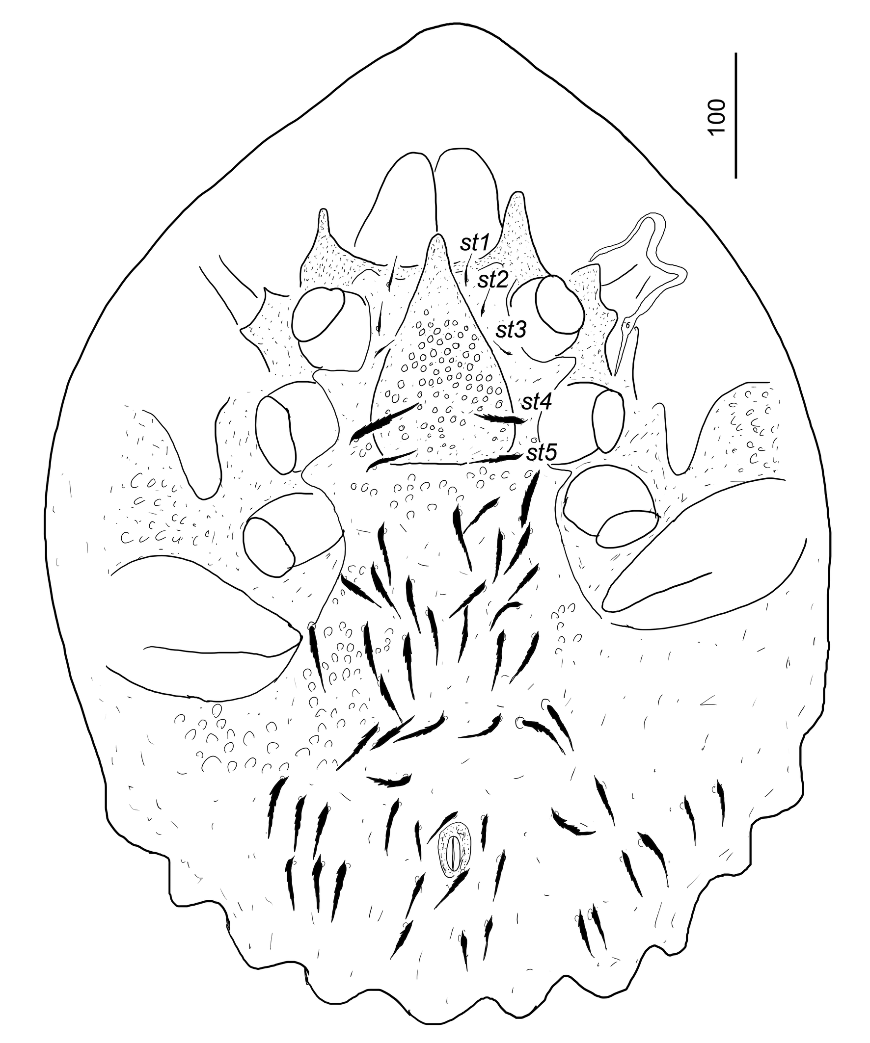
Ventral idiosoma (Figure 2). Anterior part of sternal shield smooth, close to st4 bearing some oval pits. All sternal setae T-shaped. Setae st1–st3 with short stem, smooth and short crossbar (ca 17–21), st4 and st5 with short stem and long (ca 40–57), serrate crossbar. Setae st1 situated close to anterior margin of sternal shield, st2 and st3 at level of midcoxae II, st4 at level of midcoxae III, st5 near basal edges of genital shield.
Ventral shield covered by oval pits (7–8×6–8) and bearing more the 18 pairs of T-shaped setae. Ventral setae with a short stem and a long and serrate crossbar (ca 39–47). Anal shield 26–28 long and 9–11 wide.
Genital shield scutiform with narrow apical part, 177–180 long and 106–108 wide at level of st4. Surface of genital shield covered by small oval pits (ca 4–5×4–5).
Pedofossae deep, smooth and separate furrow for tarsi IV absent. Stigmata situated between coxae II and III. Prestigmatid part of peritreme m-shaped, poststigmatid part short and straight.
Tritosternum with narrow basis, its laciniae trifurcated for two short lateral and one long central branches. Margins of trifurcated branches pilose (Figure 3b)
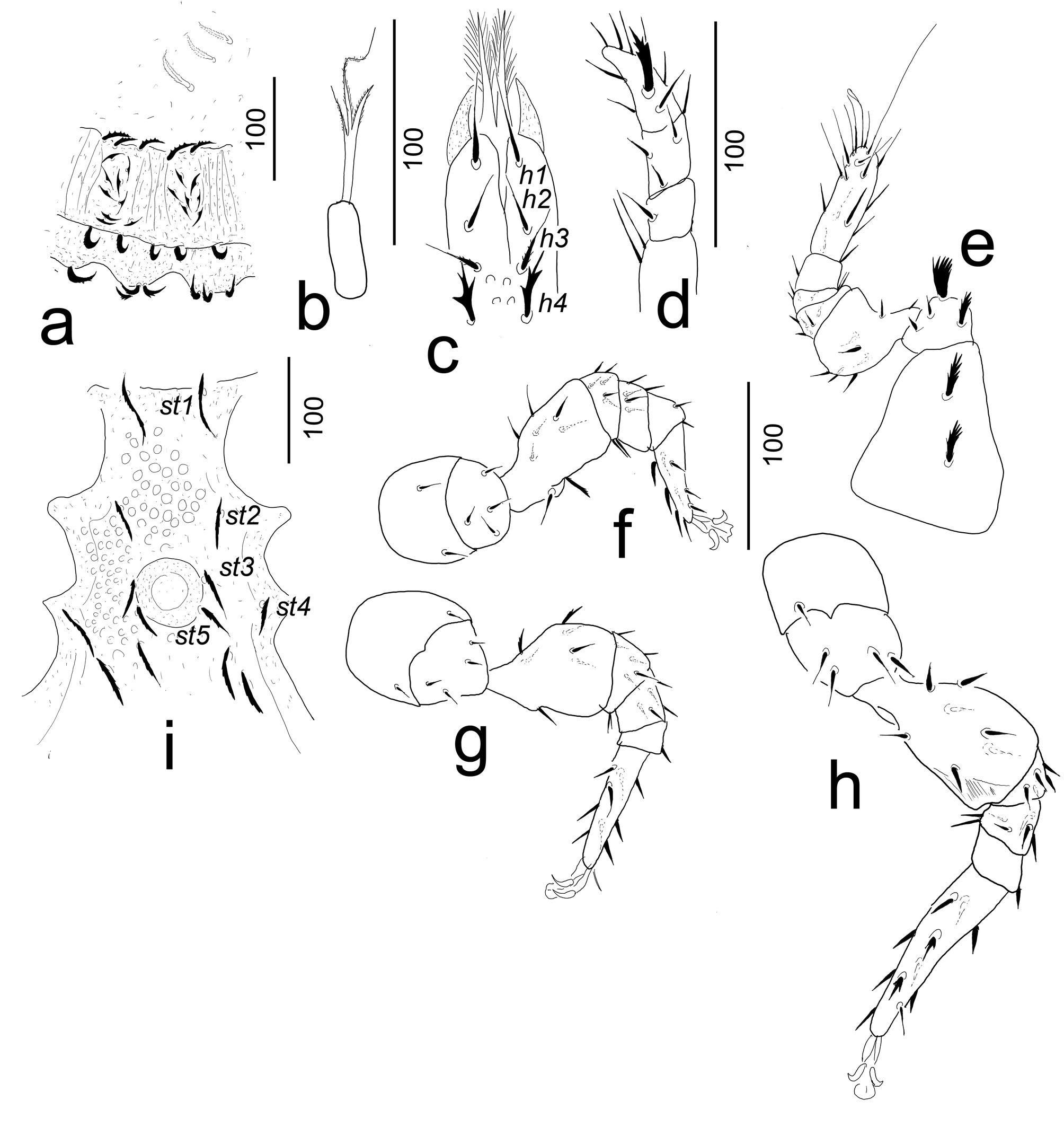
Gnathosoma (Figure 3c). Corniculi horn-like, internal malae longer than corniculi and with long hairs on margins. Hypostomal setae h1 long (ca 38–42) and smooth, h2, h3 and h4 shorter (ca 25–27), h2 smooth, h3 pilose, h4 marginally serrate. Palp with two smooth ventral setae on trochanter, other setae on palp smooth, except a robust seta (ca 30–32 long) on palp tarsus (Figure 3d). Epistome and chelicerae not visible without breaking of idiosoma.
Legs (Figures 3e–h). Leg I without ambulacral claws, majority of setae on legs smooth and needle-like setae; except coxae and trochanter of leg I, femora of legs II–III and tarsi of leg IV, bearing serrate setae. Legs length: I 300–312, II 258–264, III 290–302, IV 355–360.
Male. Length of idiosoma 610–630, width 390–405 (n=1).
Dorsal idiosoma. Ornamentation and chaetotaxy as in adult female.
Ventral idiosoma. Surface of sternal shield covered by oval pits. Five pairs of long (ca 38–42 µm) and serrate sternal setae present. Setae st1 situated close to anterior margin of sternal shield, st2 at level of posterior margin of coxae II, st3 and st5 close to lateral margin of genital opening, st4 at posterior margin of coxae III. Genital shield circular, without sculptural pattern and without eugenital setae, ca 64 long and ca 62 wide. Shield situated between coxae III–IV (Figure 3i). Position and shape of ventral setae and ornamentation of ventral shield as in adult female.
Nymphs and larvae. Unknown.
Etymology
The name of the new species refers to the country, where the type materials were collected.
Remarks
The T-shaped sternal and ventral setae are unique within this family. The new species has marginal prolongations situated on the caudal area. This character state is visible only in the Japanese P. iriomotensis Hiramatsu, 1979 (see Kontschán and Ermilov 2023). The distinguishing characteristic between those two species are summarized in Table 1.
Download as
Characters
P. bruneiensis n. sp.
P. iriomotensis
Length of dorsal setae
reaching the insertion of next setae
not reaching the insertion of next setae
Marginal prolongations with pilose setae on dorsal shield
absent
present
T-shaped sternal and ventral setae
present
absent
Setae st4 and st5
longer than st1–st3
as long as st1–st3
Length of ventral setae
reaching the insertion of next setae
not reaching the insertion of next setae
Sculptural pattern on female genital shield
with oval pits
with web-like
Position of oval pits on male sternal shield
anterior and posterior to genital opening
anterior to genital opening
Prestigmatid part of peritreme
m-shaped
harpin-bend like with a lateral bend
Phymatodiscus hiramatsui n. sp.
ZOOBANK: 79D88108-5B65-4D18-8444-DA684CF6BC1E
(Figures 4–6, 7d–f)
Diagnosis
Marginal prolongations absent. Dorsal shield with two central elevated area. Non-elevated part of dorsal shield covered by oval pits. Anterior part of elevated area with very long smooth setae. Female genital shield with web-like sculptural pattern.
Material examined
Holotype. Female. Bru-88/21. Brunei, Belait District, Andulau Forest Reserve, 3.5 km S de Sungai Liang (39.5 km de Labit), forêt primaire (mixed dipterocarp forest), K-7 (Kompartment 7), prélèvement de sol au pied de grands arbres, 50 m; 19.XI.1988; leg. B. Hauser. Paratypes. Two females and one male. Locality and date same as in holotype.
Description
Female (n=3) Length of idiosoma 540–555, width 430–442. Shape of idiosoma oval, posterior margin rounded. Colour yellowish-brown.

Dorsal idiosoma (Figures 4, 7d–f). Dorsal shield completely separated from marginal shield. Two large (109–122 long and 80–133 wide) elevated area situated on central part of the dorsal shield. Posterior margin of anterior elevated part with smooth and long setae (ca 37–47). Two pairs of longer setae (ca 70–94) situated on lateral part of the anterior elevated area. Anterior margin of posterior elevated area with smooth and long setae (ca 35–44), dorsal part with three pairs of shorter (ca 20–22) and one pair of very long (ca 85–90) and smooth setae. Dorsal shield with eleven pairs of long (ca 34–45) and serrate setae on margins, and two pairs of short (ca 17–22) and smooth setae situated close to posterior margin of dorsal shield. Surface of non-elevated central part of dorsal shield covered by oval pits (ca 8–9×10–11). Margins of dorsal shield with more than 22 smooth and long setae (ca. 17–20) situated in two rows, anterior margin of dorsal shield with several serrate setae (ca 20–22 in length). Marginal shield without sculptural pattern and bearing more than 40 pairs of serrate setae (ca 17–22 long).

Ventral idiosoma (Figure 5). Anterior part of sternal shield with oval pits, other part smooth. All sternal setae smooth, st1–st4 smooth and short (ca 12–14), st5 longer (ca 30–32). Setae st1 situated close to anterior margin of sternal shield, st2 at level of posterior margin of coxae II, st3 at level of anterior margin of III, st4 at level of posterior margin of coxae III, st5 near basal edges of genital shield.
Ventral shield covered by irregular pits (9–11×10–14) on central area and web-like sculptural pattern posterior to pedofossae IV and anal opening. Central part of ventral shield bearing 16 pairs of long (ca 24–36) and serrate setae. Anal shield ca 18–20 long and ca 11–12 wide. Genital shield scutiform, 159–162 long and 100–104 wide at level of st4. Surface of genital shield covered by web-like sculptural pattern.
Pedofossae deep, smooth and separate furrow for tarsi IV absent. Stigmata situated between coxae II and III. Prestigmatid part of harpin-bend-like with a triangular hoop between stigmata and hairpin-bend part, poststigmatid part short and straight.

Tritosternum with narrow basis, its laciniae trifurcated for two short lateral and one long central branch. Margins of trifurcated branches pilose (Figure 6a)
Gnathosoma (Figure 6b). Corniculi horn-like, internal malae longer than corniculi and with long hairs on margins. Hypostomal setae h1, h2 and h3 smooth, h1 long (ca 24–26), h2 short (ca 10–12), h3 long (ca 20–21), h4 marginally serrate and ca 15–16 long. Epistome with serrate base and pilose apical part (Figure 6c). Movable digit of chelicerae as long as fixed digit, both digits bearing one-one triangular tooth, internal sclerotized node associated with levator tendon present (Figure 6d).
Legs (Figures 6e–h). Leg I without ambulacral claws, majority of setae on legs smooth and needle-like setae; except some pilose setae on genua and tibiae I, trochanter II. One robust ventral seta situated on trochanter I. Legs length: I 290–301, II 261–263, III 230–3235, IV 300–305.
Male. Length of idiosoma 610–630, width 390–405 (n=1).
Dorsal idiosoma. Ornamentation and chaetotaxy as in adult female.
Ventral idiosoma. Surface of sternal shield covered by web-like sculptural pattern and some oval pits. Setae st1 smooth, ca 11 long and situated close to anterior margin of sternal shield, st2 smooth, ca 17–18 long and situated at level of posterior margin of coxae II, st3 smooth, ca 27–29 long and situated at level of posterior margin of coxae III. Setae st4 and st5 long (ca 27–29) and serrate, situated close to lateral margin of genital opening. Genital shield circular, without sculptural pattern and without eugenital setae, ca 44 long and ca 34 wide. Genital shield situated between coxae IV (Figure 6i). Position and shape of ventral setae and ornamentation of ventral shield as in adult female.
Nymphs and larvae. Unknown.

Etymology
The new species is dedicated to Dr. Nabou Hiramatsu (Keya-myo Tarami-cho, Nisisonogi-gun, Nagasaki-ken, Japan), the noted Japanese Uropodina researcher, who gave a huge contribution to the knowledge of the Southeast Asian Uropodina.
Remarks
The new species do not have marginal prolongations. This character state is visible only in the New Guinean P. polyglottis Hirschmann, 1977 (see Kontschán and Ermilov 2023). The distinguishing characteristic between these two species are summarized in Table 2.
Download as
Characters
P. hiramatsui n. sp.
P. polyglottis
Elevated part of dorsal shield
narrower
wider
Setae on elevated part of dorsal shield
smooth
pilose
Very long and smooth setae on lateral area of anterior elevated part of dorsal shield
present
absent
Sculptural pattern of elevated areas
smooth
with very small oval pits
Sculptural pattern on lateral part of dorsal shield
with oval pits
smooth
Web-like sculptural pattern on male sternal shield
present
absent
Setae st4 and st5 on male
longer than st1–st3 and serrate
similar in shape and length of st1–st3
Zoogeographical notes
Members of the family Phymatodiscidae were not found in the soil and neighbouring habitats in Brunei (Kontschán and Ermilov 2023) until now. The members of this family occur only in New Guinea, Indonesia, Malaysia, Vietnam and Singapore (Kontschán and Ermilov 2023), so the first occurrence of this family and genus in Brunei was expected. We need to assume that many other new species will be discovered from the Oriental and Australasian regions in the near future.
Acknowledgements
We are very grateful to Dr. Peter Schwendinger (MHNG) for his kind hospitality during the first author's visit to Geneva. We would also like to thank the two anonymous reviewers for their useful comments.
References
- Berlese A. 1905. Acari nuovi. Manipulus IV. (Acari di Giava). Redia. 2: 154-176.
- Berlese A. 1917. Intorno agli Uropodidae Fasc. 1. Redia. 113: 7-16.
- Canestrini G 1897. Nuovi Acaroidei della N. Guinea. Természetrajzi Füz. 20: 401-462.
- Hiramatsu N. 1979. Gangsystematik der Parasitiformes. Teil 333. Stadien einer neuen Phymatodiscus-Art aus Japan (Dinychini, Uropodinae). Acarologie. 25: 108-109.
- Hiramatsu N. 1985. Zwei neue Phymatodiscus-Arten (Acari, Uropodidae) aus Borneo. Kontyu. 53(2): 270-276.
- Hirschmann W. 1977. Gangsystematik der Parasitiformes. Teil 254. Die Gattung Phymatodiscus (Berlese, 1917), Teilgang and Stadien von 4 neuen Phymatodiscus-Arten aus Neuguinea (Dinychini, Uropodinae). Acarologie. 23: 6-65.
- Kontschán J., Ermilov SG. 2023. Remarks on the genus Phymatodiscus Berlese, 1917, with the description of Phymatodiscidae fam. nov. and Bardizon eotvosi gen. nov., sp. nov. from Indonesia (Acari, Mesostigmata). ZooKeys. 1182: 223-235. https://doi.org/10.3897/zookeys.1182.109744
- Kontschán J., Ripka G. 2016. Three new Uropodina mites (Acari: Mesostigmata) from Singapore. Rev. Suisse Zool. 123(2): 291-301. https://doi.org/10.5281/zenodo.155303
- Kontschán J., Starý J. 2011. Uropodina species from Vietnam (Acari: Mesostigmata). Zootaxa. 2807: 1-28. https://doi.org/10.11646/zootaxa.2807.1.1
- Kontschán J., Starý J. 2012. New Uropodina species and records from Malaysia (Acari: Mesostigmata). Acta Zool. Acad. Sci. Hung. 58(2): 177-192. http://doi.org/10.5281/zenodo.5735765
to post any comment or correction.


2026-03-09
Date accepted:
2026-05-22
Date published:
2026-05-27
Edited by:
Faraji, Farid

This work is licensed under a Creative Commons Attribution 4.0 International License
2026 Kontschán, Jenő and Ermilov, Sergey G.
 Download article
Download the citation
RIS with abstract
(Zotero, Endnote, Reference Manager, ProCite, RefWorks, Mendeley)
RIS without abstract
BIB
(Zotero, BibTeX)
TXT
(PubMed, Txt)