


Phoretic, trophic and fungal associations of Histiogaster arborsignis (Acari: Acaridae) with Ips typographus: a study case from the Czech Republic
Milosavljević, Marija  1
; Žurovcová, Martina
2
; Toušková, Hana
3
; Žurovec, Michal
4
; Brož, Václav
5
and Kolesnikov, Vasiliy B.
6
1
; Žurovcová, Martina
2
; Toušková, Hana
3
; Žurovec, Michal
4
; Brož, Václav
5
and Kolesnikov, Vasiliy B.
6
1Biology Centre of the Czech Academy of Sciences, Institute of Entomology, Ceske Budejovice, 37005, Czech Republic.
2Biology Centre of the Czech Academy of Sciences, Institute of Entomology, Ceske Budejovice, 37005, Czech Republic.
3Biology Centre of the Czech Academy of Sciences, Institute of Entomology, Ceske Budejovice, 37005, Czech Republic & Faculty of Science, University of South Bohemia, Ceske Budejovice, 37005, Czech Republic.
4Centre of the Czech Academy of Sciences, Institute of Entomology, Ceske Budejovice, 37005, Czech Republic & Faculty of Science, University of South Bohemia, Ceske Budejovice, 37005, Czech Republic.
5Biology Centre of the Czech Academy of Sciences, Institute of Entomology, Ceske Budejovice, 37005, Czech Republic.
6Papanin Institute for Biology of Inland Waters, Russian Academy of Sciences, Borok, Russia.
2026 - Volume: 66 Issue: 2 pages: 317-326
https://doi.org/10.24349/17bh-a0ybOriginal research
Keywords
Abstract
Introduction
Histiogaster arborsignis Woodring, 1963 (Acari: Acaridae) is a cosmopolitan fungivorous mite widely associated with wood-boring insects, particularly bark and ambrosia beetles (Moser and Bogenschütz 1984; Okabe 1993; Okabe 1993a; Hulcr and Dunn 2011; Vissa and Hofstetter 2017; Klimov and Khaustov 2018; Berto et al. 2025). Like many other acarid mites, it possesses the characteristic astigmatid phoretic deutonymph (hypopus), a heavily sclerotised, non-feeding dispersal stage equipped with a ventral sucker plate that enables firm attachment to insect hosts during transport (Walter and Proctor 2013; Seeman and Walter 2023; Bajerlein et al. 2024). Hypopus formation is environmentally induced and allows the mite to exploit the patchy, ephemeral fungal habitats typical of subcortical insect galleries, where active stages resume feeding and reproduction once suitable fungal resources are encountered (Okabe 1993; Okabe 1993a; Berto et al. 2025). Through this life-history strategy, H. arborsignis integrates into the complex ecological networks of beetles and their fungal symbionts.
It is recognised as a generalist fungivore inhabiting galleries rich in symbiotic and opportunistic fungi (Mori et al. 2011). Experimental studies have demonstrated its ability to develop on multiple beetle-associated fungi, including ophiostomatoid and ambrosia fungi, highlighting its ecological compatibility with subcortical microbial communities (Okabe 1993; Okabe 1993a; Berto et al. 2025). Related acarid taxa have also been shown to utilise major bark beetle fungal associates, contributing to microbial turnover and influencing competitive interactions among fungi in the gallery environment (Bridges and Moser 1983 Cardoza et al. 2008; Vissa and Hofstetter 2017).
Across Europe, Ips typographus (Linnaeus, 1758) (Coleoptera: Curculionidae: Scolytinae), the Eurasian spruce bark beetle, hosts one of the most diverse known assemblages of phoretic mites. (Moser and Bogenschütz 1984; Moser et al. 1997; Paraschiv and Isaia 2020). A recent synthesis documented 97 mite species associated with I. typographus across 12 European countries, with Russia, Germany, and Finland exhibiting the highest richness (Milosavljević et al. 2022). The most widespread species are Dendrolaelaps quadrisetus, Trichouropoda polytricha, Uroobovella ipidis, Proctolaelaps fiseri, and Histiostoma piceae (Khaustov et al. 2018; Milosavljević et al. 2022). Within this extensive fauna, H. arborsignis had previously been confirmed only in Germany (Milosavljević et al. 2022), with no verified records from the Czech Republic. The newly documented occurrence presented here therefore represents a significant regional range extension and suggests that H. arborsignis may be an overlooked and underestimated component of Central European I. typographus–associated mite communities.
The ecological relevance of the species is further emphasised by the broader evolutionary conservatism of the genus Histiogaster. Eocene Rovno amber contains fossil congeners that show striking morphological similarity to extant species, indicating that these mites have occupied comparable ecological niches for more than 30 million years (Kolesnikov et al. 2025). This evolutionary stability suggests that tri-trophic interactions involving wood-boring insects, fungi, and acaroid mites have been persistent features of forest ecosystems across deep geological time (Walter and Proctor 2013; Klimov 2018).
In this study, we examine the relationship between H. arborsignis and I. typographus in greater detail, providing an integrated overview of the mite's ecology and biological interactions. Molecular DNA barcoding was used to confirm the taxonomic identity of the species, enabling clear differentiation from closely related taxa. During the investigation, the entomopathogenic fungus Beauveria bassiana (Balsamo-Crivelli, 1835) Vuillemin, 1912 was isolated and identified as an organism associated with the mite and potentially transported through its phoretic activity. Special attention is given to the mite's trophic role, including its feeding on larvae and pupae of I. typographus and its reliance on adult beetles for dispersal. The findings offer new insight into the complexity of the bark beetle–mite–fungus system and highlight the potential ecological and biocontrol significance of H. arborsignis within European forest ecosystems.
Material and methods
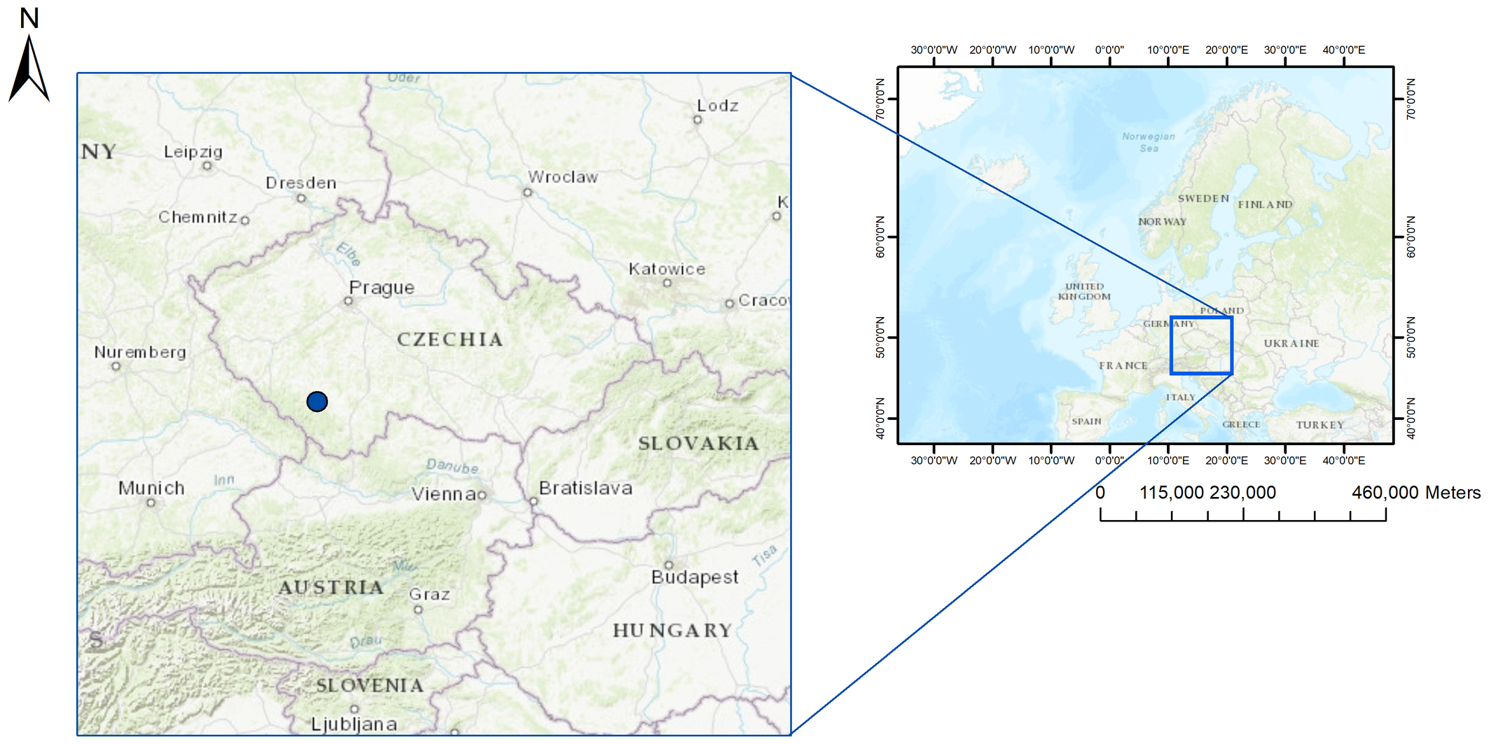
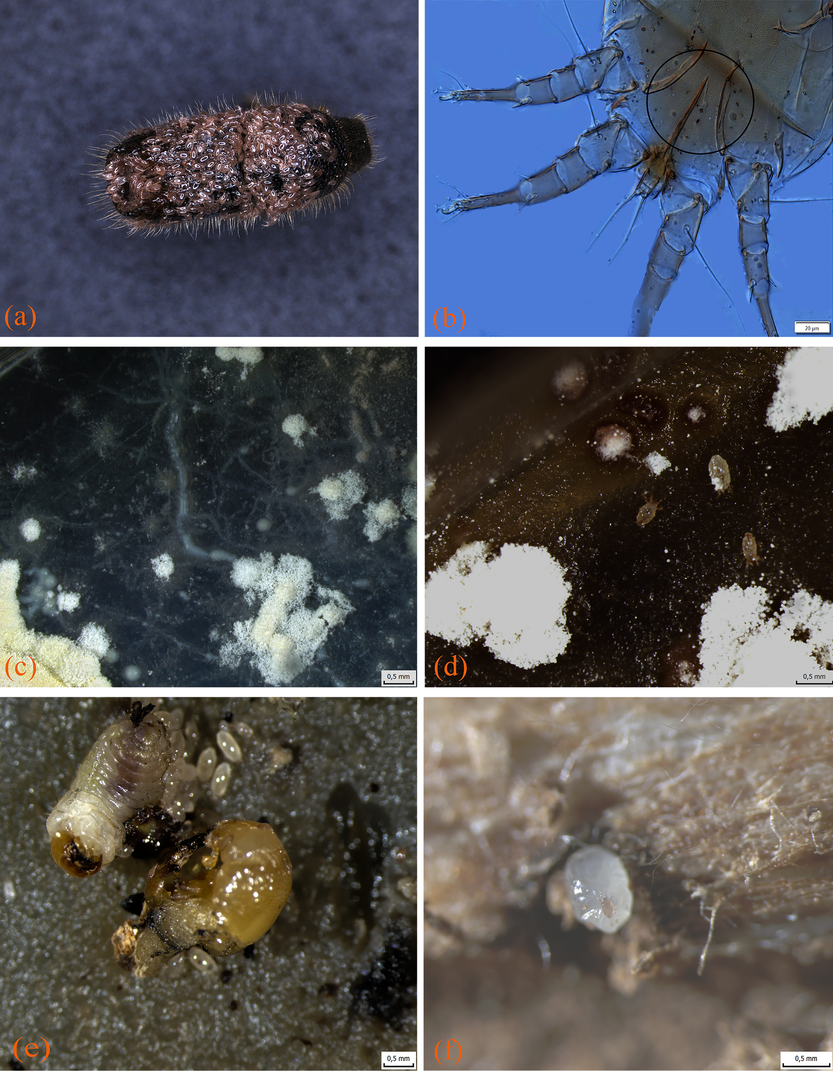
An infested spruce tree bolt (0.5 m long, 15 cm diameter) containing I. typographus was collected in spring 2021 at a forest site in the Czech Republic (48.621471, 14.265567) (Figure 1). Emerging adult beetles were obtained under controlled laboratory conditions and examined under a binocular microscope. Many individuals carried exceptionally high numbers of phoretic mites on the pronotum, elytral base, disc and declivity. In total, 64 beetles were collected, with mite loads ranging from a few individuals to more than 200 deutonymphs per host. Mites were removed and identified morphologically using established taxonomic keys (Woodring 1966, Klimov et al. 2022). Microscopic inspection showed that deutonymphs frequently carried fungal spores on their bodies. To assess potential predatory behaviour, a larva and a pupa of I. typographus were introduced into a Petri dish containing live deutonymphs and observed for 24 h.
Fungal cultivation and DNA extraction
According to the recommendations of Raja et al. (2017), three markers were used to identify the isolated fungus. In addition to the standard ITS marker for fungal DNA barcoding, two complementary markers, Tef1 and RPB1, were also used. PCR amplification of the markers was performed separately using the Taq Unis system (Top-Bio sr.o.). ITS was amplified using primers ITS1f/ITS4 (White 1990) with the following thermal profile: 94 °C for 2 min; 35 cycles of 94 °C for 30 sec, 50 °C for 45 sec, and 72 °C for 1 min; final extension at 72 °C for 2 min. Tef1α was amplified using primers EF1-983F/EF1-2218 (Rehner and Buckley 2005) with the following thermal profile: 94 °C for 2 min; 35 cycles of 94 °C for 30 sec, 45 °C for 45 sec, and 72 °C for 1 min; final extension at 72 °C for 2 min. RPB1 was amplified using primers RPB1 af/RPB1 cr (Matheny et al. 2002) with the following thermal profile: 94 °C for 2 min; 35 cycles of 94 °C for 30 sec, 50 °C for 30 sec, and 72 °C for 1 min; final extension at 72 °C for 2 min. After electrophoretic verification of the products, they were purified with a mixture of Exo I and FastAP (Thermo Fisher Scientific Inc.) and sent to Eurofins Genomics for Sanger sequencing. The obtained sequences were manually edited in the Chromas program (removal of 5′; and 3′ ends). For all markers, sequences of the expected lengths were obtained (ITS: 632 bp; Tef1: 949 bp; RPB1: 722 bp). Sequence identity was assessed using GenBank BLASTn. A subsample of 20 phoretic deutonymphs was transferred onto PDA medium (potato dextrose agar, Merck KGaA, Germany) supplemented with antibiotics, and incubated at 26 ± 2 °C under a 16:8 h photoperiod for 14 days. Fungal growth emerging from mite-transferred spores was reisolated onto fresh PDA plates for subsequent DNA extraction and identification.
Mite identification
Specimens of H. arborsignis were morphologically identified under a compound microscope prior to molecular analysis. Individuals were used either fresh or after preservation in 96% ethanol. For DNA isolation, both single-mite extractions and pooled extractions were performed. In pooled extractions, three to five individuals per sample were combined. All specimens selected for DNA extraction were pierced with a sterile mounting needle to rupture the cuticle and facilitate lysis.
Total genomic DNA was isolated using the DNeasy Blood & Tissue Kit (QIAGEN) following a modified version of the Matthews (2018) single-mite protocol, adapted for small arthropods with tough cuticles. All buffers were used at half the prescribed volume for pooled extractions.
Molecular analysis
DNA extraction
For single-mite extractions, mites were first placed on a sterile surface with 5–10 µL of Buffer ATL. A small incision was made in the cuticle with a sterile needle, and the mite along with the drop of ATL was transferred into a 1.5 mL tube containing 180 µL ATL. Proteinase K (20 µL) was added, and samples were incubated at 56 °C for 24 h without vortexing to maximize lysis. After incubation, 200 µL Buffer AL was added, the sample was gently inverted, heated at 70 °C for 5 min, and subsequently mixed with 200 µL chilled 96–100% ethanol. The entire lysate was loaded onto a DNeasy column and centrifuged according to kit recommendations. Wash steps consisted of 500 µL AW1 followed by 500 µL AW2, with centrifugation at 14,000 rpm to dry the membrane completely.
Elution was carried out with pre-heated (60–70 °C) Buffer AE. A low-volume elution of 22 µL was applied directly to the membrane, incubated for 5 min, and centrifuged at 14,000 rpm for 2 min. A second elution of 22 µL, followed by a final 50 µL elution, was performed when higher yields were required. For pooled extractions, the same protocol was used but with all buffer volumes reduced by half, and individuals lysed together in 5 µL ATL before adding Proteinase K.
PCR Amplification
A fragment of the mitochondrial cytochrome c oxidase subunit I (COI) gene was amplified using the Taq Unis PCR system (Top-Bio s.r.o.) and standard barcode primers LepF1/LepR1 (Hebert et al. 2004). PCR conditions were as follows: initial denaturation at 94 °C for 1 min; 35 cycles of 94 °C for 30 s, 47 °C for 40 s, and 72 °C for 1 min; final extension at 72 °C for 2 min. Amplification success was verified by electrophoresis on agarose gels.
Purification and Sequencing
Successful PCR products were enzymatically purified using Exonuclease I and FastAP (Thermo Fisher Scientific) and subsequently sent to Eurofins Genomics for Sanger sequencing. Bidirectional reads were assembled and inspected for quality, and sequences were translated to check for stop codons indicative of pseudogenes. The obteind sequences (576 – 629 bp) were manually edited in the Chromas program (removal of 5′; and 3′ends). Sequence identity was assessed using GenBank BLASTn.
The generated sequences were deposited in NCBI GenBank (Benson et al. 2017) under the accession numbers PX830422 (ITS), PX842437 (tef1), and PX842438 (RPB1) for B. bassiana, and PX836890 (COXI) for H. arborsignis.
Imaging was performed using an Olympus SZX16 stereomicroscope and an Olympus BX63 compound microscope. The images were processed using QuickPHOTO MICRO 3.2, Adobe Photoshop 2021, and Adobe Illustrator 2021. Prior to imaging, specimens were prepared following standard acarological techniques (Saito et al. 1993). Prepared mite specimens are deposited in the authors' reference collection at the Biology Centre of the Czech Academy of Sciences, České Budějovice, Czech Republic. Isolated fungal cultures obtained during this study are likewise maintained in the authors' culture collection at the same institution.
Results
The collected specimens were identified as belonging to H. arborsignis. A detailed morphological description of the deutonymphs, females and males of this species, will be presented later in a separate work. During the photographic study, deutonymphs were frequently observed carrying fungal spores on their bodies. Subsequent laboratory experiments confirmed that these spores belonged to B. bassiana (Balsamo-Crivelli, 1835) Vuillemin, 1912. When the deutonymphs were transferred to an agar medium, they successfully transferred the fungal spores, which germinated and grew, indicating that the mites act as effective carriers. In addition, the deutonymphs were observed to moult into adults in the presence of fungal colonies, often in close interaction with the growing mycelium, suggesting that they may also consume the fungus as a food source. The feeding experiments were conducted by offering the deuteronymphs different developmental stages of I. typographus. Within 24 hours, the deutonymphs actively attacked and consumed larvae and pupae, while the adults remained unimpressed (Figure 2). In natural beetle galleries, deutonymphs were also observed in close association with beetle eggs, indicating a possible predatory effect at this stage as well. Overall, these observations suggest that the deutonymphs of H. arborsignis are multifunctional partners of I. typographus: they are phoretic agents on adult beetles, vectors of entomopathogenic fungal spores (B. bassiana), and opportunistic predators of susceptible beetle stages such as larvae, and pupae. The transition from deutonymphs to adults in fungal environments is further evidence of their adaptability as dispersers and consumers in bark beetle habitats.
Discussion
This study provides new evidence of the ecological versatility of H. arborsignis in association with the spruce bark beetle I. typographus. Although previous European records identified this species only from Germany (Moser and Bogenschütz 1984; Milosavljević et al. 2022), our material, confirmed independently by morphology and COI barcoding, demonstrates its presence in the Czech Republic, significantly extending its known Central European distribution. This finding supports the growing recognition that phoretic mites associated with bark beetles are often much more widespread and diverse than previously recorded, reflecting both sampling limitations and the high dispersal potential of bark beetle phoretic assemblages (Klimov and Tolstikov 2011; Cilbircioğlu et al. 2021).
The exceptionally high deutonymphal loads observed on some I. typographus adults, reaching over 200 individuals per beetle in some cases, fall within the upper range reported for scolytine-associated phoretic mites, where tens to hundreds of deutonymphs may accumulate on a single dispersing adult (Moser and Bogenschütz 1984; Moser et al. 1997; Paraschiv and Isaia 2020). Such densities are expected to influence both beetles and mites, potentially affecting beetle flight performance, energetic cost of dispersal, and probability of pathogen transmission (Reynolds et al. 2014; Seeman and Walter 2023). For mites, aggregation on dispersing beetles provides an efficient mechanism for reaching new fungal patches while maximizing colonization success (Walter and Proctor 2013; Bajerlein and Błoszyk 2024).
One of the key findings of this study is that H. arborsignis acts as an efficient vector of viable fungal propagules. Deutonymphs carried visible spores, and when transferred to PDA medium, they successfully deposited and initiated rapid germination of B. bassiana. Comparable patterns of fungal vectorship have been well documented in other bark beetle systems, particularly among tarsonemid and acarid mites known to transport bluestain fungi such as Ophiostoma and Ceratocystis (Bridges and Moser 1983; Moser 1985; Klepzig and Hofstetter 2011). Likewise, Japanese populations of I. typographus japonicus harbor phoretic mites capable of carrying multiple hyperphoretic fungi (Moser et al. 1997). Reviews of bark beetle–mite–fungus systems consistently emphasize that mite-mediated spore transport is a central mechanism structuring fungal community composition within galleries (Hofstetter et al. 2015; Vissa and Hofstetter 2017).
The novelty of our findings lies in the identification of B. bassiana, a well-known entomopathogenic fungus, as a viable passenger transported by H. arborsignis. Previous studies have shown that soil microarthropods, such as collembolans and mites, are often not susceptible to entomopathogenic fungi and may even consume and inactivate fungal propagules without adverse effects (Broza et al. 2001). Recent work further highlights that mites can participate in complex tri-trophic interactions, functioning simultaneously as fungal grazers and vectors (Touray et al. 2025). In this context, fungal grazing by mites may influence the persistence and effectiveness of entomopathogenic fungi used in biological control, potentially reducing their efficacy in natural environments. While mite vectorship has traditionally been associated with nutritional mutualists or blue-stain pathogens, our results demonstrate that entomopathogens may also exploit phoresy as a dispersal route (Hulcr and Dunn 2011; Hofstetter and Moser 2014). The rapid growth of Beauveria colonies within one week after transfer indicates that this pathway is functional and potentially ecologically significant. If such processes occur in natural galleries, H. arborsignis could facilitate the within-tree spread of entomopathogenic fungi, increasing infection pressure on bark beetle populations and potentially influencing beetle fitness. Berto et al. (2025) showed that H. arborsignis develops successfully on several ambrosia beetle, associated fungi, particularly Graphium spp., and remains unaffected by exposure to B. bassiana, suggesting that the mite may persist in environments treated with fungal biopesticides and potentially contribute to their local redistribution. In contrast to several phytophagous and parasitic mite species, such as Tetranychus urticae, Polyphagotarsonemus latus and Dermanyssus gallinae, for which B. bassiana causes high mortality and is widely applied as a biological control agent, H. arborsignis exhibits a pronounced tolerance to this entomopathogenic fungus (Chandler et al. 2000; Steenberg and Kilpinen 2003; Shi and Feng 2004).
In addition to functioning as a fungal vector, H. arborsignis displayed opportunistic predatory behavior. Deutonymphs readily attacked and consumed live larvae and pupae of I. typographus, leaving only chitinized remains within 24 hours; however, the possibility of opportunistic feeding on weakened or dead individuals (scavenging) cannot be excluded. Predation by phoretic mites on scolytine eggs and larvae has been widely documented, particularly for genera such as Dendrolaelaps and Iponemus (Kinn 1967; Kinn 1980; Kinn 1982; Raffa et al. 2015). Our observations demonstrate that H. arborsignis occupies a similar ecological role, acting simultaneously as fungivore, predator and microbial vector. These combined effects may have measurable consequences for bark beetle reproductive success and gallery community dynamics (Klepzig and Hofstetter 2011; Hofstetter and Moser 2014).
The transition of deutonymphs to adults when exposed to fungal colonies, along with the emergence of both sexes, further highlights the central importance of fungal resources in the development of H. arborsignis. Previous experimental work has demonstrated that Histiogaster species can develop successfully on a wide range of beetle-associated fungi, with growth and fecundity dependent on fungal species and quality (Okabe 1993; Okabe 1993a). Berto et al. (2025) similarly showed that H. arborsignis can complete several generations on ambrosia fungi such as Graphium spp., and interacts with fungal biopesticides. Our results extend this versatility to include B. bassiana, underscoring the mite's trophic plasticity and adaptability to diverse fungal environments. Altogether, these findings align with contemporary views that phoresy is a dynamic ecological strategy, not a simple transport mechanism (Walter and Proctor 2013; Seeman and Walter 2023). In the H. arborsignis/I. typographus system, the interaction is multifaceted: beetles provide transport and access to resource-rich patches, whereas mites alter the gallery environment through fungivory, predation and microbial introduction. Such relationships can produce both beneficial and detrimental outcomes for beetles, depending on ecological context (Raffa et al. 2015; Hulcr and Dunn 2011; Vissa and Hofstetter 2017). Finally, establishing the presence of H. arborsignis in the Czech Republic links modern distribution patterns to the deep evolutionary history of the genus. This underscores the long-term stability of tri-trophic associations among bark beetles, fungi and acarid mites an interaction network that continues to shape conifer forest dynamics today (Klimov 2018; Klimov and Khaustov 2018).
Conclusion
This study documents the first confirmed occurrence of H. arborsignis associated with I. typographus in the Czech Republic, expanding the known European range of this species and revealing its presence as an overlooked but ecologically significant component of the spruce bark beetle gallery community. Our combined morphological and molecular analyses demonstrate that H. arborsignis is not merely a passive phoretic associate but a multifunctional participant within the bark beetle system. The species disperses on adult beetles, transports viable propagules of the entomopathogenic fungus B. bassiana, develops successfully in fungal-rich substrates and exhibits opportunistic predation on beetle larvae and pupae. These findings indicate that H. arborsignis may potentially influence bark beetle population processes through both direct and indirect mechanisms, including mortality of immature stages and modification of microbial assemblages within galleries. The observed interactions illustrate the complexity and ecological importance of mite-mediated processes in conifer forest ecosystems and highlight the need to incorporate phoretic mites more fully into conceptual and applied models of bark beetle dynamics. Further research should quantify the frequency and ecological consequences of mite-driven fungal transfer, assess the impact of predation under natural conditions and explore how forest management practices and environmental change modulate these multi-trophic associations.
Project funding
This work was funded by the European Community's Interreg Czech–Austria Program (BIPC, registration no. ATCZ00189), supporting M.M., M.Ž., M.Ž., V.B. and H.T. Research conducted by V.B.K. was carried out within the framework of State Assignment No. 124032500016-4.
Acknowledgements
We would like to thank Dr. Pavel Klimov (Lilly Hall of Life Sciences, Purdue University, USA) for his valuable advice and support throughout the development of the genetic methodology, as well as for his assistance during the analysis and interpretation of the results. We are also grateful to Dr. Hana Sehadová (Biology Centre of the Czech Academy of Sciences, České Budějovice, Czech Republic) for her kind help imaging procedures. The authors used the services of the Czech-BioImaging research infrastructure, specifically the Laboratory of Microscopy and Histology, Biology Centre CAS, supported by project LM2023050 funded by the Ministry of Education, Youth and Sports of the Czech Republic, with instrumental equipment co-financed by the European Union.
References
- Bajerlein D., Błoszyk J., Halliday R.B., Konwerski S. 2024. Hitchhiking through life: review of phoresy in Uropodina mites (Parasitiformes: Mesostigmata). Eur. Zool. J., 91: 31-63. https://doi.org/10.1080/24750263.2023.2288847
- Baumann J. 2018. Tiny mites on a great journey: a review on scutacarid mites as phoronts and inquilines. Acarologia, 58: 192-251. https://doi.org/10.24349/acarologia/20184238
- Berto M.M., Cruz L.F., Avery P.B., Cloonan K.R., Dunlap C.A., Carrillo D. 2025. Beyond phoresy: interactions between Histiogaster arborsignis (Acari: Acaridae), ambrosia beetles and their fungal symbionts in Florida avocados. Symbiosis, 96: 91-104. https://doi.org/10.1007/s13199-025-01063-0
- Benson D.A., Cavanaugh M., Clark K., Karsch-Mizrachi I., Lipman, D.J. Ostell J., Sayers E. 2017. GenBank. Nucleic Acids Res. 45(D1): D37-D42. https://doi.org/10.1093/nar/gkw1070
- Bridges J.R., Moser J.C. 1983. Role of two phoretic mites in transmission of bluestain fungus Ceratocystis minor. Ecol. Entomol., 8: 9-12. https://doi.org/10.1111/j.1365-2311.1983.tb00476.x
- Broza M., Pereira R.M., Stimac J.L. 2001. The nonsusceptibility of soil Collembola to insect pathogens and their potential as scavengers of microbial pesticides. Pedobiologia, 45: 523-534. https://doi.org/10.1078/0031-4056-00104
- Cardoza Y.J., Moser J.C., Klepzig K.D., Raffa K.F. 2008. Multipartite symbioses among fungi, mites, nematodes and the spruce beetl Dendroctonus rufipennis. Environ. Entomol., 37: 956-963. https://doi.org/10.1093/ee/37.4.956
- Cilbircioğlu C., Kovač M., Pernek M. 2021. Associations of phoretic mites on bark beetles of the genus Ips in the Black Sea Mountains of Turkey. Forests, 12(5): 516. https://doi.org/10.3390/f12050516
- Chandler D., Davidson G., Pell J.K., Ball B.V., Shaw K., Sunderland K.D. 2000. Fungal biocontrol of Acari. Biocontrol Sci. Technol., 10(4): 357-384. https://doi.org/10.1080/09583150050114972
- Hajek A.E., St. Leger R.J. 1994. Interactions between fungal pathogens and insect hosts. Annu. Rev. Entomol., 39: 293-322. https://doi.org/10.1146/annurev.en.39.010194.001453
- Hebert P.D.N., Penton E.H., Burns J.M., Janzen D.H., Hallwachs W. 2004. Ten species in one: DNA barcoding reveals cryptic diversity. Proc. Natl. Acad. Sci. USA, 101: 14812-14817. https://doi.org/10.1073/pnas.0406166101
- Hofstetter R.W., Dinkins-Bookwalter J., Davis T.S., Klepzig K.D. 2015. Symbiotic associations of bark beetles. In: Vega F.E., Hofstetter R.W. (Eds.). Bark Beetles: Biology and Ecology of Native and Invasive Species. Academic Press, San Diego: 209-245. https://doi.org/10.1016/B978-0-12-417156-5.00006-X
- Hulcr J., Dunn R.R. 2011. The sudden emergence of pathogenicity in insect-fungus symbioses. Proc. R. Soc. B., 278: 2866-2873.
- Khaustov A.A., Klimov P.B., Trach V.A., Bobylev A.N., Salavatulin V., Khaustov V.A., Tolstikov A.V. 2018. Review of mites (Acari) associated with the European spruce bark be-tle Ips typographus in Asian Russia. Acarina, 26: 3-35. https://doi.org/10.21684/0132-8077-2018-26-1-3-79
- Kinn D.N. 1967. Notes on the life cycle and habits of Digamasellus quadrisetus. Ann. Entomol. Soc. Am., 60: 862-865. https://doi.org/10.1093/aesa/60.4.862a
- Klimov P.B., Khaustov A.A. 2018. A review of acarid mites (Acariformes: Acaridae) associated with bark beetles, with description of Ipsoglyphus bochkovi gen. et sp. nov. Syst. Appl. Acarol., 23: 875-901. https://doi.org/10.11158/saa.23.5.13
- Klimov P.B., Kolesnikov V.B., Khaustov A.A., Ermilov S.G., Tolstikov A.V. 2022. The mite genus Histiogaster (Acari: Acaridae) in the Eastern Palaearctic, with a description of two new species and report of pre-copulatory guarding. Acarina (Russ. J. Acarol.), 30(2): 121-147. https://doi.org/10.21684/0132-8077-2022-30-2-121-147
- Kolesnikov V.B., Vorontsov D.D., Perkovsky E.E., Klimov P.B. An exceptionally well-preserved Eocene fossil mite, Histiogaster altilis sp. n. (Acari: Astigmata), from tree sap: Evidence of morphological and ecological niche conservatism, with a review of fossil Astigmata. Acarologia 65(1): 213-241. https://doi.org/10.24349/c35e-8bmj
- Matheny P.B., Liu Y.J., Ammirati J.F., Hall B.D. 2002. Using RPB1 sequences to improve phylogenetic inference among mushrooms (Inocybe, Agaricales). Am. J. Bot., 89(4): 688-698. https://doi.org/10.3732/ajb.89.4.688
- Matthews A.E., Klimov P.B., Proctor H.C., Dowling A.P.G., Diener L., Hager S.B., Larkin J.L., Raybuck D.W., Fiss C.J., McNeil D.J., Boves T.J. 2018. Cophylogenetic assessment of New World warblers (Parulidae) and their symbiotic feather mites (Proctophyllodidae). J. Avian Biol., 49(3): e01580. https://doi.org/10.1111/jav.01580
- Milosavljević M., Tabaković-Tošić M., Pernek M., Rakonjac L., Lučić A., Eremija S., Rindos M. 2022. Mites associated with the European spruce bark beetle Ips typographus (Linnaeus, 1758) in Europe, with new evidence for the fauna of Serbia. Forests, 13: 1586. https://doi.org/10.3390/f13101586
- Moser J.C. 1985. Use of sporothecae by phoretic Tarsonemus mites to transport ascospores of coniferous bluestain fungi. Trans. Br. Mycol. Soc., 84: 750-753. https://doi.org/10.1016/S0007-1536(85)80138-8
- Moser J.C. 1995. Mite associates and hyperphoretic fungi of the southern pine beetle. Proc. Entomol. Soc. Wash., 97: 539-550.
- Moser J.C., Bogenschütz H. 1984. Mites associated with flying Ips typographus. Z. Angew. Entomol., 97: 437-442. https://doi.org/10.1111/j.1439-0418.1984.tb03774.x
- Moser J.C., Perry T.J., Furuta K. 1997. Phoretic mites and their hyperphoretic fungi associated with flying Ips typographus japonicus Niijima (Coleoptera: Scolytidae) in Jpan. J. Appl. Entomol., 121: 425-430. https://doi.org/10.1111/j.1439-0418.1997.tb01429.x
- Mori B.A., Proctor H.C., Walter D.E., Evenden M.L. 2011. Phoretic mite associates of mountain pine beetle at the leading edge of an infestation in northwestern Alberta, Canada. Can. Entomol., 143(1): 44-55. https://doi.org/10.4039/n10-043
- Okabe K. 1993. Population growth and dispersal behavior of Histiogaster sp. (Acari: Acaridae) on economically important fungi. Appl. Entomol. Zool., 28(1): 11-18. https://doi.org/10.1303/aez.28.11
- Okabe K. 1993a. Developmental period and fecundity of Histiogaster sp. (Acari: Acari-dae) on three fungi. Appl. Entomol. Zool., 28(4): 479-487. https://doi.org/10.1303/aez.28.479
- Paraschiv M., Isaia G. 2020. Disparity of phoresy in mesostigmatid mites upon their specific carrier Ips typographus (Coleoptera: Scolytinae). Insects, 11(11): 771. https://doi.org/10.3390/insects11110771
- Raffa K.F., Grégoire J.-C., Lindgren B.S. 2015. Natural history and ecology of bark beetles. In: Vega F.E., Hofstetter R.W. (Eds.). Bark Beetles: Biology and Ecology of Native and Invasive Species. Elsevier Academic Press, San Diego: 1-40. https://doi.org/10.1016/B978-0-12-417156-5.00001-0
- Raja H.A., Miller A.N., Pearce C.J., Oberlies N.H. 2017. Fungal identification using molecular tools. J. Nat. Prod., 80: 756-770. https://doi.org/10.1021/acs.jnatprod.6b01085
- Rehner S.A., Buckley E. 2005. A Beauveria phylogeny inferred from nuclear genes. Myco-logia, 97: 84-98. https://doi.org/10.1080/15572536.2006.11832842
- Reynolds D.R., Reynolds A.M., Chapman J.W. 2015. Non-volant modes of migration in terrestrial arthropods. Anim. Migr., 2(1): 8-28. https://doi.org/10.2478/ami-2014-0002
- Saito Y., Osakabe Mh., Sakagami Y., Yasui Y. 1993. A Method for Preparing Permanent Specimens of Mites with Canada Balsam. Appl. entomol. Zool. 28: 593-597. https://doi.org/10.1303/aez.28.593
- Schebeck M., Schopf A., Ragland G.J., Stauffer C., Biedermann P.H.W. 2023. Evolutionary ecology of the bark beetles Ips typographus and Pityogenes chalcographus. Bull. Entomol. Res., 113(1): 1-10. https://doi.org/10.1017/S0007485321000353
- Seeman O.D., Walter D.E. 2023. Phoresy and mites: more than just a free ride. Annu. Rev. Entomol., 68: 69-88. https://doi.org/10.1146/annurev-ento-120220-013329
- Shi W.B., Feng M.G. 2004. Lethal effect of Beauveria bassiana, Metarhizium anisopliae and Paecilomyces fumosoroseus on the eggs of Tetranychus cinnabarinus (Acari: Tetranychidae) with a description of a mite egg bioassay system. Biol. Control, 30(2): 165-173. https://doi.org/10.1016/j.biocontrol.2004.01.017
- Steenberg T., Kilpinen O. 2014. Synergistic interaction between the fungus Beauveria bassiana and desiccant dusts applied against poultry red mites Dermanyssus gallinae (Acari: Dermanyssidae). Exp. Appl. Acarol., 62: 511-524. https://doi.org/10.1007/s10493-013-9757-8
- Touray M., Cimen H., Cakmak I., Hazir S. 2025. Tri-trophic interactions of soil mite Sancassania polyphyllae with entomopathogenic fungi and insect hosts. Ecosphere, 16: e70469. https://doi.org/10.1002/ecs2.70469
- Vissa S., Hofstetter R.W. 2017. The role of mites in bark and ambrosia beetle-fungal interac-tions. In: Shields V.D.C. (Ed.). Insect Physiology and Ecology. InTechOpen: 135-156. https://doi.org/10.5772/67106
- Walter D.E., Proctor H.C. 2013. Mites: Ecology, Evolution & Behaviour: Life at a Microscale. Springer, Dordrecht. https://doi.org/10.1007/978-94-007-7164-2
- White T.J., Bruns T., Lee S., Taylor J. 1990. Amplification and sequencing of fungal ribosomal RNA genes. In: PCR Protocols. Academic Press: 315-322. https://doi.org/10.1016/B978-0-12-372180-8.50042-1
- Woodring J.P. 1966. North American Tyroglyphidae (Acari): III. The genus Histiogaster, with description of four new species. Proc. Louisiana Acad. Sci., 29: 113-136.


2026-01-15
Date accepted:
2026-04-09
Date published:
2026-04-14
Edited by:
Baumann, Julia

This work is licensed under a Creative Commons Attribution 4.0 International License
2026 Milosavljević, Marija; Žurovcová, Martina; Toušková, Hana; Žurovec, Michal; Brož, Václav and Kolesnikov, Vasiliy B.
 Download article
Download the citation
RIS with abstract
(Zotero, Endnote, Reference Manager, ProCite, RefWorks, Mendeley)
RIS without abstract
BIB
(Zotero, BibTeX)
TXT
(PubMed, Txt)