


Redescription of Cunaxa vulgaris Shiba (Acariformes: Cunaxidae), a new record from China
Chen, Jian-Xin  1
; Yao, Mao-Yuan
2
; Wu, You-Fang
3
; Yi, Tian-Ci
4
; Guo, Jian-Jun
5
; Jin, Dao-Chao
6
and Peng, Pei-Ying
7
1
; Yao, Mao-Yuan
2
; Wu, You-Fang
3
; Yi, Tian-Ci
4
; Guo, Jian-Jun
5
; Jin, Dao-Chao
6
and Peng, Pei-Ying
7
1College of Agriculture, Anshun University, Anshun, 561000, P. R. China.
2College of Agriculture, Anshun University, Anshun, 561000, P. R. China.
3College of Agriculture, Anshun University, Anshun, 561000, P. R. China.
4Institute of Entomology, Guizhou University; Guizhou Provincial Key Laboratory for Agricultural Pest Management of the Mountainous Region; Scientific Observing and Experimental Station of Crop Pest in Guiyang, Ministry of Agriculture and Rural Affairs of the P. R. China, Guiyang 550025. P. R. China.
5Institute of Entomology, Guizhou University; Guizhou Provincial Key Laboratory for Agricultural Pest Management of the Mountainous Region; Scientific Observing and Experimental Station of Crop Pest in Guiyang, Ministry of Agriculture and Rural Affairs of the P. R. China, Guiyang 550025. P. R. China.
6✉ Institute of Entomology, Guizhou University; Guizhou Provincial Key Laboratory for Agricultural Pest Management of the Mountainous Region; Scientific Observing and Experimental Station of Crop Pest in Guiyang, Ministry of Agriculture and Rural Affairs of the P. R. China, Guiyang 550025. P. R. China.
7Institute of Microbiology, Qujing Medical College, Qujing 655100, Yunnan, P. R. China.
2026 - Volume: 66 Issue: 1 pages: 282-295
https://doi.org/10.24349/8mza-grdjZooBank LSID: 2957DAAE-DC81-4F5A-BA6E-D46FC141E9AC
Original research
Keywords
Abstract
Introduction
Cunaxid mites are cosmopolitan and found in most terrestrial habitats where they feeding on microarthropods and nematodes, their eggs etc., and play an important role in the agricultural ecosystems (Skvarla et al. 2014, Hernandes et al. 2015, Al-Azzazy & Al-Rehiayani 2022).
The genus Cunaxa contains 80 known species described in the world (Skvarla et al. 2014, Chen et al. 2023, Kalúz & Ermilov 2023, Khaustov & Khaustov 2024, Mirza et al. 2025). Currently, seven species have been recorded from China: C. capreola (Berlese, 1887), C. hermanni Den Heyer, 1979, C. mageei Smiley, 1992, C. setirostris (Hermann, 1804), C. womersleyi Baker & Hoffmann, 1948, C. papilla Chen & Jin, 2023, and C. striata Chen & Jin, 2023 (Hu 1997, Bei et al. 2003, Lin & Zhang 2010, Chen et al. 2023).
This paper reports a newly recorded species C. vulgaris Shiba, 1984 from China and provides the first description of males.
Materials and methods
Mites sample were extracted using modified Berlese-Tullgren funnels for 8–12 hours and preserved in 75% alcohol and then mounted in Hoyer's medium on slides (Walter & Krantz 2009). Line drawings were prepared with the aid of a drawing tube attached to a phase contrast and DIC Nikon Ni E microscope. Photographs were taken using a camera (Nikon DS-Ri 2) attached to the Nikon Ni E microscope and figures were edited with Adobe Photoshop CC 2019. The length of the gnathosoma was measured from the base to the tip of the subcapitulum, the length of the idiosoma, from the suture between the gnathosoma and idiosoma to the posterior margin of the idiosoma, the width of the idiosoma at its broadest level and the length of the legs from the base of trochanters to the tip of the claw. All measurements were taken in micrometers (µm) using the software Nikon NIS Elements AR 4.50. The dorsal and ventral setal nomenclature follows Skvarla et al. (2014) and Chen et al. (2023), legs setal notation follows Den Heyer (1981).
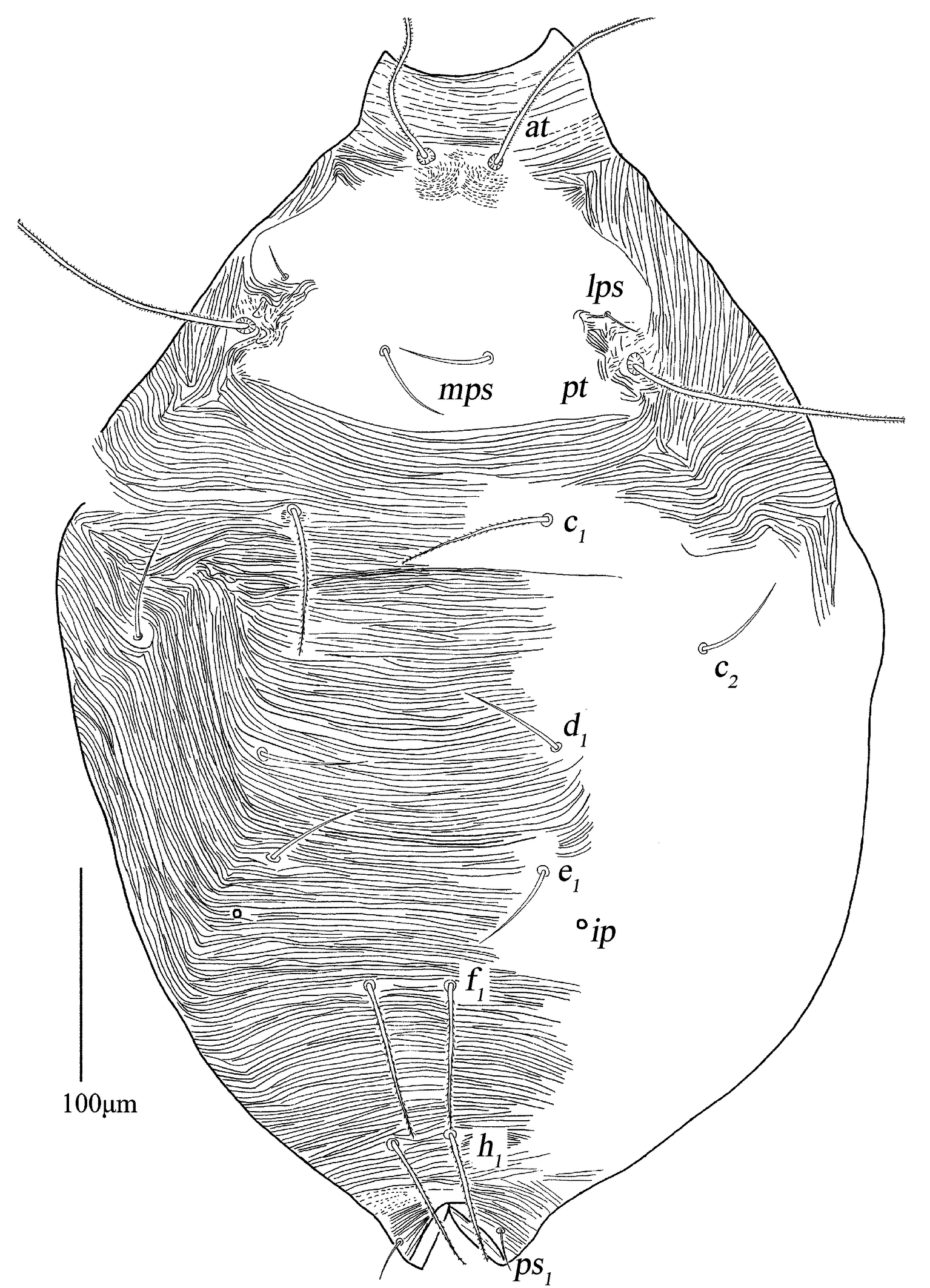
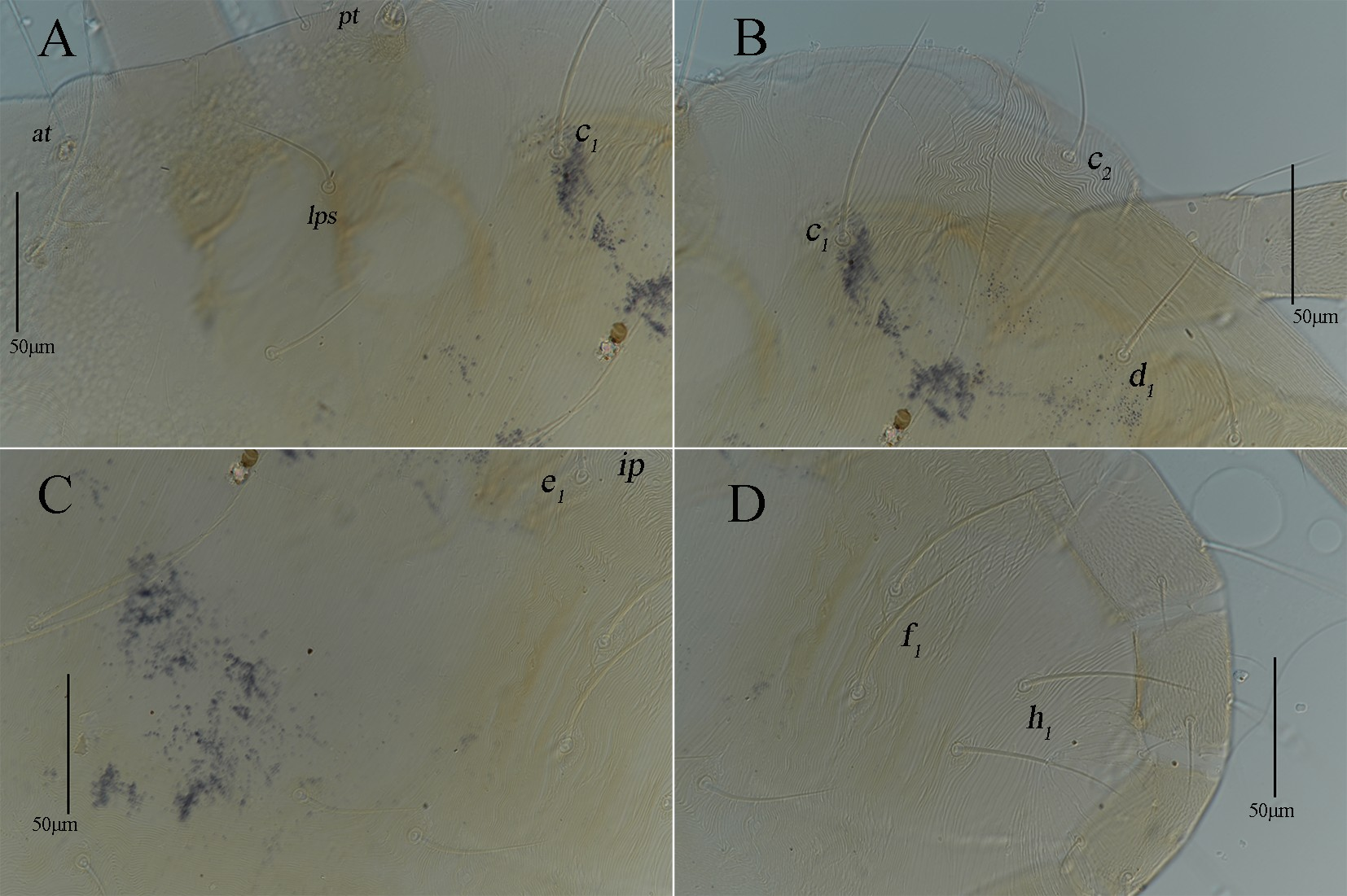
The following abbreviations are used: prodorsum: anterior trichobothria (at), posterior trichobothria (pt), lateral proterosomal (lps), median proterosomal (mps); hysterosoma: internal humerals (c1 ), external humerals (c2 ), internal dorsals (d1 ), internal lumbals (e1 ), internal sacrals (f1 ), internal clunals (h1 ), external clunals (h2 ); venter: propodogastral seta (ppgs), hysterogastral seta (hgs); anal region: pseudanal (ps); genital region: aggenitals (ag), genitals (g); gnathosoma: hypognathals (hg); leg: attenuate (sharply) solenidion (asl), blunt-pointed rod-like solenidion (bsl), famulus (fam), trichobothria (T), simple tactile seta (sts), microseta (mst), dorsoterminal solenidion (dtsl).
Results
Family Cunaxidae Thor, 1902
Subfamily Cunaxinae Thor, 1902
Genus Cunaxa Von Heden, 1826
Generic diagnosis: see Skvarla et al. (2014).
Type species: Scirus setirostris Hermann, 1804; now Cunaxa setirostris (Hermann, 1804).
Scirus Hermann, 1804, 62; Berlese, 1887, 34–40; Hull 1918, 37.
Cunaxa Von Heden, 1826, 609; Smiley, 1992,153; Corpuz-Raros & Garcia, 1995, 605; Den Heyer & Sergeyenko, 2009, 63, neotype designation.
Cunaxa vulgaris Shiba, 1984
Cunaxa vulgaris Shiba, 1984, 111
(Figs 1–13)
Diagnosis
Palp telofemur with one pointed apophysis; propodosomal shield smooth, except area between at and at, lps and pt with a few broken striae; hysterosomal shield absent, but a shield-like structure is present and covered with transverse striae on the central region of hysterosoma; c1 , f1 and h1 spiculate; f1 length not extending past base of h1 ; three pairs of hysterogastral setae (hgs1 –hgs3 ); one pair of aggenital setae (ag); genital setae g1 and g4 located laterally, g2 and g3 arranged in longitudinal row; basifemora I–IV 4-4-2-1 sts. Male resembling the female, setae c1 longer than f1 and h1 ; two pairs of hysterogastral setae (hgs1 –hgs2 ); without aggenital setae; basifemora I–IV 4-4-2-0 sts; genua I–II, tibia I, tarsi I–II with long bsl, respectively.
Redescription
Female (n=51, 6 specimens measured).
Idiosoma 519–566 long, 368–398 wide.
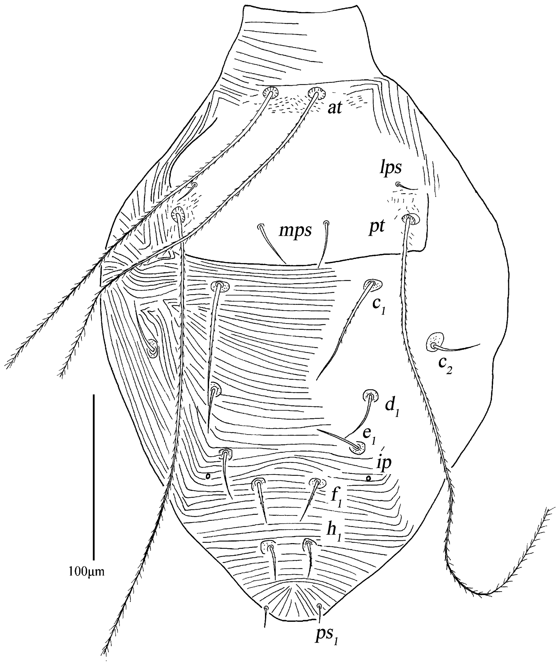
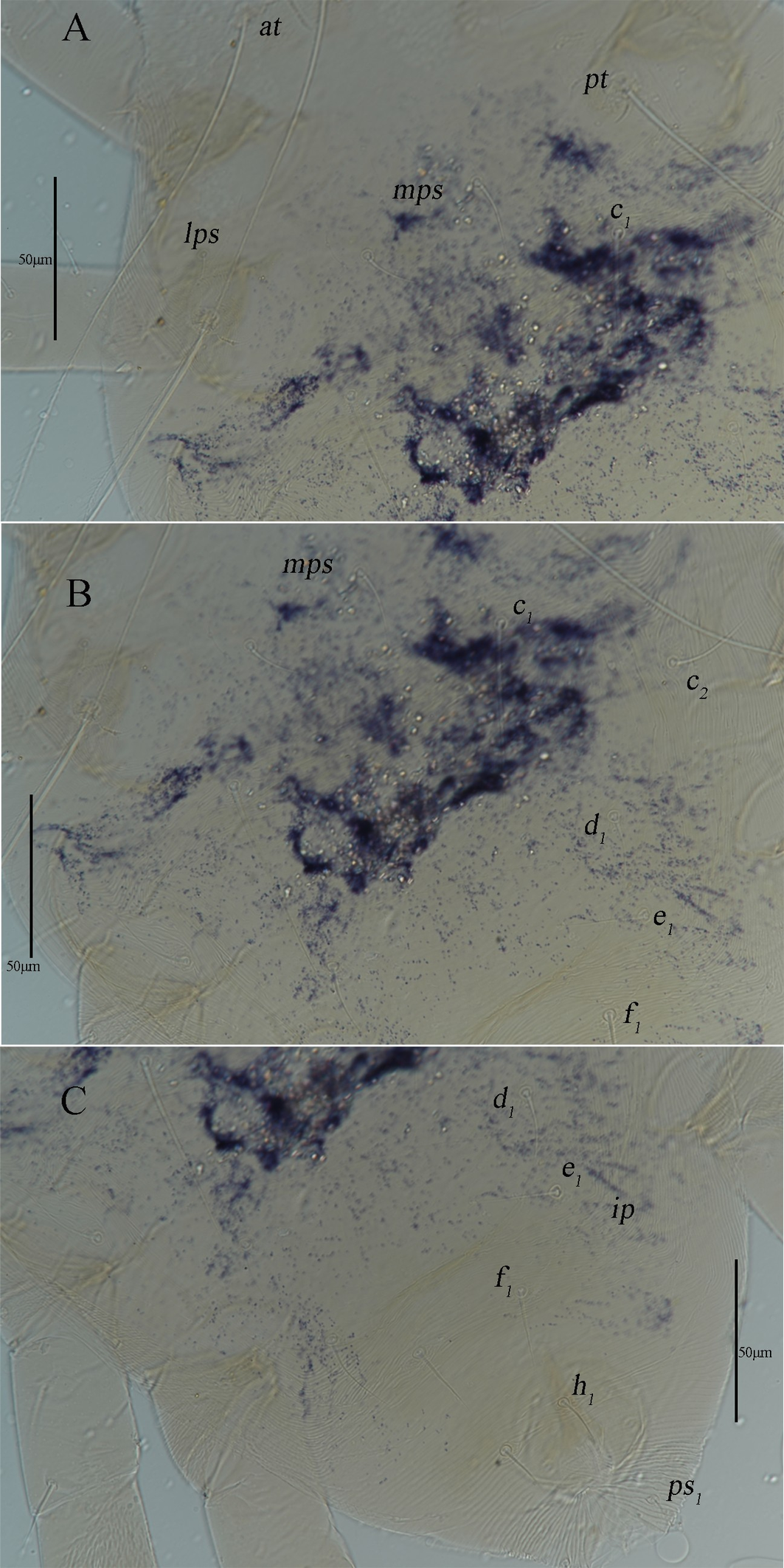
Dorsum – (Figs 1–2). Propodosomal shield smooth, except area between at and at, lps and pt with a few broken striae, 146–154 long, 200–210 wide, with two pairs of trichobothria (at and pt), two pairs of tactile setae (lps and mps). lps near pt, pt longer than at, mps longer than lps. Hysterosoma with six pairs of setae (c1 , c2 , d1 , e1 , f1 , h1 ) and one pair of lyrifissures (ip), setae c1 , f1 and h1 spiculate; f1 length not extending past base of h1 . Area between propodosomal shield and h1 with transverse striae, but a shield-like structure is observed on the central region of the hysterosoma, which is also adorned with transverse striae; area between c2 and e1 with lengthwise striae. Setal lengths and distances: at 307–312, pt 418–425, lps 13–14, mps 48–52, c1 90–101, c2 48–50, d1 52–57, e1 52–56, f1 88–94, h1 70–80; at–at 37–40, pt–pt 194–196, lps–lps 173–190, mps–mps 70–71, lps–mps 59–63, at–lps 95–100, pt–mps 64–72, pt–lps 27–33, at–mps 92–93, at–pt 123–127, c1 –c1 126–138, c2 –c2 265–288, d1 –d1 134–161, e1 –e1 137–145, f1 –f1 35–43, h1 –h1 28–37, c1 –c2 86–95, c1 –d1 110–117, c2 –d1 78–88, d1 –e1 39–55, e1 –f1 66–74, f1 –h1 78–83.
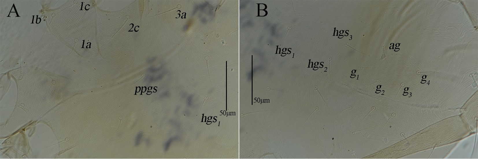
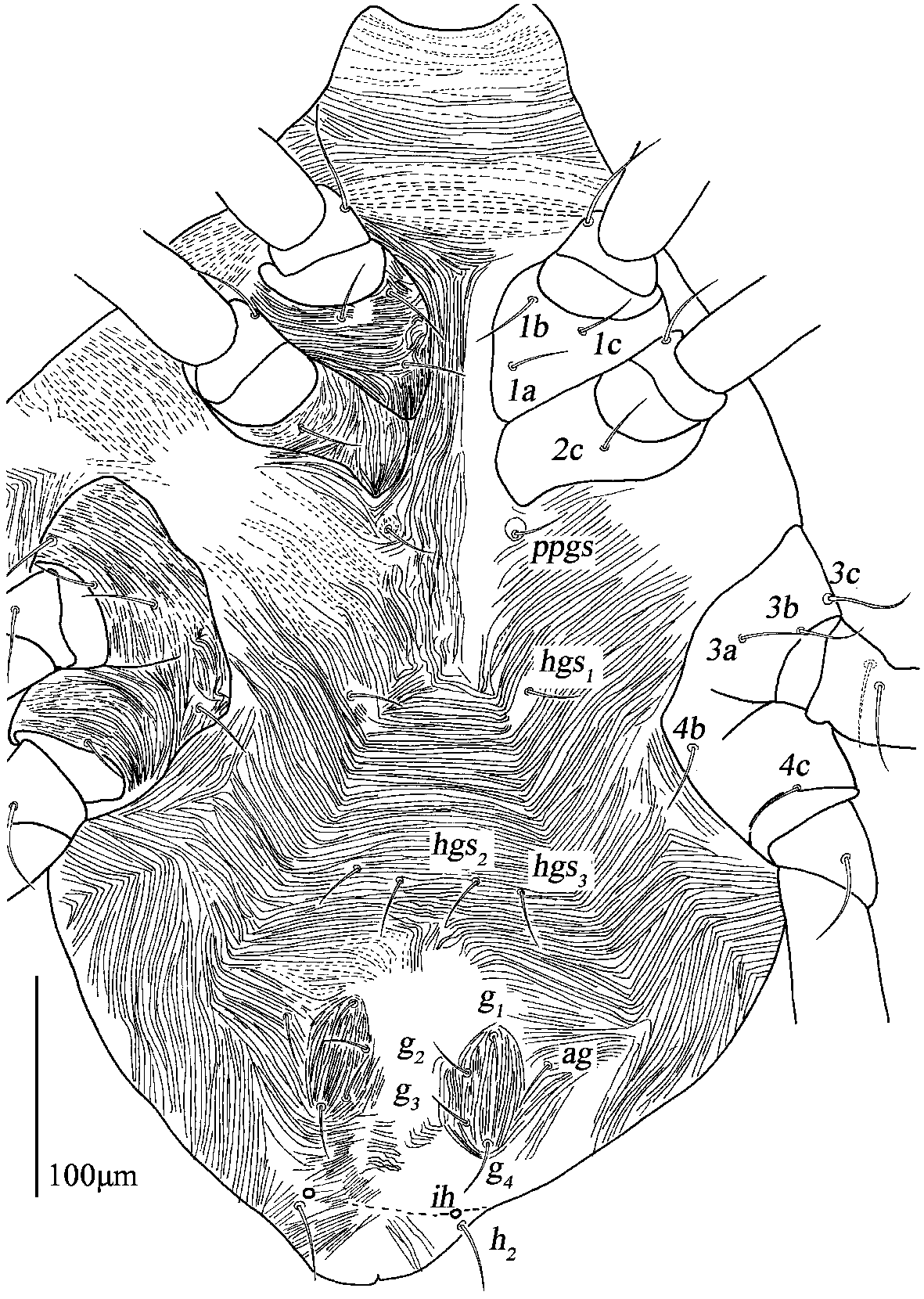
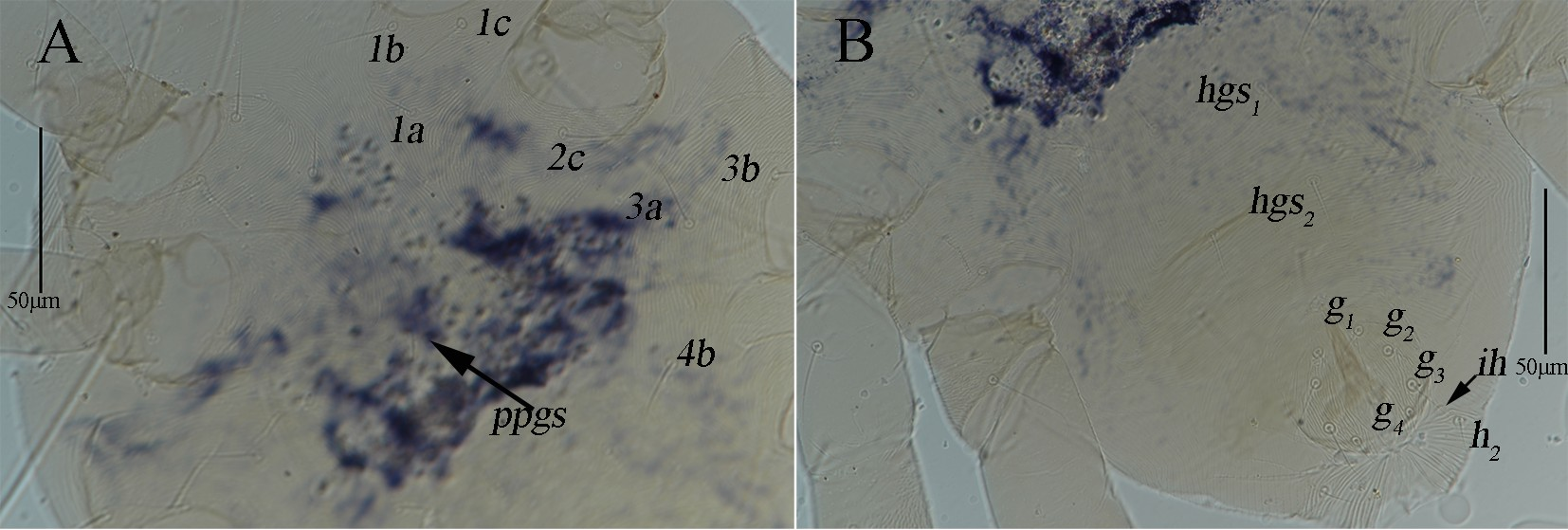
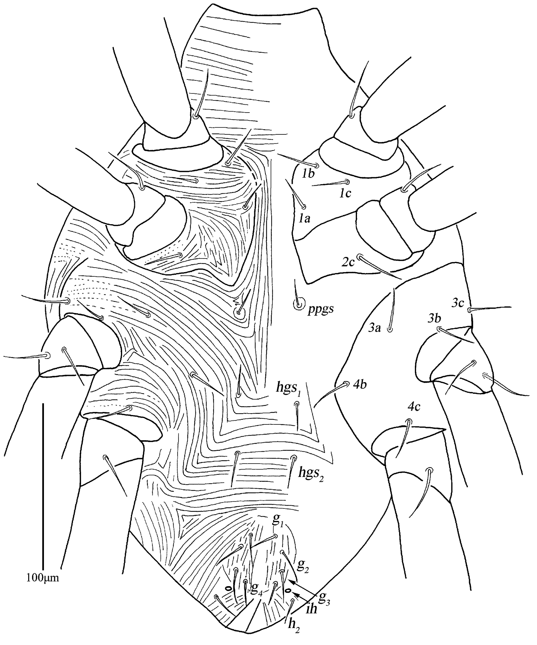
Venter – (Figs 3–4). Area between coxal fields I and gnathosoma with transverse striae; coxal fields I and II fused and with striae, and present obvious suture; coxal fields III and IV fused and with striae. Area between coxal fields I–II plate groups, between ppgs and hgs1 , lateral area outside between ppgs and hgs3 with longitudinal striae; areas between hgs1 and genital plates with transverse striae. One pair of propodogastral setae (ppgs) situated on small platelets, 24–26 in length; three pairs of hysterogastral setae (hgs1 –hgs3 ), 18–20, 19–20 and 21–23 in length, respectively. Setal formula of coxal fields I–IV: 3(1a–c)-1(2c)-3(3a–c)-2(4b–c) sts. Genital plates 60–75 long, 36–38 wide, invisible genital papillae and with longitudinal striae, four pairs of genital setae (g1 –g4 ), 21–22, 22–24, 22–23, and 22–26 in length, respectively, g1 and g4 located laterally, g2 and g3 arranged in longitudinal row; one pair of aggenital setae (ag), 23–27 in length. Anal region with longitudinal striae, one pair of pseudanal setae (ps1 ), 20–24 in length; a pair of h2 , 26–33 in length, and one pair of lyrifissures (ih) close to h2 .
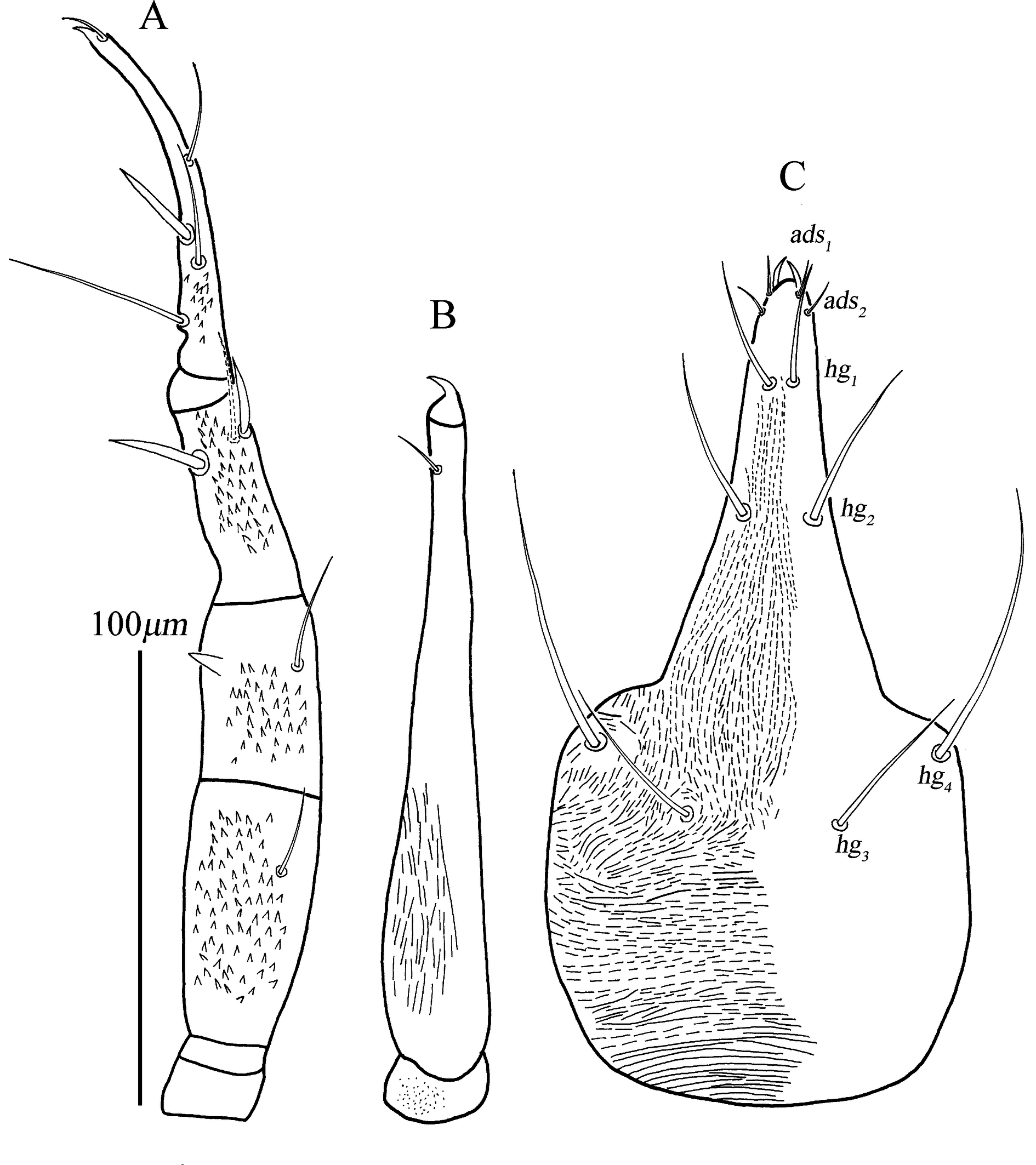
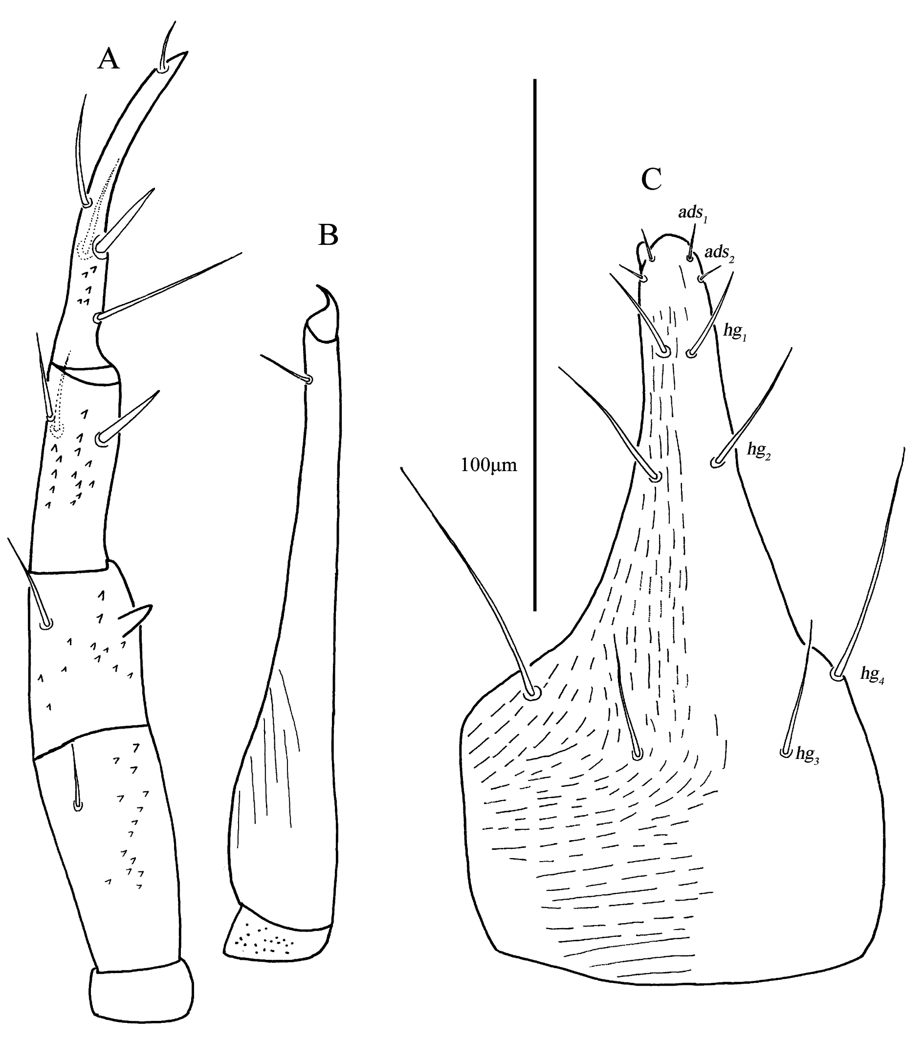
Gnathosoma – Palp (Fig. 5A). five-segmented, 215–240 long, with fine denticles. Palp chaetotaxy: trochanter none; basifemur with one simple seta; telofemur with one simple seta and one pointed apophysis; genu with two simple setae and one spine-like seta; tibiotarsus with three simple setae, of which proximal one the longest, one spine-like seta and one distal eupathidium, claw well-developed. Chelicera (Fig. 5B) 172–174 long, segment I with sparse fine papillae, segment II with broken longitudinal striae, cheliceral seta 13–15 in length; chela developed. Subcapitulum (Fig. 5C) 187–196 long, 103–107 wide, two pairs of adoral setae (ads1–ads2 ), 8–9 and 4–7 in length, respectively; four pairs of hypognathal setae (hg1 –hg4 ), 23–25, 31–33, 32–36, and 70–75 in length, respectively; areas between hg1 and hg3 with broken longitudinal striae, between hg3 and basal area with broken transverse stria. Distances of hg setae: hg1 –hg1 7–8, hg2 –hg2 12–16, hg3 –hg3 32–35, hg4 –hg4 80–86, hg1 –hg2 32–34, hg2 –hg3 71–75, hg3 –hg4 31–33.
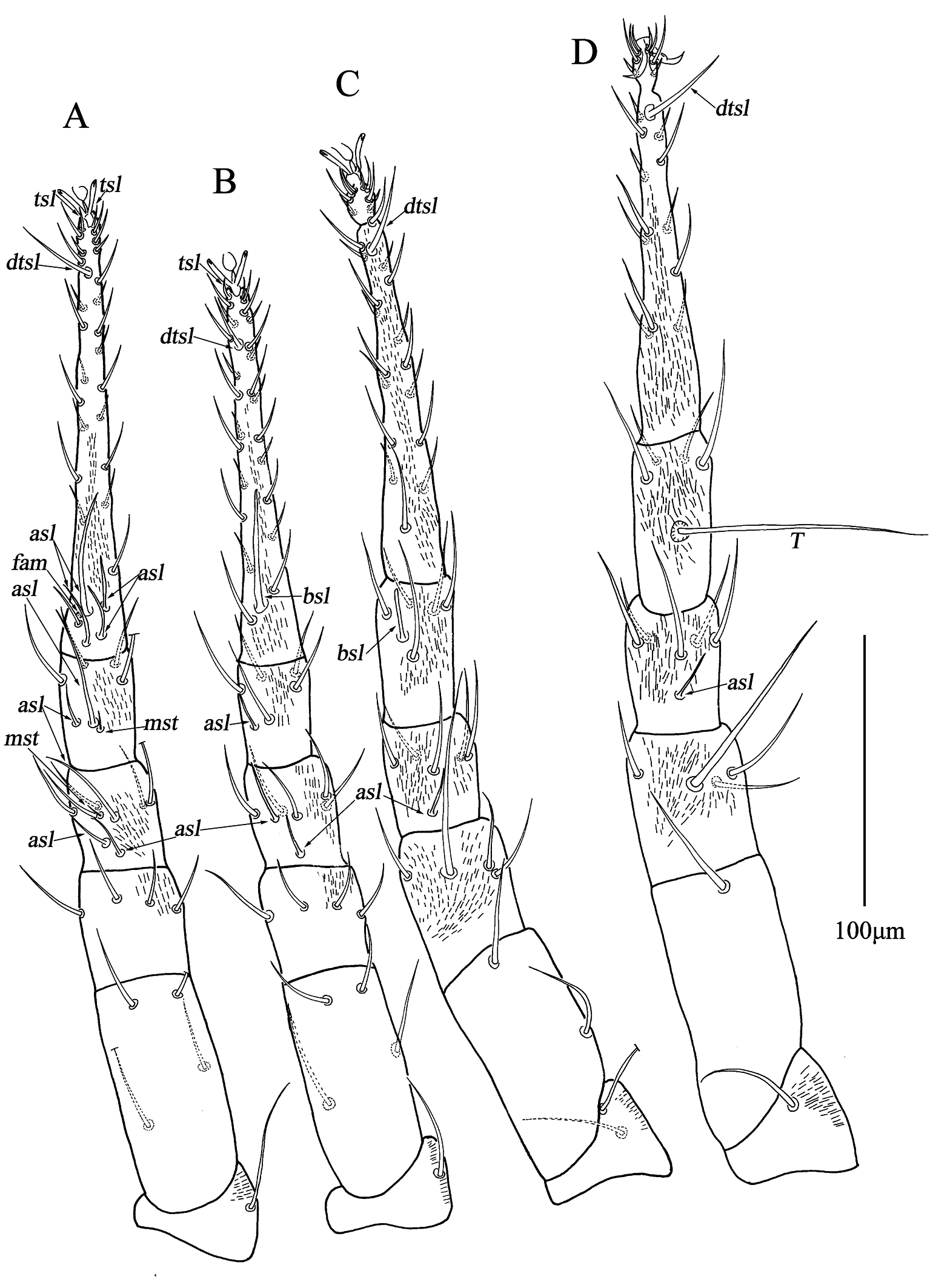
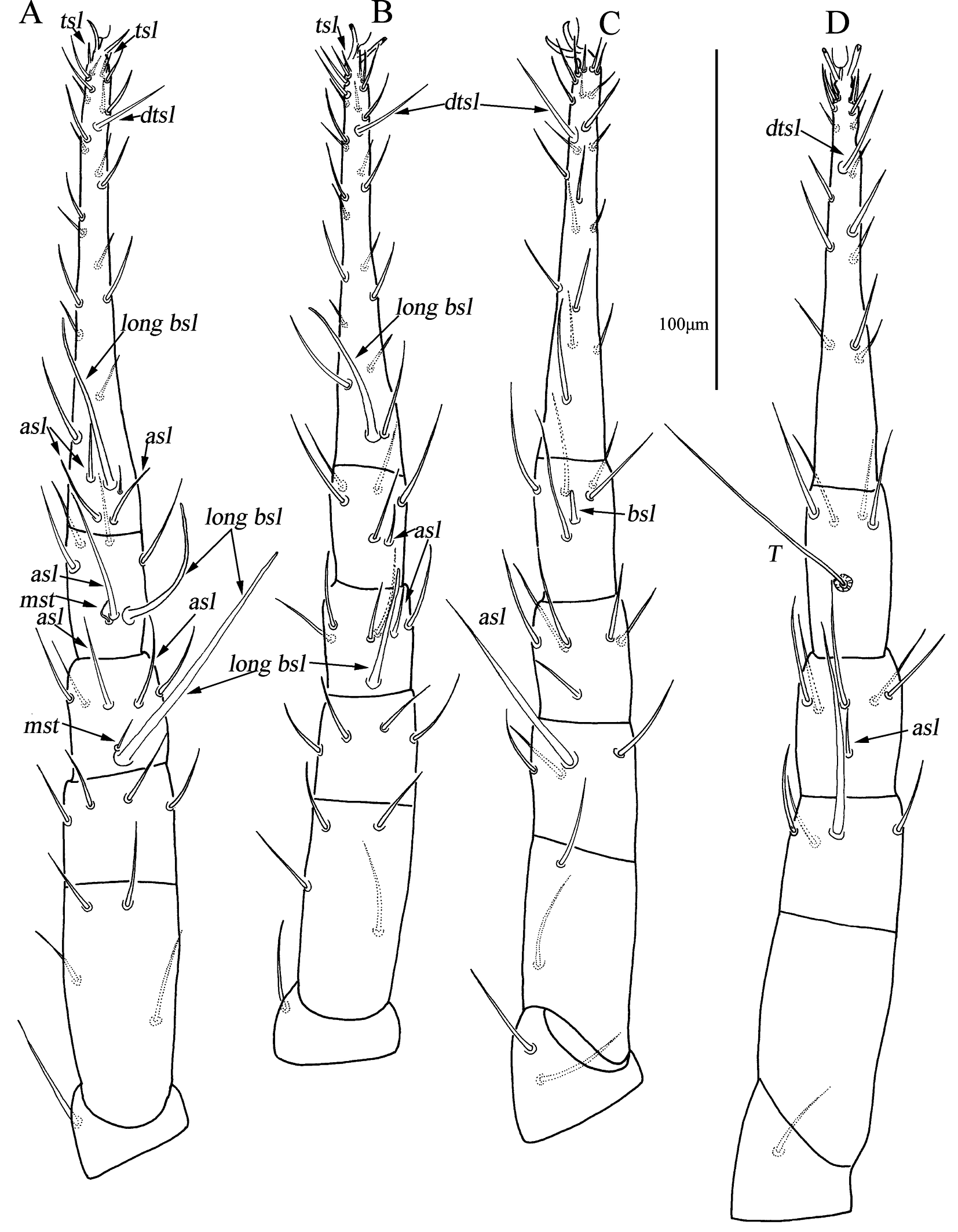
Legs – (Fig. 6). Leg I–IV with broken striae, lengths of legs I–IV: 400–407, 368–377, 393–398, and 316–425. Lengths of tarsi I–IV: 168–179, 144–151, 146–150, and 150–150. T smooth on tibia IV 94–99 in length. Legs I–IV chaetotaxy as follows: trochanters 1-1-2-1 sts; basifemora 4-4-2-1 sts; telofemora 4-4-4 (among which one thick and long)-4(among which one thick and long) sts; genua 3 asl, \{1 asl, 1 mst\}, 4 sts - 1 asl, 5 sts - 1 asl, 5 sts - 1 asl, 5 sts; tibiae 1 asl, \{1 asl, 1 mst\}, 4 sts - 1 asl, 5 sts - 1 bsl, 5 sts - 1 T, 4 sts; tarsi 4 asl, 1 fam, 1 dtsl, 2 tsl, 23 sts - 1 bsl, 1 dtsl, 1 tsl, 24 sts - 1 dtsl, 25 sts - 1 dtsl, 20 sts.
Male (n=2).
Idiosoma 369–382 long, 259–267 wide.
Dorsum – (Figs 7–8). Resembling that of female; propodosomal shield 102–124 long, 160–180 wide; f1 and h1 shorter; f1 not extending past h1 . Setal lengths and distances: at 232–235, pt 285–316, lps 10–12, mps 27–31, c1 62–68, c2 32–34, d1 26–30, e1 20–28, f1 22–26, h1 20–22; at–at 25–30, pt–pt 135–148, lps–lps 105–129, mps–mps 40–41, lps–mps 50–52, at–lps 68–75, pt–mps 49–55, pt–lps 19–22, at–mps 68–81, at–pt 90–96, c1 –c1 92–97, c2 -c2 178–180, d1 –d1 98–98, e1 –e1 83–86, f1 –f1 38–46, h1 –h1 24–28, c1 –c2 56–63, c1 –d1 59–70, c2 –d1 47–48, d1 –e1 32–38, e1 –f1 28–33, f1 –h1 39–42.
Venter – (Figs 9–10). Resembling that of female; propodogastral setae (ppgs) 13–19 in length, two pairs of hysterogastral setae (hgs1 –hgs2 ), 15–17 and 18–18 in length; genital plates 30–40 long, 17–20 wide, genital setae (g1 –g4 ), 14–16, 18–18, 17–18, and 15–16, respectively. ps1 and h2 , 10–12 and 14–14 in length.
Gnathosoma – Palp (Fig. 11A). resembling that of female; 180–186 long. Chelicera (Fig. 11B) resembling that of female; 123–129 long, cheliceral seta 7–9 in length. Subcapitulum (Fig. 11C) resembling that of female; 137–144 long, 75–81 wide; two pairs of adoral setae (ads1–ads2 ), 7–8 and 5–6 in length, lengths of hg1 –hg4 18–22, 24–24, 20–27, and 50–54, respectively. Distances of hg setae: hg1 –hg1 5–6, hg2 –hg2 13–14, hg3 –hg3 31–31, hg4 –hg4 58–60, hg1 –hg2 24–25, hg2 –hg3 50–53, hg3 –hg4 23–23.
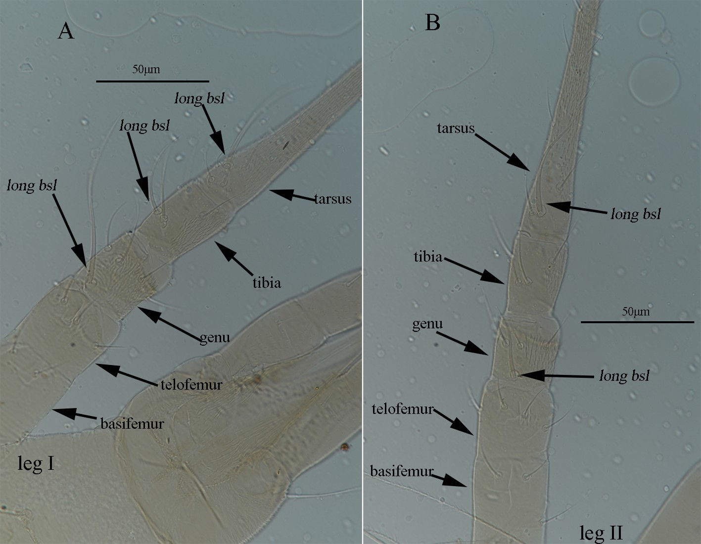
Legs – (Figs 12–13). Resembling that of female, lengths of leg I–IV: 318–335, 281–299, 302–320, and 323–338. Lengths of tarsi I–IV: 134–148, 112–124, 120–122, and 120–120. T 65–71 in length. Legs I–IV chaetotaxy as follows: basifemur IV: 0 sts; genu I 2 asl, \{1 long bsl, 1 mst\}, 3 sts; genu II 1 asl, \{1 long bsl, 1 mst\}; tibia I \{1 asl, 1 long bsl, 1 mst\}; tarsus I 3 asl, 1 long bsl, 1 fam (invisible), 1 dtsl, 2 tsl, 20 sts; tarsus II 1 long bsl, 22 sts; tarsus III 19 sts; tarsus IV 15 sts.
Other developmental stages. Unknown.
Remarks
This species was described from Japan (Shiba 1984). However, his description lacks measurements of setae and distances between setae, as well as a description of males. This species is new for the Chinese fauna. C. vulgaris is also very similar to C. heterostriata described from Crimea (Khaustov & Kuznetsov 1998) in general appearance. However, both species could be separated on the structure of cheliceral dorsum. In C. vulgaris cheliceral dorsum with broken striae while in C. heterostriata it with characteristic longitudinal ridges.
Material examined
Two ♀ and 1 ♂, were collected from soil, Jiuzhai Valley (N22°17′22.32″, E111°13′13.57″, elevation 1400 m), Aba Prefecture, Sichuan Province, China, on 16 August, 2018, by Guo-Ru Ren and Mao-Fa Yang, slides No., SC-CU-201808160101–SC-CU-201808160103; 1 ♀, was collected from fallen leaves, Mantingling Mountain (N32°36′25.98″,E104°50′14.31″, elevation 1555 m), Tangjiahe National Nature Reserve, Guangyuan County, Sichuan Province, China, on 9 May, 2019, by Yun Long, slide No., SC-CU-201905091801; 1 ♀, was collected from moss, Qianfoshan Nature Reserve (N31°40′52.64″, E104°16′20.51″, elevation 841 m) Anxian County, Mianyang City, Sichuan Province, China, on 9 May, 2019, by Yun Long, slide No., SC-CU-201905141501; 13 ♀ and 1 ♂, were collected from fallen leaves, Huaxi District (N26°25′, E106°36′, elevation 1100 m), Guiyang City, Guizhou Province, China, on 17 November, 2017, by Jian-Xin Chen, slides No., GZ-CU-2017111701–GZ-CU-2017111713; 3 ♀, were collected from fallen leaves, Huaxi District (N26°25′, E106°36′, elevation 1135 m), Guiyang City, Guizhou Province, China, on 16 November, 2016, by Mao-Yuan Yao and Yan Shen, slides No., GZ-CU-2016111601–GZ-CU-2016111603; 7 ♀, were collected from fallen leaves, Huaxi District (N26°25′, E106°36′, elevation 1100 m), Guiyang City, Guizhou Province, China, on 12 November, 2017, by Mao-Yuan Yao and Yan Shen, slides No., GZ-CU-2017111201–GZ-CU-2017111207; 1 ♀, was collected from fallen leaves, Xiaoxi Nature Reserve (N28°47′32″, E110°16′35″, elevation 644 m), Yongshun County, Hunan Province, China, on 20 August, 2016, by Jian-Xin Chen, slide No., HN- CU-201608200901; 2 ♀, were collected from moss, Xingdushan (N30°2′59″, E109°8′2″, elevation 1150 m), Lichuan City, Hubei Province, China, on 5 August, 2018, by Jian-Xin Chen, slides No., HB-CU-201808050401–HB-CU-201808050402; 3 ♀, were collected from moss, Dapanshan Nature Reserve (N29°00′36.12″, E120°28′3.8″, elevation 416 m), Pan′an County, Jinhua City, Zhejiang Province, China, on 31 July, 2018, by Mao-Yuan Yao and Yun Long, slides No., ZJ-CU-201807311801– ZJ-CU-201807311803; 4 ♀, were collected from fallen leaves, Nanling National Forest Park (N24°56′45.78″, E113°0′49.78″, elevation 903 m), Ruyuan Yao Autonomous County, Shaoguan City, Guangdong Province, China, on 28 April, 2019, by Jian-Xin Chen, slides No., GD-CU-201904280201–GD-CU-201904280204; 3 ♀, were collected from fallen leaves, Nanling National Forest Park (N24°53′46.37″, E113°1′52.70″, elevation 1100 m), Ruyuan Yao Autonomous County, Shaoguan City, Guangdong Province, China, on 29 April, 2019, by Jian-Xin Chen, slides No., GD-CU-201904290901–GD-CU-201904290903; 3 ♀, were collected from fallen leaves, Nanling National Forest Park (N24°53′44.62″, E113°1′47.71″, elevation 1123 m), Ruyuan Yao Autonomous County, Shaoguan City, Guangdong Province, China, on 29 April, 2019, by Jian-Xin Chen, slides No., GD-CU-201904291001–GD-CU-201904291003; 2 ♀, were collected from fallen leaves, Nanling National Forest Park (N24°53′48.25″, E113°2′29.22″, elevation 920 m), Ruyuan Yao Autonomous County, Shaoguan City, Guangdong Province, China, on 29 April, 2019, by Jian-Xin Chen, slides No., GD-CU-201904291201–GD-CU-201904291202; 1 ♀, was collected from fallen leaves, Dinghu Mountain National Nature Reserve (N23°10′23″, E112°32′50″, elevation 159 m), Zhaoqing City, Guangdong Province, China, on 27 August, 2019, by Min Ao, slide No., GD-CU-2019082701; 1 ♀, was collected from fallen leaves, Hanshuiyuan Forest Park (N32°45′54″, E116°13′40″, elevation 1300 m), Ningqiang County, Shanxi Province, China, on 15 July, 2019, by Jian-Xin Chen and Xue-Song Zhang, slide No., SX-CU-201907151401; 1 ♀, was collected from fallen leaves, Yuhe Town (N32°58′56″, E105°30′2″, elevation 895 m), Wudu District, Longnan City, Gansu Province, China, on 19 July, 2019, by Jian-Xin Chen and Xue-Song Zhang, slide No., GS-CU-201807190601; 1 ♀, was collected from fallen leaves (Pinus massoniana), Huangshan Scenic Area (N30°8′33.36″,E118°9′21.95″, elevation 1590 m), Anhui Province, China, on 23 May, 2018, by Mao-Yuan Yao, slide No., AH-CU-201805231301. All types are deposited in the Institute of Entomology, Guizhou University, Guiyang, P. R. China (GUGC).
Distribution
China (Sichuan, Guizhou, Hunan, Hubei, Zhejiang, Guangdong, Shanxi, Gansu and Anhui Province); Japan.
Acknowledgements
This work was supported by Guizhou Provincial Science and Technology Projects (QKHJC [2024] youth 289, 410 and QKHJC-ZK[2023]023).
References
- Al-Azzazy, M. M. & Al-Rehiayani, S. M. 2022. The soil mite Cunaxa capreolus (Acari: Cunaxidae) as a predator of the root-knot nematode, Meloidogyne incognita and the citrus Nematode, Tylenchulus semipenetrans: Implications for biological control. Acarologia, 62(1):174-185. https://doi.org/10.24349/lo4p-42kf
- Baker, E.W. & Hoffmann, A. 1948. Acaros de la familia Cunaxidae. An. Esc. Nac. de Cienc. Biol. Mex., 5(3-4): 229-273.
- Berlese, A. 1887. Acari Italiani Myriapoda et Scorpiones hucusque in Italia reperta. Redia, 14: 78-105.
- Bei, N.-X., Shi, C.-M. & Yin, S.-G. 2003. Cunaxa mageei Smiley, 1992 (Acari: Cunaxidae), a new record from China. Entomotaxonomia, 25(1): 34. [in Chinese]
- Chen, J.-X., Yao, M.-Y., Yi, T.-C., Guo, J.-J. & Jin, D.-C. 2023. Two new species of Cunaxa (Acariformes: Cunaxidae) from China. Syst. Appl. Acarol., 28(3): 508-520. https://doi.org/10.11158/saa.28.3.8
- Corpuz-Raros, L.A. & Garcia, R.C. 1995. Philippine predatory mites of the family Cunaxidae (Acari). 1. Genus Cunaxa Von Heyden. The Philippine Entomologist, 9(6): 605-624.
- Den Heyer, J. 1979. Five new African species of Cunaxa (Actinedida: Acarida). Phytophylactica, 11: 159-171.
- Den Heyer, J. 1981. Systematics of the family Cunaxidae Thor, 1902 (Actinedida: Acarida). Publ. Univ. of the North Ser. A., 24: 1-19.
- Den Heyer, J. & Sergeyenko, A. L. 2009. Neotype designation for Cunaxa setirostris (Hermann, 1804) (Acari: Prostigmata: Cunaxidae). Zootaxa, 2106(1): 61-68. https://doi.org/10.11646/zootaxa.2106.1.5
- Hermann, J.F. III. 1804. Ciron (Scirus). Mem. Apterologique, 60-62; pl. 3; fig. 12; pl. 6; fig. 12.
- Hernandes, F.A., Castro, T.M.M.G. & Venancio, R. 2015. Prostigmata (Acari: Trombidiformes) as biological control agents. pp. 151-184, Chapter 6 in Carrillo, D., Moraes, G.J. & Peña, J.E. (Eds.), Prospects for Biological Control of Plant Feeding Mites and Other Harmful Organisms. Springer, pp. 151-184. https://doi.org/10.1007/978-3-319-15042-0_6
- Hu, S.-J. 1997. Cunaxid mites recorded in China. Journal of Ninbo Teachers College, 15(1): 56-59. [in Chinese]
- Hull, J.E. 1918. Terrestrial Acari of the Tyne Province. Transactions of the Natural History Society of Northhumberland, Durham and Newcastle-upon-Tyne, N. Ser. 5(1): 1-88.
- Kalúz, S. & Ermilov, S.G. 2023. Two new species of Cunaxa (Acari, Prostigmata, Cunaxidae) from South-East Asia with a world key to the genus. Zootaxa, 5239(4): 521-536. https://doi.org/10.11646/zootaxa.5239.4.4
- Khaustov, A.A. & Khaustov, V.A. 2024. New faunistic data on Cunaxidae (Acari: Bdelloidea) of Asian Russia. Syst. Appl. Acarol., 29(8): 1058-1090. https://doi.org/10.11158/saa.29.8.3
- Khaustov, A.A. & Kuznetzov, N.N. 1998. Four new species of the genus Cunaxa (Acariformes, Cunaxidae). Zool. Zhurnal., 77(11): 1332-1341.
- Lin, J.-Z. & Zhang, Z.-Q. 2010. Bdelloidea of China: a review of progress on systematics and biology, with a checklist of species. Zoosymposia, 4, 42-50. In: Zhang, Z.-Q., Hong, X.-Y., Fan, Q.-H. (Eds) Xin, J.-L. Centenary: Progress in Chinese Acarology. Zoosymposia, 4: 1-345. https://doi.org/10.11646/zoosymposia.4.1.3
- Mirza, J. H., Kamran, M. & Alatawi, F. J. 2025. New species and a new record of the genus Cunaxa Von Heyden (Acari: Prostigmata: Cunaxidae) from Saudi Arabia. J. Nat. Hist., 59(13-16): 929-939. https://doi.org/10.1080/00222933.2025.2474740
- Shiba, M. 1984. The mites of the family Cunaxidae (Acarina: Prostigmata) in Japan I. Genus Cunaxa von Heyden. Matsuyama Shinonome Tanki Daigaku kenkyū ronsō, 15: 103-118.
- Skvarla, M.J., Fisher, J.R. & Dowling, A.P.G. 2014. A review of Cunaxidae (Acariformes, Trombidiformes): Histories and diagnoses of subfamilies and genera, keys to world species, and some new locality records. ZooKeys, 418: 1-103. https://doi.org/10.3897/zookeys.418.7629
- Smiley, R.L. 1992. The predatory mite family Cunaxidae (Acari) of the world with a new classification. West Bloomington, Michigan, Indira Publishing House, pp. 356.
- Von Heyden, C. 1826. Versuch einer systematischen Eintheilung der Acariden. ''Isis» von Oken, 18(6): 19.
- Walter, D.E. & Krantz, G.W. 2009. Collecting, rearing, and preparing specimens. In: Krantz, G.W. & Walter, D.E. (Eds.), A Manual of Acarology, 3rd edition. Lubbock, Tex., Texas Tech University Press, pp. 83-96.


2025-11-28
Date accepted:
2026-03-21
Date published:
2026-03-31
Edited by:
Akashi Hernandes, Fabio

This work is licensed under a Creative Commons Attribution 4.0 International License
2026 Chen, Jian-Xin; Yao, Mao-Yuan; Wu, You-Fang; Yi, Tian-Ci; Guo, Jian-Jun; Jin, Dao-Chao and Peng, Pei-Ying
 Download article
Download the citation
RIS with abstract
(Zotero, Endnote, Reference Manager, ProCite, RefWorks, Mendeley)
RIS without abstract
BIB
(Zotero, BibTeX)
TXT
(PubMed, Txt)