


Key to the world species of Anchigamasus Athias-Henriot, 1971 (Parasitiformes: Parasitidae)
Witaliński, Wojciech  1
and Podkowa, Dagmara
2
1
and Podkowa, Dagmara
2
1✉ Department of Comparative Anatomy, Institute of Zoology and Biomedical Research, Jagiellonian University, Gronostajowa 9, 30–387 Kraków, Poland.
2Department of Comparative Anatomy, Institute of Zoology and Biomedical Research, Jagiellonian University, Gronostajowa 9, 30–387 Kraków, Poland.
2026 - Volume: 66 Issue: 1 pages: 187-206
https://doi.org/10.24349/pix6-ywofOriginal research
Keywords
Abstract
Introduction
The Parasitidae family is a taxon comprising 47 genera in two subfamilies, i.e. Pergamasinae (24) and Parasitinae (22), as well as the genus Erithosoma (Hrúzová & Fenďa 2018; Juvara-Balş 2019; Makarova 2019; Yao et al. 2020; Yao et al. 2022). The genus Anchigamasus Athias-Henriot, 1971 (Pergamasinae) has previously been designated as one of eight subgenera in the genus Paragamasus Hull, 1918 sensu lato (Athias-Henriot 1971), subsequently considered as genera (see Hrúzová & Fenďa 2018). Genus Anchigamasus Athias-Henriot, 1971 with a type species Pergamasus geileri Karg, 1968, comprised to date 13 species (Athias-Henriot 1967, 1968, 1979; Karg 1968; Willmann 1954; Witaliński 2024a, b, c; Witaliński 2025, 2026). Anchigamasus mites are rather scarce European predators, encountered primarily in forest litter in Central and East Europe (from Austria and E. Germany to Ukraine and Romania), feeding on small arthropods, oligochaetes and nematodes. The current paper brings an updated list of the Anchigamasus species, along with the keys to both females and males.
Some useful notes on the collection and preparation of parasitid mites for taxonomic identification can be found in Witaliński (2017).
Methods
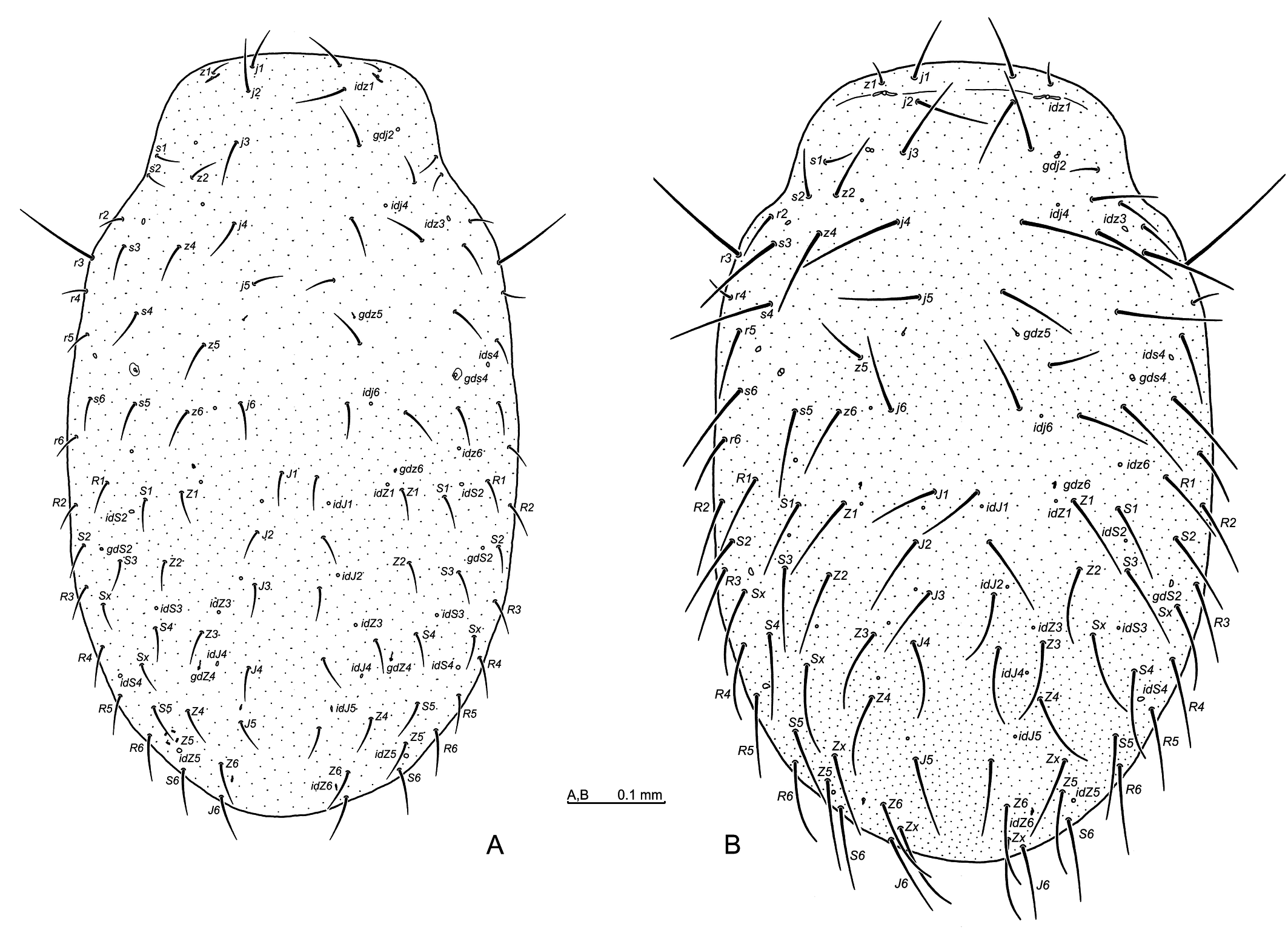
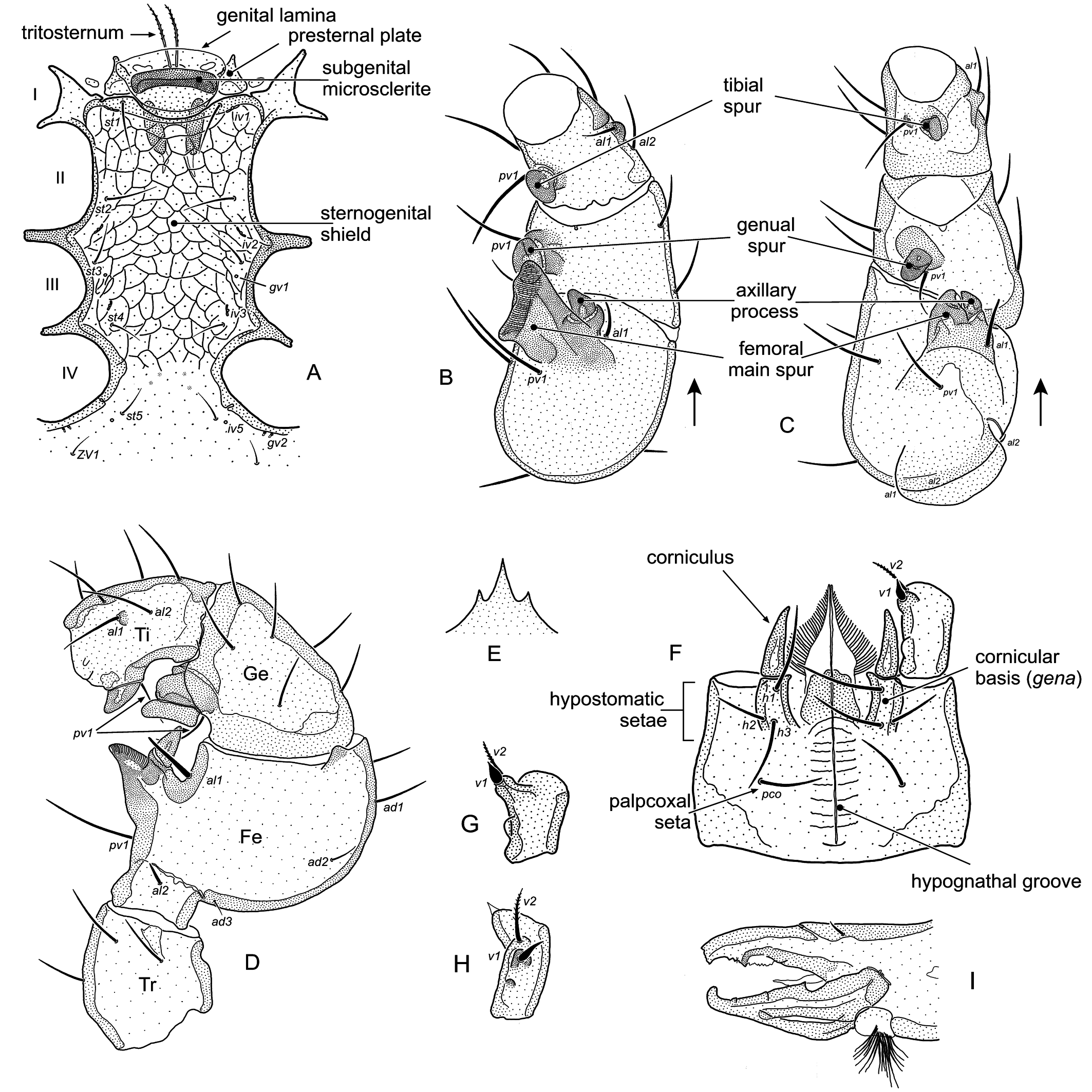
The illustrations have been either original or re-drawn from the original descriptions of species; the authors of any such drawings were duly acknowledged. The drawings were made with the aid of Corel Draw X8 and a Wacom Intuos Graphic Tablet. The system of dorsal, ventral, palpal, and leg setal notations was based on Evans and Till (1979), whereas poroidotaxy and adenotaxy on Moraza and Peña (2005), with some adjustments for Parasitidae. The scale bars were provided, whenever available.
Captions to figures
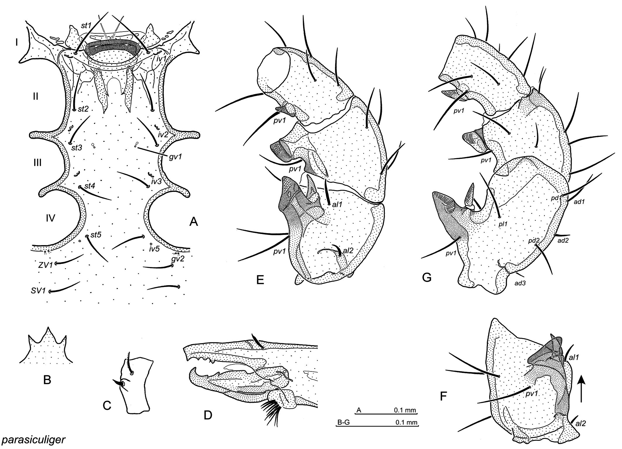
Abbreviations: I–IV the openings for coxae I to IV.
Dorsal side: podosomal setae (j, z, s, r), opisthosomal regular setae (J, Z, S, R) and supplementary setae (Sx, Zx), podosomal pores (idj, idz, ids) and opisthosomal pores (idJ, idZ, idS), podosomal gland openings (gdj, gdz, gds) and opisthosomal gland openings (gdS, gdZ);
Ventral side: st1–st3 sternal setae, st4, st5 paragynial and epigynial setae, respectively (female), st1–st5 sternal setae (male), JV, ZV, SV opisthogastral setae, Aa adanal seta, Pa postanal seta, gv1, gv2, gv3 gland openings, iv1–iv5, ivo2, ivo3, ivp pore openings.
Female genital orifice region: ax axial element of the inverted, T-shaped subapical thickening, lat subapical lateral thickening on epigynium, mscl paragynial metasclerite, ppm posterior paragynial margin, sub subapical thickening on central epigynial prong.
Palptrochanter ventral setae (v1, v2), hypostomatic setae (h1–h3), palpcoxal seta (pco).
Leg setation: al, ad, pd, pl, pv anterolateral, anterodorsal, posterodorsal, posterolateral and posteroventral setae, respectively.
Systematics
Family Parasitidae Oudemans, 1901
Subfamily Pergamasinae Juvara-Balş, 1972
Genus Anchigamasus Athias-Henriot, 1971
Paragamasus (Anchigamasus) Athias-Henriot, 1971: 172
Type species Pergamasus geileri Karg, 1968, designated by Witaliński 2024b: 412
Diagnosis of Anchigamasus (adults) (based on Witaliński 2024a, 2026)
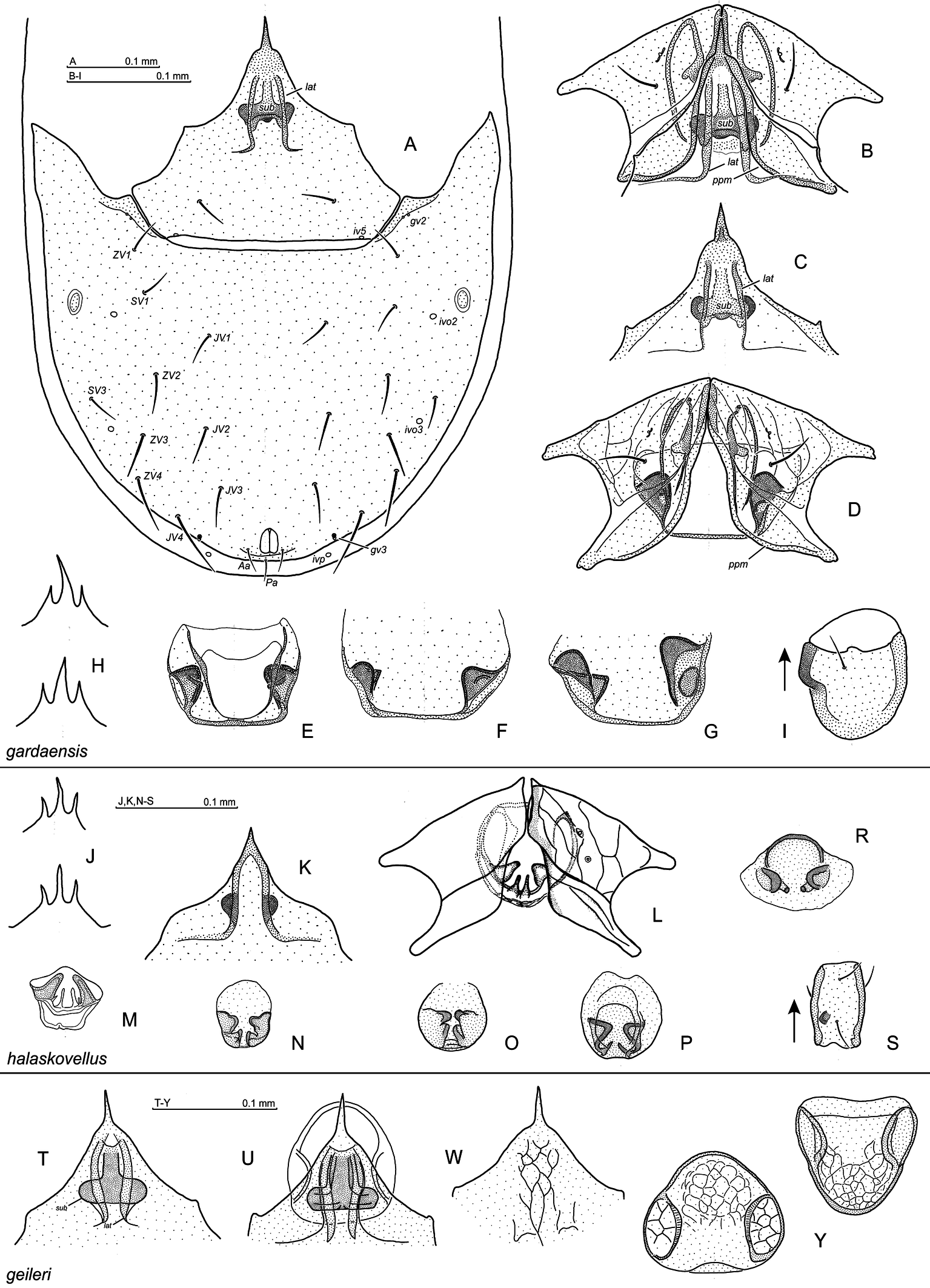
In the females and males, podosomal setae (22 pairs) and opisthosomal setae (24 regular pairs, but up to four pairs of supplementary setae can be encountered); in most species dorsal setae (especially the opisthonotal ones) short, terminating well short of the next setae row (Fig. 1A), or long, terminating at, or behind the next setae row bases (Fig. 1B); opisthogaster with 9 or 10 seta pairs (comp. e.g. Fig 2A and Fig. 3M); central prong of gnathotectum usually much longer than the lateral ones, or only slightly longer; palpfemur al seta pectinate, palpgenu al1 and al2 setae spatulate.
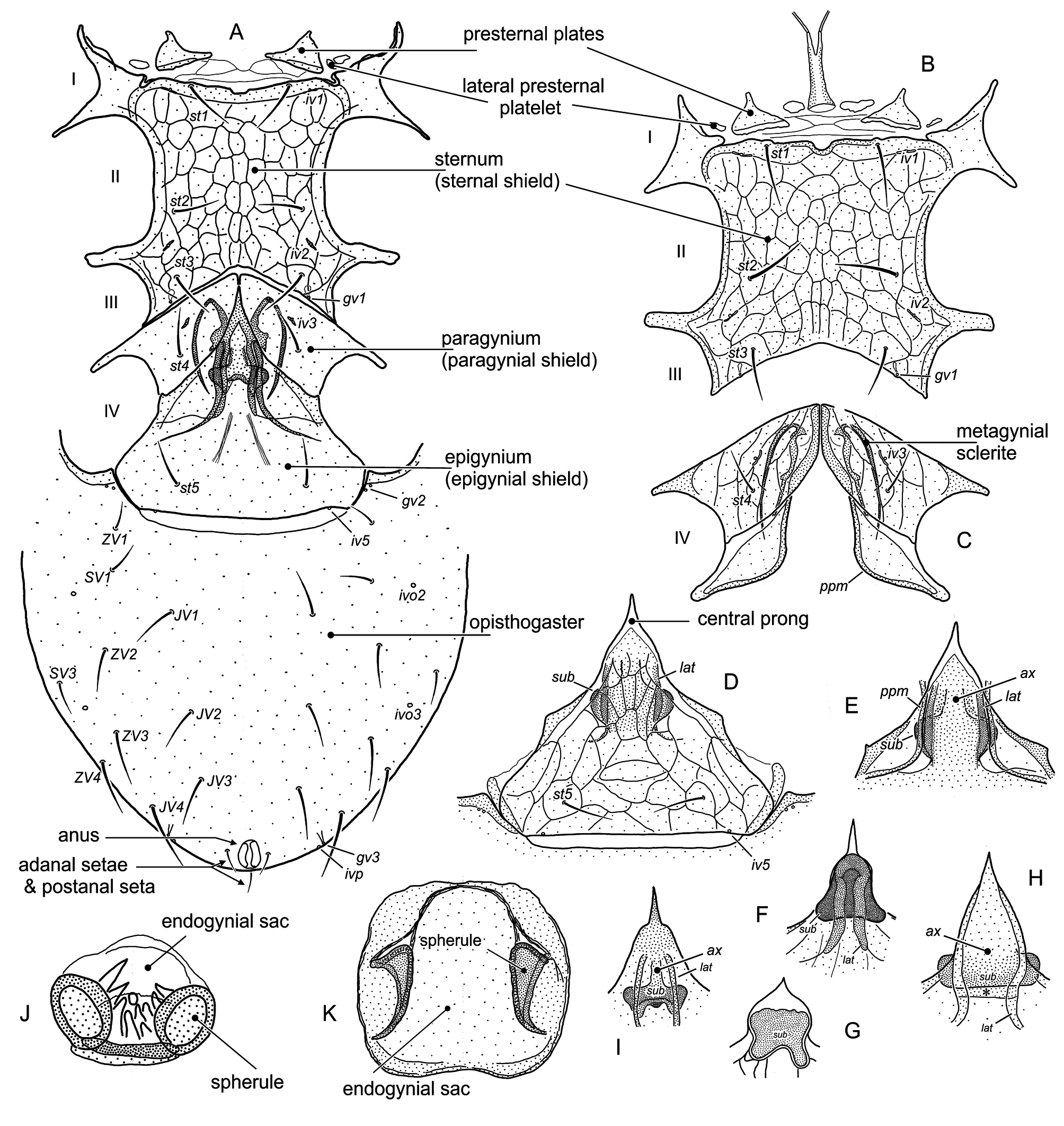
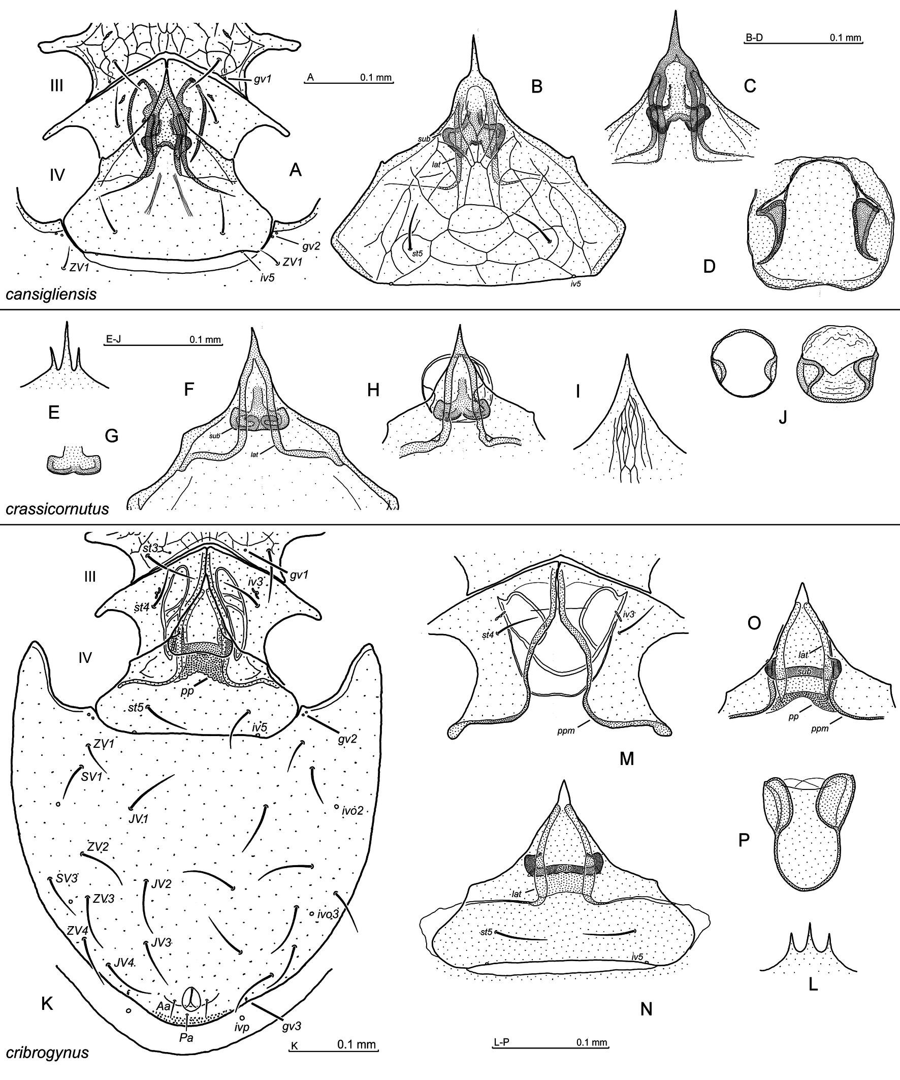
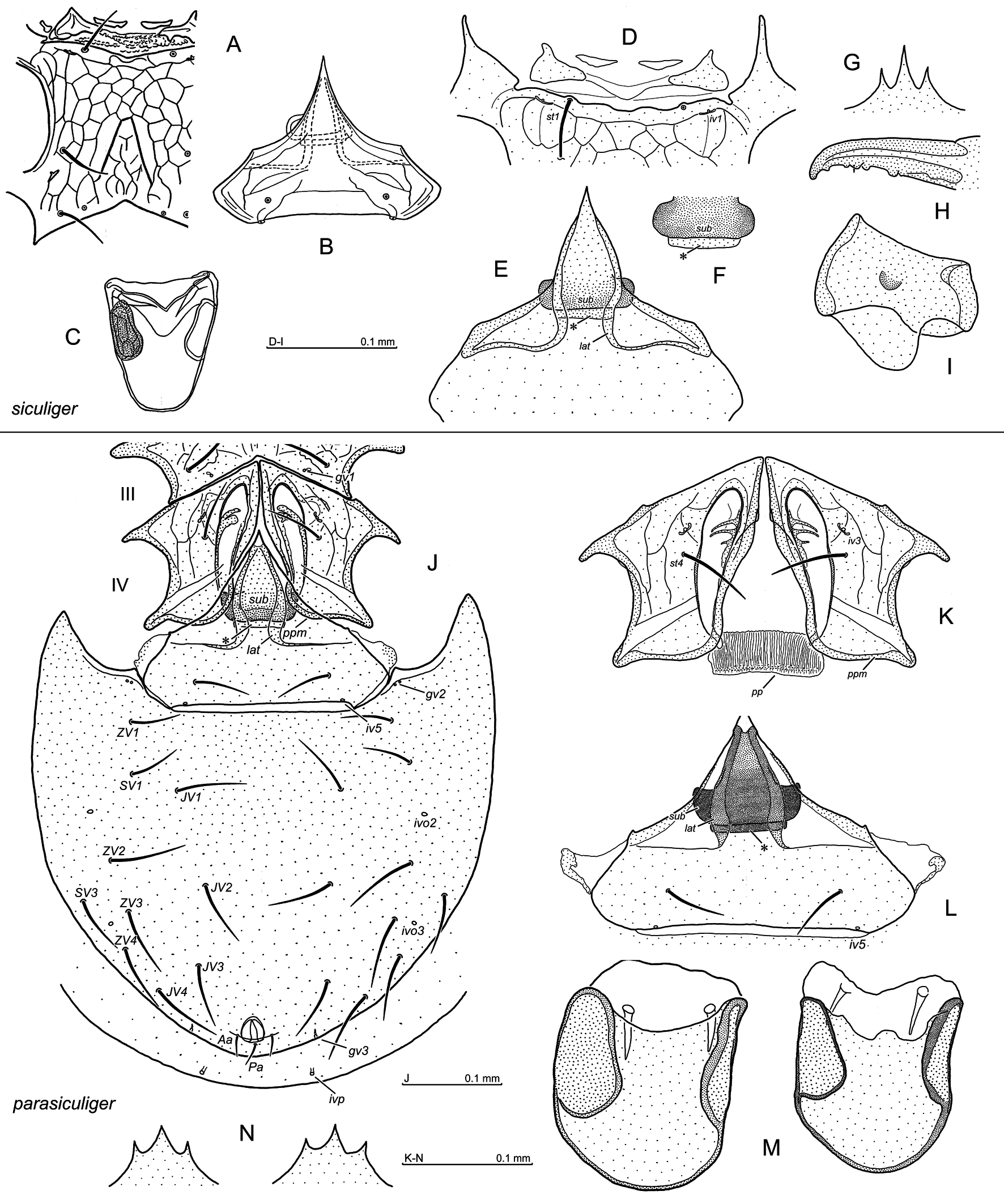
In the females (Fig. 2A–K), presternal plates subtriangular and positioned far from each other; the peritrematal shield posteriorly free, but anteriorly connected to the podonotal shield; peritreme ending anteriorly at the leg I level; gland pores gv1 present; epigynium with a distinct subapical thickening: in most species in the shape of inverted T (Fig. 2E, I), but in some species the lateral arms of the T are well pronounced, whereas the axial element of T is less distinct (Fig. 2H); in some species subapical thickening can be in more compact, bell-shape (Fig. 2F) or subquadrangle shape (Fig. 2G); posterior paragynial margins are locked between the arms of the T-shaped subapical thickening and the epigynial shield; the endogynial sac cup-shaped, circular or quadrangle shape, with lateral spherules, additionally protrusions, denticles or stipule-like structures can be encountered; in the Anchigamasus parasiculiger-group, behind the metagynial sclerites, a seemingly perforated plate may be encountered.
In the males (Fig. 8A–I), transverse suture between podonotal and opisthonotal areas; cheliceral movable digit with two teeth, followed by a tooth-like protrusion located proximally, fixed digit edentate, paucidente or pluridente; two rows of anterior most hypostomal denticles transverse, or moderately arcuate anteriorly; genae (cornicular bases) distinct and arcuate; seta v1 on palp trochanter, usually short and on a tubercle.
List of Anchigamasus species
In alphabetic order, known sex, the original name, and general locality.
- Anchigamasus alportus (Athias-Henriot, 1968); female
- Anchigamasus apuseniensis Witaliński, 2024; female, male
- Anchigamasus campanulatus Witaliński, 2025; female, male
- Anchigamasus cansigliensis Witaliński, 2024; female, male
- Anchigamasus crassicornutus (Willmann, 1954); female, male
- Anchigamasus cribrogynus Witaliński, 2026; female, male
- Anchigamasus gardaensis Witaliński, 2024; female, male
- Anchigamasus geileri (Karg, 1968); female, male
- Anchigamasus halaskovellus (Athias-Henriot, 1967); female, (male Witaliński 2024c: 1277)
- Anchigamasus parasiculiger Witaliński, 2026; female, male
- Anchigamasus siculiger (Athias-Henriot, 1967); female, male
- Anchigamasus stipularis Witaliński, 2025; female
- Anchigamasus tortulatus (Athias-Henriot, 1979); female
Pergamasus alportus Athias-Henriot, 1968: 194; Austrian Alps
Anchigamasus apuseniensis Witaliński, 2024c: 1264; Romania
Anchigamasus campanulatus Witaliński, 2025: 855; Romania
Anchigamasus cansigliensis Witaliński, 2024b: 412; Northen-Italian Prealps
Pergamasus crassicornutus Willmann, 1954: 219; Czech Rep., S-E Poland, N-E Slovakia (see Witaliński 2024a)
Anchigamasus cribrogynus Witaliński, 2026: 57; S Austria
Anchigamasus gardaensis Witaliński, 2024c: 1271; N Italy
Pergamasus geileri Karg, 1968: 335; E. Germany, S-W Poland, Czech Rep., Austria, N Hungary (see Witaliński 2024a)
Pergamasus halaskovellus Athias-Henriot, 1967: 48; N. Czech Rep., S-W Poland
Anchigamasus parasiculiger Witaliński, 2026: 51; Romania
Pergamasus siculiger Athias-Henriot, 1967: 42; Hungary
Anchigamasus stipularis Witaliński, 2025: 862; Romania
Paragamasus (Anchigamasus) tortulatus Athias-Henriot, 1979: 1152; Ukraine
Taxonomic/morphologic remarks
Genus Anchigamasus is most similar to Anidogamasus, and both genera may well be misidentified. The main differences are as follows: in the Anchigamasus females, the central prong of the epigynial plate possesses a subapical, inverted T-shaped thickening protruded dorsally, flanked with two lateral subapical thickenings. More laterally, in the intact specimen, a posterior paragynial margins can be seen, which are locked in the space between the epigynial plate and the arms of T-shaped thickening. In the Anidogamasus females, such inverted T-shaped thickening on central epigynial prong is absent, but lateral subapical thickenings may be evident.
In the males, the main differences relate to the hypostomal rows of denticles, form of cornicular bases (genae), and cheliceral movable digit. In Anchigamasus, two anterior hypostomal rows can be arcuate anteriorly (Fig. 8F), whereas others are transversal. In Anidogamasus, four anterior rows are usually curved far anteriorly. The genae in Anchigamasus are distinct and evidently arcuate, whereas in the Anidogamasus they are less pronounced and straight. Cheliceral movable digit in the Anchigamasus male, despite the regular two teeth also shows a tooth-shaped protrusion located proximally, whereas no such protrusion is to be found in Anidogamasus.
In the Anchigamasus females and males less evidently characteristic is the anterior sternal margin, frequently showing two incisions laterally to iv1 pores, which are not encountered in Anidogamasus., In the females of some Anchigamasus (parasiculuger species-group), however, the incisions are not evident.
During the study of Anchigamasus material, it has become apparent that all species can be segregated into four species-groups, i.e. campanulatus, apuseniensis, crassicornutus and parasiculiger group.
Proposed newly formed and defined species-groups and their diagnoses
Only the key characteristics are presented
Anchigamasus campanulatus species-group
Diagnosis — dorsal setae short; opisthogaster bears 9 pairs of setae; female epigynial thickening compact, bell-shaped or subquadrangle; in male, gv1 gland openings behind st3 setae, fixed digit of chelicera rounded terminally and edentate, femoral main spur sigmoidal in ventral view.
Included species
- Anchigamasus campanulatus Witaliński, 2025
- Anchigamasus tortulatus (Athias-Henriot, 1979)
Note — Anchigamasus tortulatus is included to this species-group provisionally, since the number of opisthogastral setae is not given in the original paper by Athias-Henriot, as well as a male has not yet been encountered.
Anchigamasus apuseniensis species-group
Diagnosis — dorsal setae short; opisthogaster bears 9 pairs of setae; female epigynial thickening in the shape of inverted T, with well visible arms and well or moderately pronounced axial element; in male, gv1 gland openings behind st3 setae, fixed digit of chelicera hooked terminally, femoral main spur finger-shaped.
Included species
- Anchigamasus apuseniensis Witaliński, 2024c
- Anchigamasus stipularis Witaliński, 2025
Anchigamasus crassicornutus species-group
Diagnosis — dorsal setae short; opisthogaster bears 10 pairs of setae; female epigynial thickening in the shape of inverted T; in male, gv1 gland openings behind st3 setae, fixed digit of chelicera obtuse terminally, or hooked (A. halaskovellus), femoral main spur variable or finger-shaped (A. halaskovellus).
Included species
- Anchigamasus alportus (Athias-Henriot, 1968)
- Anchigamasus cansigliensis Witaliński, 2024b
- Anchigamasus crassicornutus (Willmann, 1954)
- Anchigamasus gardaensis Witaliński, 2024c
- Anchigamasus geileri (Karg, 1968)
- Anchigamasus halaskovellus (Athias-Henriot, 1967)
Anchigamasus parasiculiger species-group
Diagnosis — dorsal setae long; opisthogaster bears 10 pairs of setae; female epigynial thickening in the shape of inverted T, with well pronounced arms, but the axial element is wide and weakly discernible; at posterior ends of the metagynial thickenings a plate, which looks like perforated one, is encountered; in male, gv1 glands at st3 setae level, male chelicera fixed digit hooked (A. cribrogynus) or obtuse (A. parasiculiger, A. siculiger) terminally, several arthrodial corona processes can be thickened, as well as forked terminally, Femoral main spur finger-shaped in ventral and lateral views or cut obliquely in ventral view (A. parasiculiger).
Included species
- Anchigamasus cribrogynus Witaliński, 2026
- Anchigamasus parasiculiger Witaliński, 2026
- Anchigamasus siculiger (Athias-Henriot, 1967)
Key to the adults of Anchigamasus species
Females
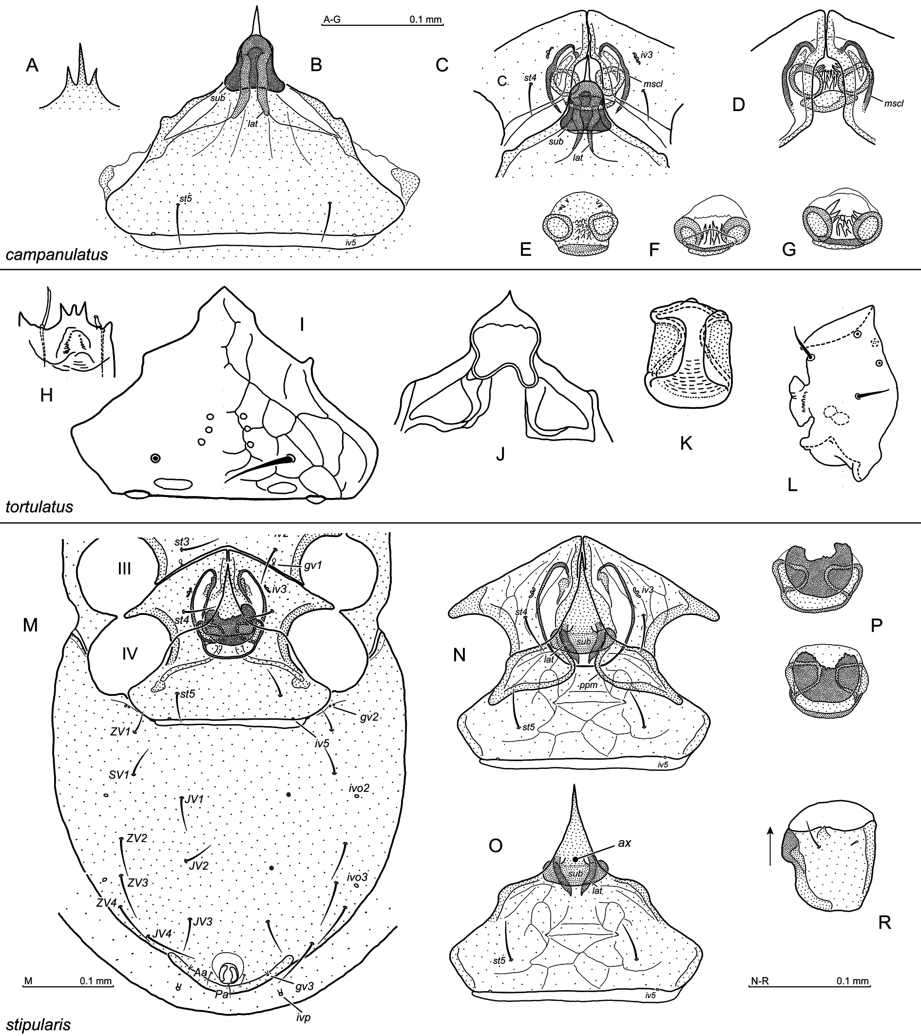
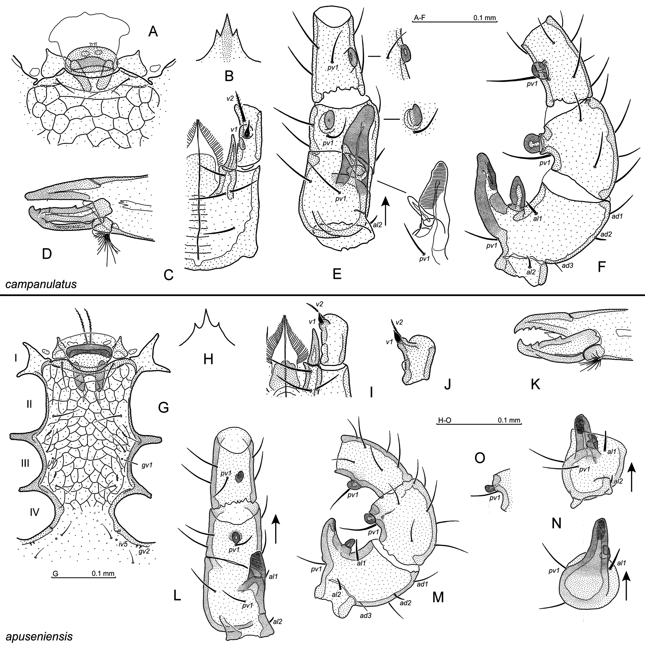
1. Opisthogaster with 9 pairs of setae (SV3 absent) (Fig. 3M)
...... 2
— Opisthogaster with 10 pairs of setae (SV3 present) (Fig. 2A)
...... 5
2. Epigynial central thickening compact, bell-shaped (Figs 2F, 3B) or subquadrangle (Figs 2G, 3J), located under the apex of central epigynial prong
...... campanulatus species-group – 3
— Epigynial central thickening in the shape of inverted T, with well visible arms and well (Fig. 2E, I) or moderately pronounced axial element (Fig. 3O)
...... apuseniensis species-group – 4
3. Epigynial central thickening bell-shaped (Fig. 2F), located under the apex of the central epigynial prong; gnathotectum central prong acute and twice longer than the lateral ones (Fig. 3A); endogynial sac with many thorns located between spherules (Figs 2J, 3E–G)
...... A. campanulatus Witaliński, 2025 (Figs 2F, 3A–G)
— Epigynial central thickening subquadrangle (Figs 2G, 3J), gnathotectum central prong shorter than the lateral ones and terminally rounded (Fig. 3H); endogynial sac without thorns or teeth
...... A. tortulatus (Athias-Henriot, 1979) (Figs 2G, 3H–L)
4. Endogynial spherules irregularly rounded and directed adaxially (Fig. 3P), posterior margin of endogynial sac smooth, a distinct thick stipule-like plate covers the endogynium ventrally (Fig. 3P)
...... A. stipularis Witaliński, 2025 (Fig. 3M–R)
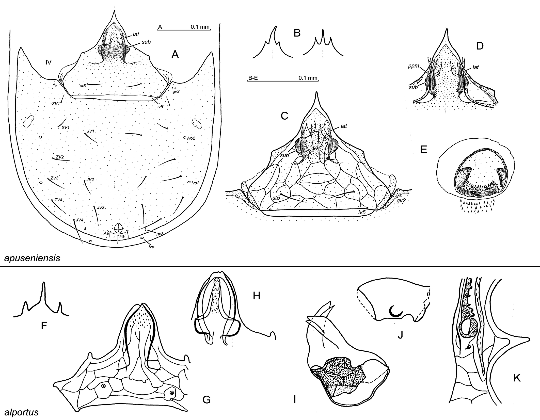
— Endogynial spherules conical and directed anteroaxially, posterior margin of the endogynial sac with many denticles (Fig. 4E), stipule-like plate covering endogynium ventrally absent
...... A. apuseniensis Witaliński, 2024c (Figs 2B–E, 4A–E)
5. Podonotal and opisthonotal setae short, ending far before next setae row (e.g. Fig. 1A); perforated plate behind the endogynium absent
...... crassicornutus species-group – 6
— Podonotal and especially opisthonotal setae long, ending well at the next setae row (e.g. Fig. 1B); perforated plate behind endogynium present (Figs 6K, N, 7K)
...... parasiculiger species-group – 11
6. Epigynial central prong blunt (Fig. 4G, H); endogynium anterior margin with two large, bifid protrusions (Fig. 4I)
...... A. alportus (Athias-Henriot, 1968) (Fig. 4F–K)
— Epigynial central prong acute and frequently elongated; bifid protrusions of endogynium anterior margin absent
...... 7
7. Endogynium with two pairs of spherule-like structures (Fig. 5E–G, M–P)
...... 8
— Endogynium with one pair of spherules (e.g. Figs 2K, 5Y)
...... 9
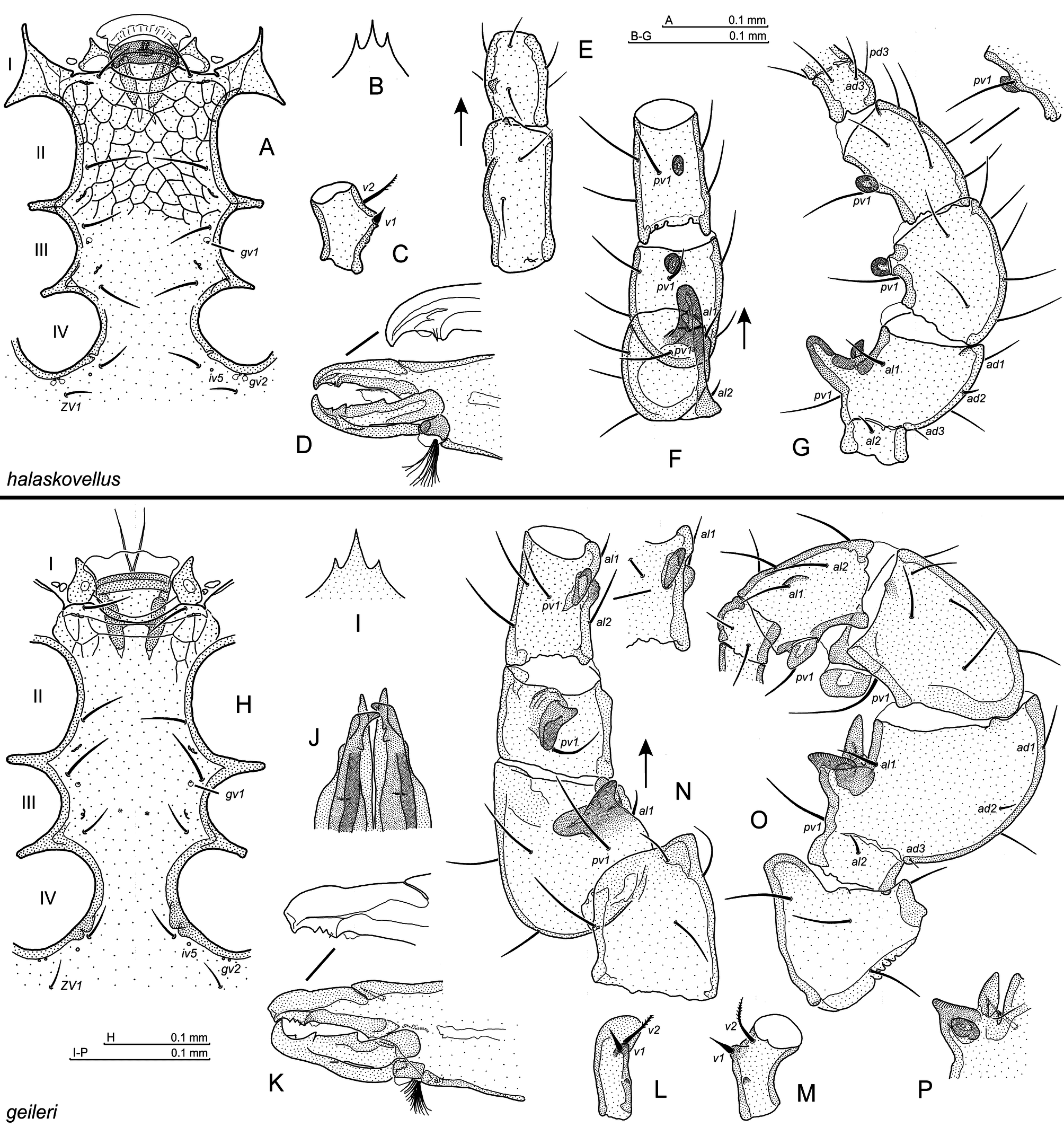
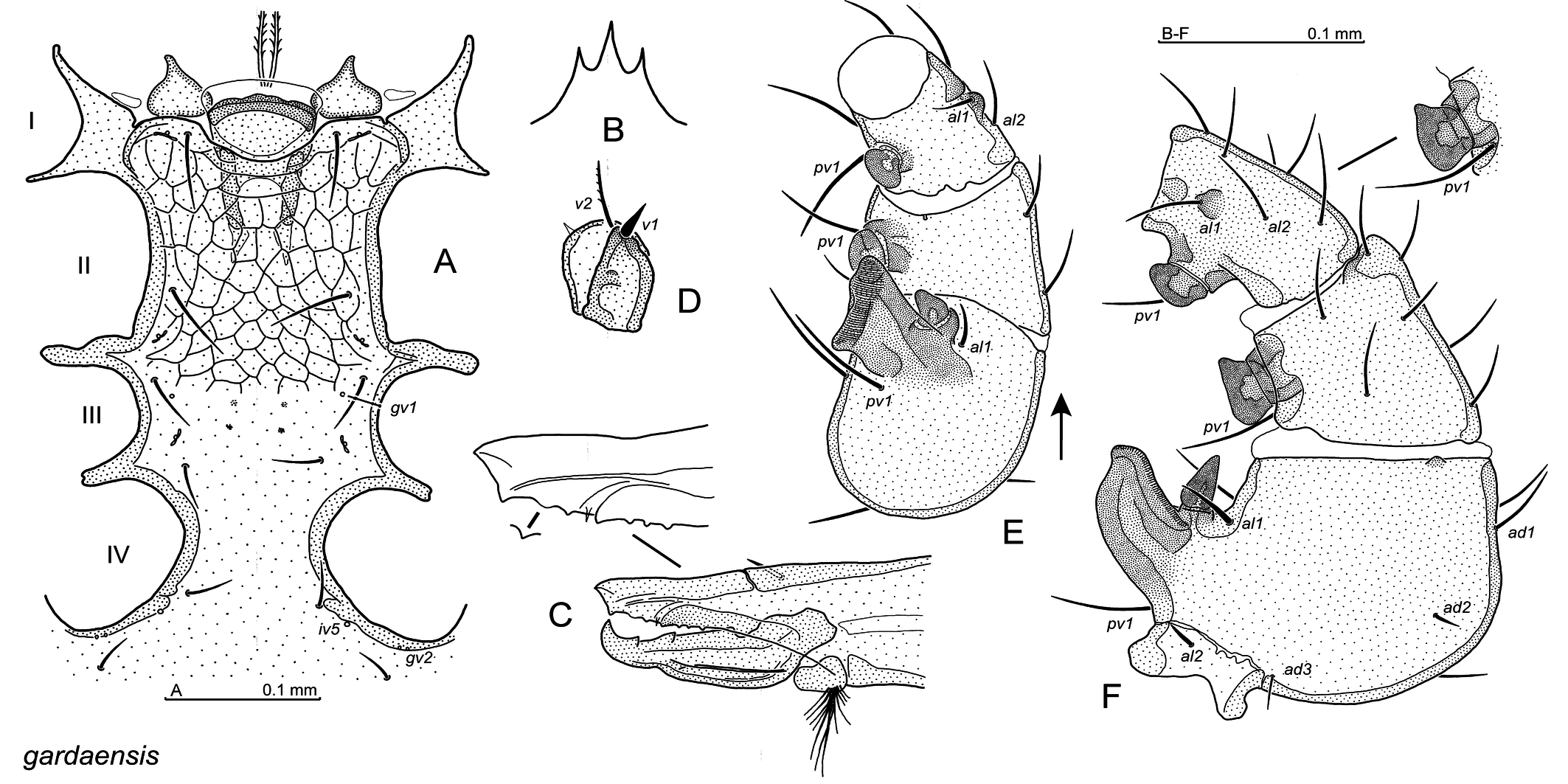
8. Anteroventral tubercle at v2 level on Tr I absent; ventral pair of spherule-like structures rounded terminally, whereas the dorsal one triangular and usually pointed apically (Fig. 5E–G)
...... A. gardaensis Witaliński, 2024 (Figs 1A, 2I, 5A–I)
— Tr I with anteroventral tubercle at v2 level (Fig. 5S); the lateral pair of spherule-like structures larger than the ventral one, which is frequently finger-shaped (Fig. 5L–P)
...... A. halaskovellus (Athias-Henriot, 1967) (Fig. 5J–S)
9. Endogynium cup-shaped, posterior part of endogynial sac reticulated (Fig. 5Y); epigynium central prong bears polygonal reticulation (Fig. 5W)
...... A. geileri (Karg, 1968) (Fig. 5T–Y)
— Endogynium circular or quadrilateral in ventral perspective (e.g. Fig. 6D, J); posterior part of endogynial sac smooth; epigynium central prong with fan-like (Fig. 6B) or longitudinal (Fig. 6I) reticulation
...... 10
10. Spherules conical, directed obliquely anteriad (Fig. 6D); epigynium central prong bears a fan-like reticulation (Fig. 6B)
...... A. cansigliensis Witaliński, 2024 (Figs 2A, I, 6A–D)
— Spherules apically rounded, directed adaxially (Fig. 6J); epigynium central prong carries a longitudinal reticulation (Fig. 6I)
...... A. crassicornutus (Willmann, 1954) (Fig. 6E–J)
11. Endogynium sac-shaped with two acute long protrusions directed posteriad at the entrance to the endogynial sac (Fig. 7C, M)
...... 12
— Endogynium sac-shaped or V-shaped, acute protrusions at the endogynial sac entrance absent (Fig. 6M, P)
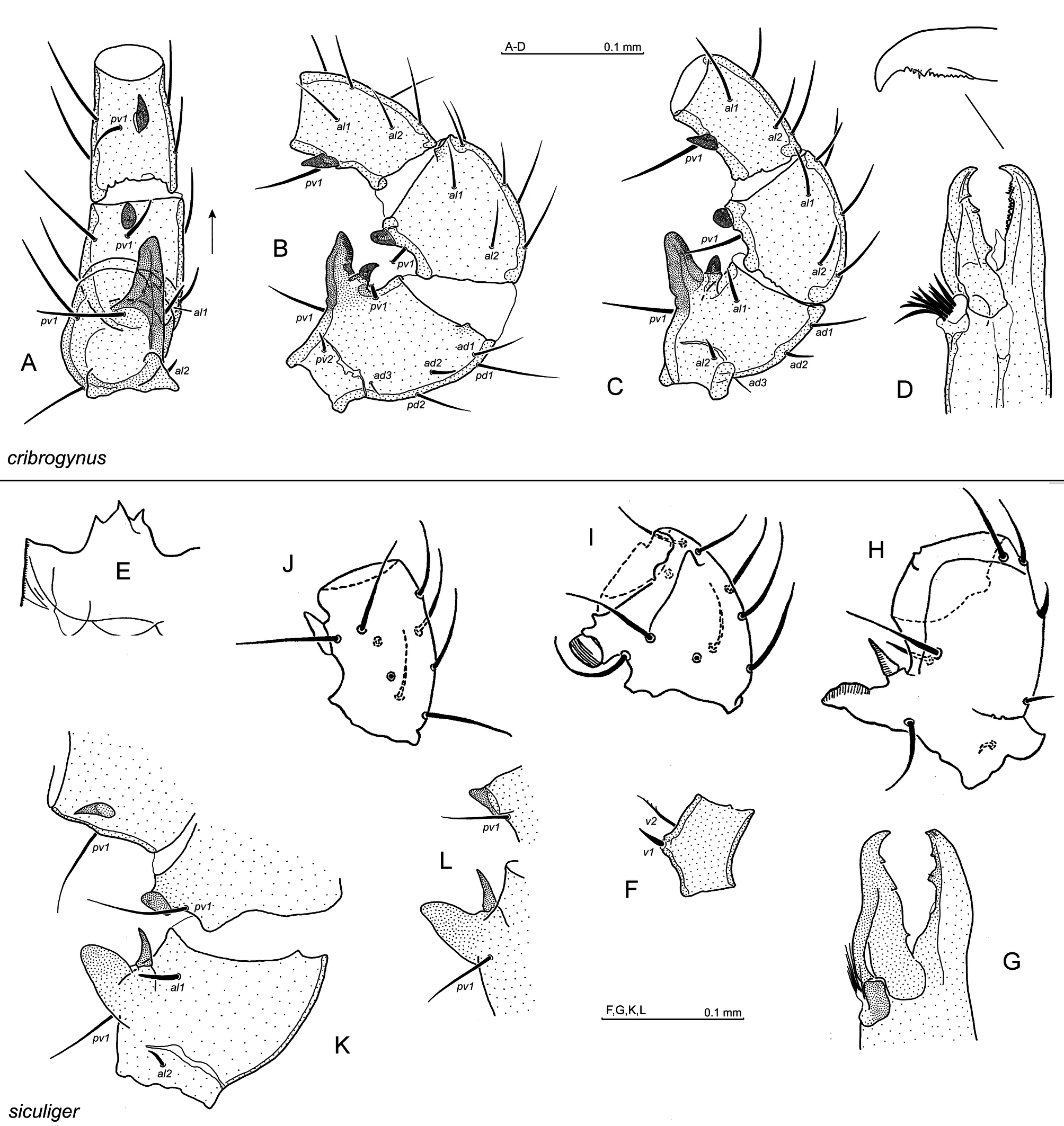
...... A. cribrogynus Witaliński, 2026 (Fig. 6K–P)
12. Lateral arms of the T-shaped thickening under central epigynial prong rounded terminally (Fig. 7B, E, F)
...... A. siculiger (Athias-Henriot, 1967) (Fig.7A–I)
— Lateral arms of the T-shaped thickening under central epigynial prong with arms acute anterolaterally (Fig. 7L)
...... A. parasiculiger Witaliński, 2026 (Figs 1B, 7J–N)
Males
1. Podonotal and opisthonotal setae short, ending far before next setae row; gv1 gland openings located behind st3 setae and apparently axially to their bases (e.g. Figs 8A, 9G)
...... 2
— Podonotal and opisthonotal setae long, ending well at the next setae row; gv1 gland openings located at st3 setae level and axially to their bases (Fig. 14I)
...... parasiculiger species-group – 8
2. Opisthogaster with 9 pairs of setae
...... 3
— Opisthogaster with 10 pairs of setae
...... crassicornutus species-group – 4
3. In ventral view, leg II femoral main spur bent in half way (Fig. 9E), axillary process long and straight (Fig. 9E, F); chelicera fixed digit edentate and rounded terminally (Fig. 9D)
...... campanulatus species-group – A. campanulatus Witaliński, 2025 (Fig. 9A–F)
— In ventral view, leg II femoral main spur straight, axillary process short and hooked (Fig. 9L, N); fixed digit of chelicera with ca. 3 teeth and pointed terminally (Fig. 9K)
...... apuseniensis species-group – A. apuseniensis Witaliński, 2024 (Figs 8A, F, G; 9G–O)
4. Tr I with anteroventral tubercle at v2 level (Fig. 10E)
...... A. halaskovellus (Athias-Henriot, 1967) (Fig. 10A–G)
— Anteroventral tubercle on Tr I absent; seta al1 on Ti II located on tubercle (e.g. Fig. 8B–D); cheliceral fixed digit obtuse terminally (e.g. Fig. 8I)
...... 5
5. Main spur on Fe II forked terminally, best visible in ventral view (Fig. 10N)
...... A. geileri (Karg, 1968) (Figs 8E, 10H–P)
— Fe II main spur not forked terminally (e.g. Fig. 8B–D)
...... 6
6. Fe II main spur ventrally very short, axillary process triangular, ending at the main spur tip level (Fig. 11E, F); laterally, Fe II main spur triangular, with its ventral margin nearly straight (Fig. 11G)
...... A. crassicornutus (Willmann, 1954) (Figs 8 C, D, H, I, 11A–G)
— Ventrally, Fe II main spur not very short, obliquely cut terminally (rhomboidal) (Fig. 11J-L) or irregular (wider terminally than basally) (Fig. 12E); axillary process ending not as above; laterally, Fe II main spur not triangular, its ventral margin distinctly arcuate (Figs 11M, 12 F)
...... 7
7. Ventrally, Fe II main spur rhomboidal (Fig. 11J-L)
...... A. cansigliensis Witaliński, 2024 (Fig. 11H–M)
— Ventrally, Fe II main spur wider terminally than basally (Fig. 12E)
...... A. gardaensis Witaliński, 2024 (Figs 8B, 12A-F)
8. Spur on Ge II located on a nearly flat cuticle (Fig. 13B, C), cheliceral fixed digit bears many small denticles (Fig. 13D)
...... A. cribrogynus Witaliński, 2026 (Fig. 13A-D)
— Spur on Ge II located on a distinct article protrusion (e.g. Figs 13I, K, 14E, G)
...... 9
9. Fe II main spur finger-shaped in lateral view and pointed or roundish terminally (Fig. 13H, K, L), cheliceral fixed digit obtuse terminally, with one tooth ahead and one by the side of pilus dentilis (Fig. 13G)
...... A. siculiger (Athias-Henriot, 1967) (Fig. 13E–L)
— Fe II main spur not finger-shaped in lateral view (Fig. 14G), in ventral view cut obliquely (Fig. 14E, F), cheliceral fixed digit obtuse terminally, with two teeth ahead and one tooth by the side of pilus dentilis (Fig. 14D)
...... A. parasiculiger Witaliński, 2026 (Fig. 14A–G)
Acknowledgements
The study was partly supported by a grant allocated by the Jagiellonian University, Kraków, Poland (Grant Ref. No N18/DBS/000005).
References
- Athias-Henriot C. 1967. Observations sur les Pergamasus. I. Sous-genre Paragamasus Hull, 1918 (Acariens anactinotriches: Parasitidae). Mém. Mus. natl. Hist. nat., Sér. A (Zool.), 49: 1-197.
- Athias-Henriot C. 1968. Observations sur les Pergamasus. V. Additions et corrections aux Paragamasus d'Europe tempérée, principalement occidentale (Acariens anactinotriches, Parasitidae). Bulletin scientifique de Bourgogne, 25: 175-228.
- Athias-Henriot C. 1971. Paragamasus (Tanygamasus) probsti (Oudemans) (systématique, géographie), avec quelques mises au point synonymiques (Arachnides, Gamasides tocospermiques, Parasitidae). Zool. Meded., 45: 167-179.
- Athias-Henriot C. 1979. A contribution to the knowledge of the family Parasitidae of the fauna of the Ukrainian SSR. Zoologicheskii Zhurnal, 58: 1148-1156 (in Russian).
- Evans G.O., Till W.M. 1979. Mesostigmatic mites of Britain and Ireland (Chelicerata: Acari Parasitiformes). An introduction to their external morphology and classification. Trans. Zool. Soc. London, 35: 139-262. https://doi.org/10.1111/j.1096-3642.1979.tb00059.x
- Hrúzová K., Fenďa P. 2018. The family Parasitidae (Acari: Mesostigmata) - history, current problems and challenges. Acarologia, 58 (Suppl.): 25-42. https://doi.org/10.24349/acarologia/20184280
- Juvara-Balş I. 2019. Occigamasus, a new genus of pergamasine mites, with description of two new species from the west coast of North America (Parasitiformes: Gamasina: Parasitidae). Acarologia, 59: 551-570. https://doi.org/10.24349/acarologia/20194354
- Karg W. 1968. Neue Arten der gattung Pergamasus Berlese, 1903 (Acarina, Parasitiformes). Deut. Entomol. Z., 15: 335-358. https://doi.org/10.1002/mmnd.4810150407
- Makarova O.L. 2019. North Pacific versus North Atlantic: a case with species of the amphiboreal littoral mite genus Thalassogamasus gen. nov. (Parasitiformes, Mesostigmata Parasitidae). Zootaxa, 4647: 457-485. https://doi.org/10.11646/zootaxa.4647.1.29
- Micherdziński W. 1969. Die Familie Parasitidae Oudemans, 1901 (Acarina, Mesostigmata). Państwowe Wydawnictwo Naukowe, Kraków, 690 pp.
- Moraza M.L., Peña M.A. 2005. The family Pachylaelapidae Vitzthum, 1931 on Tenerife Island (Canary Islands), with description of seven new species of the genus Pachylaelaps (Acari, Mesostigmata: Pachylaelapidae). Acarologia, 45: 103-129.
- Willmann C. 1954. Mährische Acari Hauptsächlich aus dem Gebiete des Mährischen Karstes. Českoslov. parasitologie, I: 213-272.
- Witaliński W. 2017. Key to the world species of Holoparasitus Oudemans, 1936 (Acari: Parasitiformes: Parasitidae). Zootaxa, 4277 (3): 301-351. https://doi.org/10.11646/zootaxa.4277.3.1
- Witaliński W. 2024a. The Anchigamasus mite species (Parasitiformes: Parasitidae), synonyms revisited. Acarologia, 64: 803-818. https://doi.org/10.24349/2r5k-fyp7
- Witaliński W. 2024b. A new mite species and a new type species designation in the Anchigamasus genus (Parasitiformes: Parasitidae). Annales Zoologici, 74: 411-422. https://doi.org/10.3161/00034541ANZ2024.74.3.007
- Witaliński W. 2024c. Two new Anchigamasus mite species and an unknown male in Anchigamasus halaskovellus (Athias-Henriot, 1967) (Parasitiformes: Parasitidae). Acarologia, 64: 1263-1282. https://doi.org/10.24349/vtyn-syp0
- Witaliński W. 2025. Two new mite species of Anchigamasus Athias-Henriot, 1971 (Parasitiformes: Parasitidae). Acarologia, 65: 854-867. https://doi.org/10.24349/v0z2-t67t
- Witaliński W. 2026. Two new and one re-described mite species of Anchigamasus Athias-Henriot, 1971 (Parasitiformes: Parasitidae). Acarologia, 66: 50-68. https://doi.org/10.24349/9vhs-j1rm
- Yao M.-Y., Yi T.-C., Jin D.-C., Guo J.-J. 2020. A new species of Psilogamasus Athias-Henriot, 1969 from China and redefinition of the genus (Parasitiformes: Parasitidae). Acarologia, 60: 831-841. https://doi.org/10.24349/acarologia/20204404
- Yao M.-Y., Chen J.-X., Yi T.-C., Jin D.-C., Guo J.-J. 2022. A new species of Dyoneogamasus Athias-Henriot, 1979 (Parasitiformes: Parasitidae) from China and redefinition of the genus. Internat. J. Acarol., 48: 575-580.


2026-01-26
Date accepted:
2026-03-04
Date published:
2026-03-18
Edited by:
Faraji, Farid

This work is licensed under a Creative Commons Attribution 4.0 International License
2026 Witaliński, Wojciech and Podkowa, Dagmara
 Download article Download low definition
Download the citation
RIS with abstract
(Zotero, Endnote, Reference Manager, ProCite, RefWorks, Mendeley)
RIS without abstract
BIB
(Zotero, BibTeX)
TXT
(PubMed, Txt)