


Two new species of the feather mite genus Dolichodectes (Acariformes: Proctophyllodidae) from flycatchers (Passeriformes: Muscicapidae) in South Africa
Mironov, Sergey V.  1
1
1✉ Zoological Institute, Russian Academy of Sciences, Universitetskaya Embankment 1, 199034, Saint Petersburg, Russia.
2026 - Volume: 66 Issue: 1 pages: 171-186
https://doi.org/10.24349/qfvs-mo92ZooBank LSID: 231741E2-EB9B-4398-A7F2-1CFF7F80C09E
Original research
Keywords
Abstract
Introduction
The feather mite genus Dolichodectes Park & Atyeo, 1971 (Analgoidea: Proctophyllodidae) with eleven currently known species is a non-specious and easily recognizable genus of the subfamily Pterodectinae (Gaud and Mouchet 1957; Park and Atyeo 1971; Mironov and Fain 2003; Mironov et al. 2010, 2012, 2015; Mironov 2023; Constantinescu et al. 2018). In the subfamily Pterodectinae, this genus belongs to the Montesauria generic complex characterized by the position of the genital papillae in males, which are situated, with the exception of a few species, at the level of the genital arch and usually are strongly reduced. This position is unusual for the most taxa of the family Proctophyllodidae, in which the genital papillae are commonly situated anterolateral to the genital arch. Based on the host associations and geographical distribution, this generic complex can be referred to as a grouping of derived pterodectine genera associated with passerines of the Old World (Mironov 2006, 2009).
Within the Montesauria generic complex, the genus Dolichodectes is easily distinguishable in the following features. In both sexes, the idiosoma is noticeably elongated (three or more times longer than wide), in males, the opisthosomal lobes are strongly elongated, setae ps3 are situated posterior or posterolateral from the adanal suckers, resulting in the bases of setae g and ps3 being arranged in a longitudinal rectangle or a very tall trapezoid (Park and Atyeo 1971; Mironov 2009, 2023). Representatives of the genus are distributed on oscine passerines of the Old World; six of eleven previously described species are associated with hosts of the family of Old World flycatchers (Muscicapidae), remaining species are erratically distributed on selected hosts from the families Acrocephalidae, Passeridae, Phylloscopidae, Platysteiridae, Stenostiridae and Turdidae (Mironov et al. 2015; Constantinescu et al. 2018). An updated checklist of known Dolichodectes species, their host associations, and a key to species were recently provided by Mironov (2023).
The present work provides the descriptions of two new Dolichodectes species collected from two muscicapid hosts in South Africa.
Material and methods
The material used in the present study was collected in the Pongola Game Reserve (KwaZulu-Natal, South Africa) in November-December 2024. Birds, captured with standard mist nets for small passerines, were identified, ringed, and also examined for the presence of feather mites and other ectoparasites, at first visually and then with a stereomicroscope. Feather mites were picked up from live birds with a preparation needle and fixed in 96% ethanol. After the procedures of ringing and examination for feather mites, the birds were released to the wild. In the laboratory conditions, the collected mites were mounted on microscope slides in Hoyer's medium according to the standard technique (Krantz and Walter 2009).
The descriptions of the new species are provided according to the standards used for Dolichodectes species and related pterodectine mites (Valim and Hernandes 2010; Mironov et al. 2010, 2012; Hernandes 2022, 2023). General morphological terms, idiosomal and leg chaetotaxies follow Gaud and Atyeo (1996) with minor corrections for coxal setation by Norton (1998). Measuring techniques used for particular structures were described in extensive taxonomic papers on pterodectines (Mironov and González-Acuña 2011; Mironov and Chandler 2017; Mironov and Galloway 2021). All the measurements are in micrometers (μm). Primary drawings and measurements were made using a Leica light microscope DM2500 (Leica Microsystems, Inc.) with DIC illumination and camera lucida; and the final digital drawings were prepared with a Wacom Cintiq 22 graphics tablet (Wacom Co., Ltd).
The taxonomic system and the scientific names of birds follow the IOC World Bird List, v 15.1 (Gill et al. 2025). Type materials are deposited in mite collections of the museum with the following abbreviations: NMB — the National Museum in Bloemfontein (Free State, South Africa); ZISP—the Zoological Institute of the Russian Academy of Sciences (Saint Petersburg, Russia); SVM —an index in primary field collection number of mite samples.
Taxonomy
Family Proctophyllodidae Trouessart & Mégnin, 1884
Subfamily Pterodectinae Park & Atyeo, 1971
Genus Dolichodectes Park & Atyeo, 1971
Type species: Proctophyllodes (Pterocolus) edwardsi Trouessart, 1885, by original designation.
Dolichodectes robertsi sp. n.
ZOOBANK: FC008BC5-79D4-4481-959A-FF2967BBF1B0 ![]()
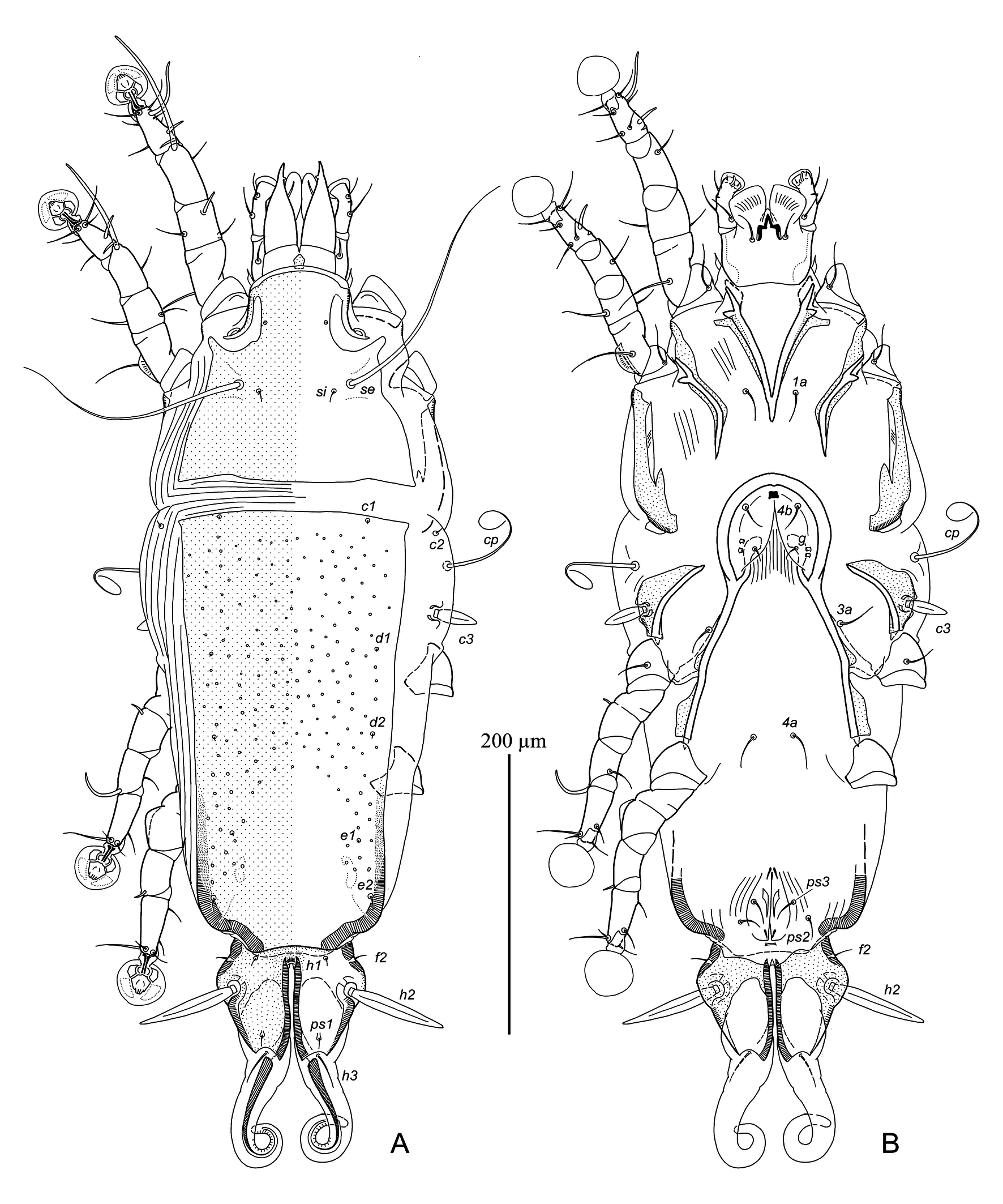
(Figures 1–3, 7A, B)
Type material
Holotype male, 9 male and 10 female paratypes from Cercotrichas leucophrys (Vieillot, 1817) (Passeriformes, Muscicapidae), SVM 24-1120-2, South Africa, KwaZulu-Natal, Pongola Game Reserve, White Elephant Bush Camp, 27°24′42.6″S 31°53′53.3″E, 20 November 2024, coll. S.V. Mironov.
Depository
Holotype, 4 male and 7 female paratypes—NMB, remaining paratypes—ZISP.
Description
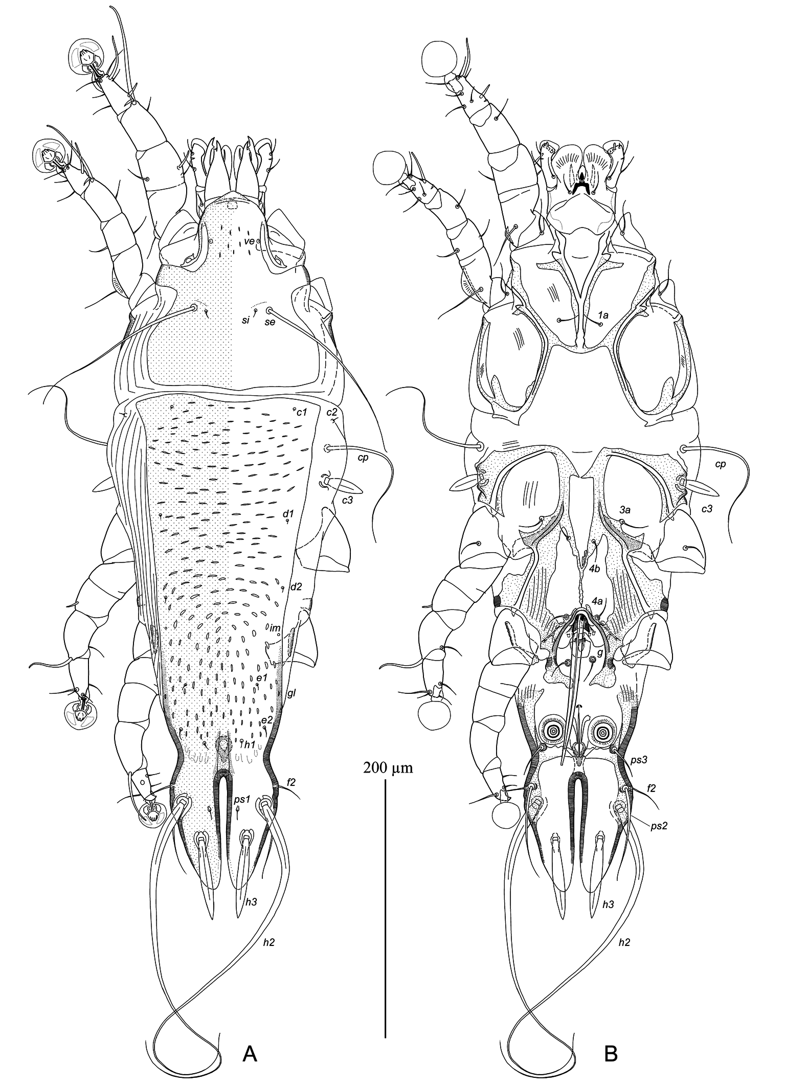
Male — (holotype, range for 9 paratypes in parentheses) (Figures 1, 3A–E, 7A). Idiosoma, length × width, 545 (535–560) × 190 (170–195), length of hysterosoma 390 (385–400). Prodorsal shield: entire, anterior margin with short and rounded rostral process 8 (5–8) long, anterolateral extensions connected to epimerites Ia, lateral margins concave, posterior margin almost straight, posterior corners rounded, surface in anterior part with few minute dash-like lacunae, length along midline including rostral process 158 (150–160), width at posterior margin 138 (125–140) (Figure 1A). Setae ve represented by microsetae. Bases of scapular setae se separated by 60 (52–60). Scapular shields narrow, barely developed dorsally. Humeral shields absent. Setae cp and c2 situated on soft tegument. Setae c3 lanceolate, 30 (28–32) × 10 (10–11). Distance between prodorsal and hysteronotal shields 10 (8–14). Hysteronotal shield: length from anterior margin to lobar apices 385 (380–390), width at anterior margin 150 (145–150), anterior margin shallowly concave medially, anterior corners nearly rectangular, area from anterior margin to level of trochanters IV with dash-like transverse striae, area from level of trochanters IV to bases of opisthosomal lobes with dash-like longitudinal striae. Metapodosomal sclerites narrow stick-like, situated at level of trochanters IV. Opisthosoma strongly attenuate from level of setae e2 to bases or opisthosomal lobes. Opisthosomal lobes about 2.5 times longer than wide at base, with slightly convex lateral margin at level of their midlength, with bases of setae f2 and h2 on these smoothed extensions; greatest width of opisthosoma at level of these extensions 88 (86–92), posterior end of lobes narrowed, semi-ovate (Figures 1B, 3A). Setae h3 large lanceolate, 60 (60–68) long and 11 (9–11) wide, situated slightly posterior to midlength of opisthosomal lobes and closer to level of macrosetae h2 than to lobar apices. Terminal cleft shaped as narrow parallel-sided slit, length 88 (85–90), greatest width 5 (5–8). Supranal concavity shaped as inverted teardrop, with heavily sclerotized border, distant from anterior end of terminal cleft. Setae f2 posterior from level of anterior end of terminal cleft; setae ps2 situated at same transverse level and mesal from them. Setae h1 at level of supranal concavity. Setae ps1 filiform, about 10 long, situated slightly posterior to levels of setae h2. Setae ps2 80 (65–80) long, not extending to lobar apices. Distances between bases of dorsal setae: c2:d2 135 (130–140), d2:e2 110 (105–115), e2:h2 63 (62–66), h2:h3 25 (25–30), d1:d2 55 (45–58), e1:e2 35 (32–38), h1:h2 58 (50–60), ps1:h3 22 (22–25), h2:h2 60 (60–68), h3:h3 35 (33–38), f2:f2 85 (85–90), ps2:ps2 78 (73–78).
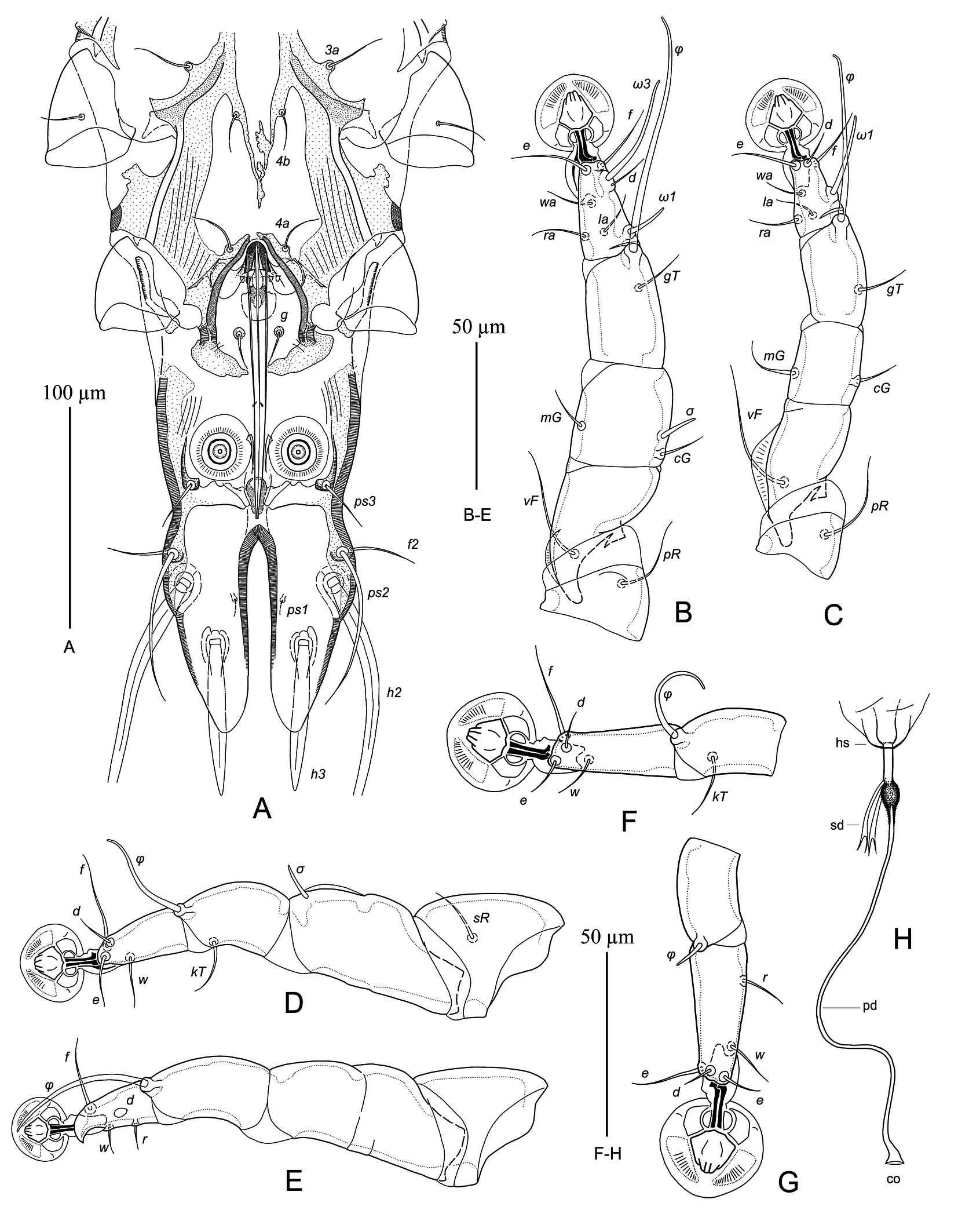
Epimerites I fused into a Y, sternum about 1/2 of total length of epimerites, posterior end of sternum with transverse extensions connected to medial part of epimerites II (Figure 1B). Epimerites II long and fused with corresponding epimerites IIa. Rudimentary sclerites rEpIIa absent. Coxal fields I–III closed, coxal fields IV almost closed. Coxal fields I, II without large sclerotized areas. Coxal fields IV with large sclerotized areas connecting epimerites IV and IVa. Genital arch of moderate size, with large rounded lateral plates, 25 (22–25) long, 38 (36–40) wide; basal sclerite of genital apparatus roughly rectangular. Aedeagus 120 (115–120) long, extending to midlength between supranal concavity and anterior end of terminal cleft. Genital papillae well distinct, situated at midlevel of genital arch. Paragenital apodemes (derivatives of epimerites IVa) fused to each other at anterior ends into arch (in some specimens fusion incomplete); pregenital apodeme represented by a long Y-shaped sclerite, anterior branches of this sclerite connected with inner margins of epimerites IIIa, posterior end extending to the arch formed by paragenital apodemes. Genital shields shaped as sclerites of irregular form and fused with bases of corresponding epimerites IVa. Paragenital apodemes fused with genital shields almost completely encircle genital apparatus. Setae 4b on anterior branches of pregenital apodemes, posterior to setae 3a; setae 4a on anterior ends of paragenital apodemes; setae g off genital shields, on small circular sclerites. Opisthoventral shields narrow, flanking nearly rectangular anal field laterally and posteriorly by transverse band-shaped apodeme (Figure 3A). Anterior margin of transverse apodeme with 2 or 3 pairs of rounded sclerotized denticles. Adanal suckers 15 (15–16) in diameter, corolla without denticles, surrounding membrane with radial striae. Setae ps3 situated on opisthoventral shield near posterior corners of anal field; setae g and ps3 arranged in tall trapezoid. Distances between ventral setae: 3a:4b 16 (16–20), 4b:4a 62 (58–62), 4a:g 35(35–40), g:ps3 70 (66–70), ps3:h3 70 (70–75), ps3:ps3 60 (50–60).
Legs I longer and thicker than legs II, femur I with small ventral crest; femora II with long ventral crests, other segments of these legs without processes (Figure 3B, C). Solenidion σ of genu I nearly at midlength of this segment; genual setae cGI, cGII and mGI setiform, seta mGII thickened in basal part and with filiform apex. Setae sR of trochanters III present. Solenidion ω1 of tarsus II elongate, extending to midlength of ambulacral disc; setae d of tarsi II, III half as long as corresponding setae f. Tarsus IV 33 (30–35) long, with apical claw-like process; seta d button-like, situated at midlength of this segment; seta e absent (Figure 3E). Solenidion φ of tibia IV almost extending to distal margin of ambulacral disc. Lengths of solenidia: ω1I 15 (15–17), ω1II 27 (26–29), σI 14 (13–14), σIII 12 (9–13), φIV 30 (28–30).
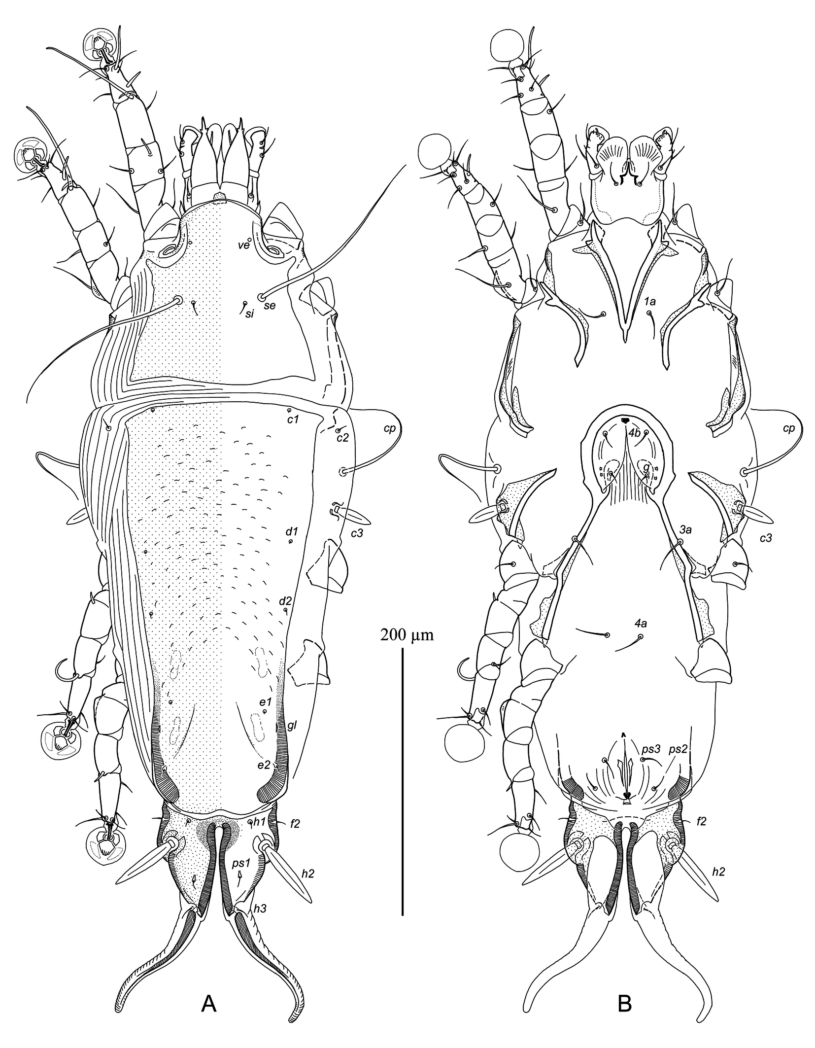
Female — (range for 10 paratypes) (Figures 2, 3F–H, 7B). Idiosoma, length × width, 520–560 × 200–215, length of hysterosoma 375–400. Prodorsal shield: anterolateral extensions angular and free from epimerites Ia, lateral margins shallowly concave at level of scapular setae, almost straight, posterior corners pointed, posterior margin straight or slightly convex, length along midline 138–148, width at posterior margin 138–145, surface without ornamentation (Figure 2A). Setae ve represented by microsetae. Bases of setae se separated by 65–70. Scapular shields not developed dorsally. Humeral shields absent. Setae cp and c2 situated on soft tegument. Setae c3 lanceolate, 23–28 × 7–8. Anterior and lobar parts of hysteronotal shields completely separated dorsally from each other by narrow transverse band of soft tegument, and weakly connected ventrolaterally. Anterior hysteronotal shield: roughly rectangular, slightly narrowed posteriorly, anterior margin straight or slightly convex, posterior margin convex with small median concavity, greatest length 300–330, width at anterior margin 148–155, width of posterior part 100–110; surface of anterior 2/3 with numerous sparsely disposed transverse dashes. Length of lobar region 80–88, greatest width 90–100; parts of lobar shield covering opisthosomal lobes connected to each other by narrow transverse bridge at anterior end of terminal cleft. Terminal cleft narrowly triangular, with lateral margins slightly divergent posteriorly, 63–68 long, 20–30 wide at level of lobar apices. Supranal concavity absent. Setae f2 present. Setae h1 distant from anterior margin of lobar shield. Setae h2 spindle-like, 50–58 long, 9–10 wide. Setae ps1 situated dorsally on opisthosomal lobes, equidistant from levels of setae h3 than to h2. Setae h3 minute filiform, about 10–12 long, much shorter than length of lobar region. Distances between dorsal setae: c2:d2 130–160, d2:e2 112–120, e2:h2 50–60, h2:h3 48–54, d1:d2 50–60, e1:e2 38–48, h1:h2 15–18, h2:ps1 25–30, h1:h1 43–50, h2:h2 70–78.
Epimerites I fused into a Y, with very short stem about 10 long (Figure 2B). Lateral parts of coxal fields I, II without sclerotized areas. Epimerites IVa absent. Translobar apodemes of opisthosomal lobes wide, not fused to each other anterior to terminal cleft. Greatest width of epigynum 70–95. Copulatory opening situated immediately posterior to anal opening. Primary spermaduct with small ball-like enlargement in 8-12 from head of spermatheca, secondary spermaducts 18–25 long (Figure 3H). Distances between pseudanal setae: ps2:ps2 42–48, ps3:ps3 25–28, ps2:ps3 20–27.
Legs I, II subequal, femur II with long ventral crest, other segments of these legs without processes. Solenidion σ of genu I shorter than half-length of this segment and situated in its anterior part. Genual setae cGI, II and mGI short filiform, setae mGII filiform with thickened basal part. Genu IV with narrow dorsal ridge. Setae sR of trochanters III present. Setae d of tarsi II–IV much shorter than corresponding setae f. Solenidion φIV about 1/4 of corresponding tarsus (Figure 3H). Lengths of solenidia: ω1I 16–18, ω1II 15–20, σI 10–12, σIII 7–10, φIII 30–35, φIV 8–9.
Remarks
The new species Dolichodectes robertsi sp. n. is most close to D. glyphonotus (Gaud & Mouchet, 1957) described from the Cassin's Flycatcher Muscicapa cassini Heine in Cameroon (Gaud and Mouchet 1957) in having the following features in males. The opisthosomal lobes are 2.5–3 times longer than wide at base, with the lobar apices rounded and the lateral margins moderately convex, the aedeagus does not extend beyond the midlength of lobar apices, setae h3 are narrowly lanceolate, situated closer to setae h2 than to the lobar apices, and the hysteronotal shields bears numerous minute lacunae. The new species differs from D. glyphonotus in having the following features: in both sexes of D. robertsi, the posterior part of the prodorsal shield lacks ornamentation; in males, the hysteronotal shield is covered with numerous dash-like lacunae, the aedeagus extends to the midlength between the supranal concavity and the anterior end of terminal cleft; in females, the posterior corners of the prodorsal shield are pointed, setae h2 are 50–58 long, and the primary spermaducts has a distinct ovate enlargement near the head of spermatheca. In both sexes of D. glyphonotus, the posterior part of the prodorsal shield bears small ovate lacunae; in males, the hysteronotal shield is covered with numerous ovate and circular lacunae up to 8 in diameter, and the tip of aedeagus extends to or beyond the anterior end of terminal cleft; in females, the posterior corners of the prodorsal shield are rounded, setae h2 are 65–70 long; and the primary spermaducts has a narrow ampuliform enlargement near the head of spermatheca.
Etymology
The species is named in honor of Dr. Lyndon F. Robert (Long Ashton, Bristol, UK), an ecologist, skillful bird-ringer, and leader of our research team in the Pongola Game Reserve.
Dolichodectes megalobus sp. n.
ZOOBANK: EDC0CF41-C648-4D91-8D7A-64002D38D244 ![]()
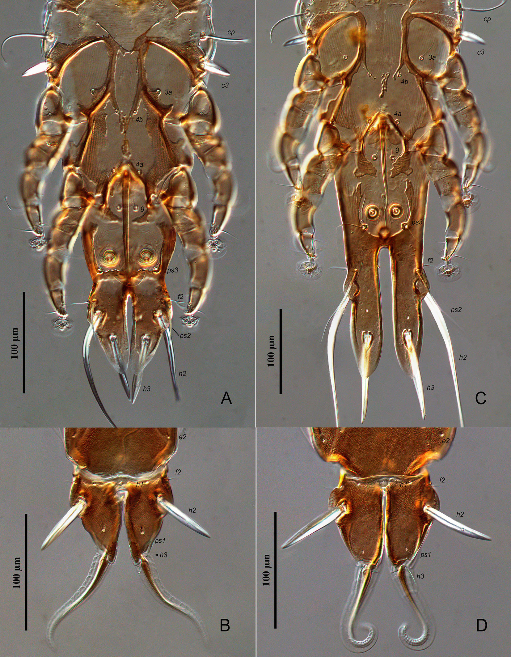
(Figures 4–6, 7C, D)
Type material
Holotype male, 20 male and 24 female paratypes from Agricola pallidus (von Müller, 1851) (Passeriformes, Muscicapidae), SVM 24-1202-4, South Africa, KwaZulu-Natal, Pongola Game Reserve, White Elephant Bush Camp, 27°24′42.6″S 31°53′53.3″E, 2 December 2024, coll. S.V. Mironov.
Depository
Holotype, 10 male and 10 female paratypes—NMB, remaining paratypes—ZISP.
Description
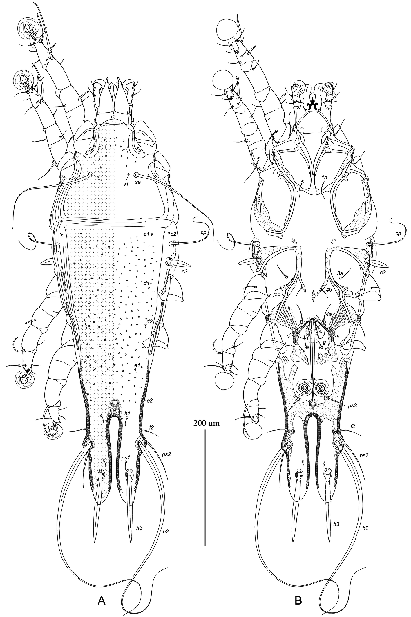
Male — (holotype, range for 10 paratypes in parentheses), (Figures 4, 6A–E, 7C). Idiosoma, length × width, 645 (600–645) × 220 (200–220), length of hysterosoma 460 (440–655). Prodorsal shield: entire, anterior margin with short and rounded rostral process 6 (4–7) long, anterolateral extensions connected to epimerites Ia, lateral margins concave, posterior margin almost straight, posterior corners rounded, surface of anterior part with sparsely disposed minute circular lacunae, length along midline from rostral apex 168 (155–165), width at posterior margin 170 (160–170) (Figure 4A). Setae ve represented by microsetae. Bases of scapular setae se separated by 78 (75–80). Scapular shields narrow, barely developed dorsally. Humeral shields present, shaped as small longitudinal sticks situated submarginally. Setae cp and c2 situated on soft tegument. Setae c3 lanceolate, 35 (30–35) × 10 (10–11). Distance between prodorsal and hysteronotal shields 13 (10–13). Hysteronotal shield: length from anterior margin to lobar apices 460 (435–460), width at anterior margin 165 (150–160), anterior margin straight, anterior corners pointed or rectangular, surface from anterior margin to bases of opisthosomal lobes with small circular lacunae less than 5 in diameter. Metapodosomal sclerites represented by two pairs of narrow stick-like sclerites, situated at levels of trochanters III and IV, respectively. Opisthosoma strongly attenuate from level of trochanters III to bases or opisthosomal lobes. Opisthosomal lobes 3.5–3.7 times longer than wide at base, with small lateral extensions bearing setae h2; greatest width of opisthosoma at level of extensions 108 (105–112); posterior end of lobes semi-ovate (Figure 4A, B). Setae h3 narrowly lanceolate, 108 (95–105) long and 9 (9–10) wide, situated at level of posterior 1/3 of opisthosomal lobes, approximately equidistant from levels of setae h2 and lobar apices. Terminal cleft shaped as a narrow, almost parallel-sided slit, length 150 (145–150), greatest width 18 (15–18). Supranal concavity small, roughly ovate, flanked posteriorly by small Y-shaped sclerotized patch, distant from anterior end of terminal cleft. Setae f2 at level of anterior 1/4 of terminal cleft; setae ps2 situated at same transverse level and mesal from them. Setae h1 situated at level of anterior end of terminal cleft. Setae ps1 filiform, about 20 long, situated closer to level of setae h3 than h2. Setae ps2 80 (80–90) long, not extending beyond lobar apices. Distances between bases of dorsal setae: c2:d2 155 (150–155), d2:e2 120 (110–125), e2:h2 77 (70–78), h2:h3 53 (52–58), d1:d2 62 (62–67), e1:e2 50 (43–50), h1:h2 88 (85–95), ps1:h3 22 (23–33), h2:h2 88 (85–95), h3:h3 48 (48–54), f2:f2 95 (95–100), ps2:ps2 75 (75–85).
Epimerites I fused into a Y, sternum about 1/2 of total length of epimerites, posterior end of sternum with transverse extensions connected to medial part of epimerites II (Figure 4B). Epimerites II long and fused with corresponding epimerites IIa. Rudimentary sclerites rEpIIa represented by oblique small sclerites. Coxal fields I–III closed, coxal fields IV open. Coxal fields I, II without large sclerotized areas. Coxal fields IV with large sclerotized areas connecting epimetites IV and IVa. Genital arch of moderate size, with large rounded lateral plates, 35 (32–35) long, 48 (40–45) wide; basal sclerite of genital apparatus almost rectangular. Aedeagus 115 (110–115) long, extending to anterior margins of adanal suckers. Genital papillae well distinct, situated at midlevel of genital arch. Paragenital apodemes (derivatives of epimerites IVa) close to each other at anterior ends but not fused. Pregenital sclerites poorly developed, represented by a pair of oblique sclerites on inner margins of epimerites IIIa and median longitudinal sclerite of irregular form, entire or split into separate fragments. Genital shields shaped as large sclerites of irregular form, fused with bases of corresponding epimerites IVa, and almost completely encircle genital apparatus (Figure 6A). Setae 4b situated on anterior branches of pregenital apodemes posterior to level of setae 3a; setae 4a on anterior parts of paragenital apodemes; setae g off genital shields. Opisthoventral shields wide, their anterior parts with oblique projections directed anteromedially, their posterior parts fused to each other and form wide transverse apodeme touching anterior end of terminal cleft. Anal field flanked laterally and posteriorly by opisthoventral shields. Adanal suckers 18 (16–18) in diameter, corolla without denticles, surrounding membrane with radial striae. Setae ps3 on small extensions on anterior margin of transverse apodeme; bases of setae g and ps3 in almost rectangular arrangement. Distances between ventral setae: 3a:4b 18 (15–17), 4b:4a 55 (50–55), 4a:g 44(40–44), g:ps3 85 (63–80), ps3:h3 128 (125–135), ps3:ps3 50 (50–55).
Legs I longer and thicker than legs II, femur I with barely distinct ventral crest; femora II with long ventral crests, other segments of this legs without processes (Figure 6B, C). Solenidion σ of genu I situated in proximal part of segment; genual setae cGI, mGI and cGII filiform, seta mGII thickened in basal part and with filiform apex. Setae sR of trochanters III present. Solenidion ω1 of tarsus II elongate, extending beyond midlength of ambulacral disc Setae d of tarsi II, III half as long as corresponding setae f. Tarsus IV 33 (30–33) long, with apical claw-like process; seta d button-like, situated in midlength of this segment; seta e absent (Figure 6E). Solenidion φ of tibia IV extending to distal margin of ambulacral disc. Lengths of solenidia: ω1I 18 (18–23), ω1II 38 (32–38), σI 22 (18–22), σIII 20 (15–20), φIV 48 (48–50).
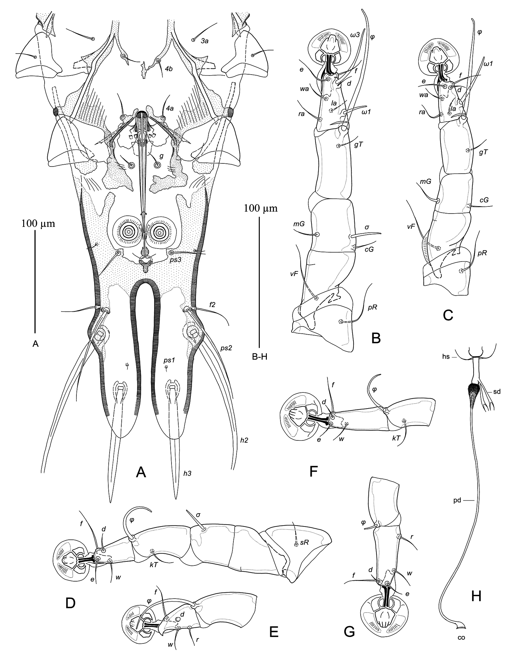
Female — (range for 10 paratypes) (Figures 5, 6F–H, 7D). Idiosoma, length × width, 535–585 × 210–225, length of hysterosoma 380–425. Prodorsal shield: anterolateral extensions angular and free from epimerites Ia, lateral margins at level of scapular almost straight, posterior corners pointed, posterior margin straight, length along midline 145–155, width at posterior margin 160–165, surface without ornamentation (Figure 5A). Setae ve represented by microsetae. Bases of setae se separated by 77–83. Scapular shields not developed dorsally. Humeral shields absent. Setae cp and c2 situated on soft tegument. Setae c3 lanceolate, 25–28 × 8–9. Anterior and lobar parts of hysteronotal shields separated dorsally from each other by narrow transverse band of soft tegument and weakly connected ventrolaterally. Anterior hysteronotal shield: roughly rectangular, slightly narrowed posteriorly, anterior margin straight or slightly convex, posterior margin with wide and short trapezoidal extension, greatest length 300–335, width at anterior margin 152–170, width of posterior part 125–140; surface of anterior 2/3 with numerous sparsely disposed minute circular lacunae. Length of lobar region 85–93, greatest width 100–115; parts of lobar shield covering opisthosomal lobes connected to each other by narrow transverse bridge at anterior end of terminal cleft. Terminal cleft narrow, parallel-sided, with lateral margins almost touching and slightly divergent posteriorly, 65–70 long, 5–10 wide at level of lobar apices. Supranal concavity absent. Setae f2 present. Setae h1 at anterior margin of lobar shield. Setae h2 spindle-like, 62–68 long, 10–11 wide. Setae ps1 situated dorsally on opisthosomal lobes, closer to levels of setae h3 than to h2. Setae h3 short filiform, about 18–25 long, 1/5–1/4 the length of lobar region. Distances between dorsal setae: c2:d2 142–160, d2:e2 112–120, e2:h2 58–70, h2:h3 50–55, d1:d2 60–68, e1:e2 40–45, h1:h2 20–25, h2:ps1 35–38, h1:h1 46–50, h2:h2 75–88.
Epimerites I fused into a Y, with very short stem about 10 long (Figure 5B). Lateral parts of coxal fields I, II without sclerotized areas. Epimerites IVa absent. Translobar apodemes of opisthosomal lobes wide and not fused to each other anterior to terminal cleft. Greatest width of epigynum 75–78. Copulatory opening situated immediately posterior to anal opening. Primary spermaduct with small ball-like enlargement in 8–12 from head of spermatheca, secondary spermaducts 22–25 long (Figure 6H). Distances between pseudanal setae: ps2:ps2 45–50, ps3:ps3 27–29, ps2:ps3 13–16.
Legs I, II subequal, femur II with long ventral crest, other segments of these legs without processes. Solenidion σ of genu I shorter than half-length of this segment and situated in its anterior part. Genual setae cGI, II and mGI short filiform, setae mGII filiform with thickened basal part. Genu IV with narrow dorsal ridge. Setae sR of trochanters III present. Setae d of tarsi II–IV much shorter than corresponding setae f. Solenidion φIV about 1/5–1/4 of corresponding tarsus (Figure 6G). Lengths of solenidia: ω1I 20–23, ω1II 16–18, σI 15–16, σIII 8–10, φIII 32–38, φIV 7–9.
Remarks
Dolichodectes megalobus sp. n. is most close to D. diplocercus (Gaud & Mouchet, 1957) described from the Fraser's Rufous Thrush, Stizorhina fraseri (Strickland), in Cameroon (Gaud and Mouchet 1957). In males of both species, the opisthosomal lobes are strongly elongated, 3–3.5 times longer than wide at base, and almost parallel-sided, the lobar apices are rounded, the aedeagus does not extend to the terminal cleft, setae h3 are narrowly lanceolate, and the hysteronotal shield has ornamentation. The new species, Dolichodectes megalobus, differs from to D. diplocercus in the following features: in males of D. megalobus, the hysteronotal shield is covered with numerous circular lacunae from the anterior margin to the level of supranal concavity, the aedeagus is 110–115 long, and setae h3 are 95–105 long that is about 2/3 the length of terminal cleft; in females, the prodorsal shield lacks ornamentation, the lobar shield is distinctly wider than long (85–93 × 100–115), and setae h2 are shorter than the distance between their bases. In males of D. diplocercus, the hysteronotal shield is covered with numerous transverse striae from the anterior margin to the level of trochanters IV, the aedeagus is 80 long, and setae h3 are about 60 long (n=1); in females, the entire surface of the prodorsal shield is covered with circular lacunae, which are noticeably larger in the anterior half (up to 5 in diameter), the length and greatest width of the lobar shield are subequal (83–90 × 88–95), and setae h2 are longer than the distance between their bases (n=5).
The paratype specimens of D. diplocercus (1 male, 5 females), examined herein for comparison, were granted by J. Gaud to the feather mite collection deposited in the Zoological Institute of the Russian Academy of Sciences.
Etymology
The specific epithet mega (L, giant) refers to very long opisthosomal lobes in males of the described species.
Acknowledgements
The author thanks Dr. Heinz Kohrs for the opportunity to carry out the field investigation in his property of the Pongola Game Reserve, and Dr. Lyndon F. Roberts (Bristol, UK) for the invitation to participate in his bird-ringing trip to South Africa. Capturing, ringing and investigation of birds was carried out under the permission OP 2529/2024 issued by Ezemvelo KZN Wildlife Permit office. The present taxonomic work was supported by the Ministry of Science and Higher Education of the Russian Federation (project No. 122031100263-1) to SVM.
References
- Constantinescu I.C., Chişamera G.B., Petrescu A., Adam C. 2018. Two new species of feather mites (Acarina: Psoroptidia) from the Oriental Magpie-Robin, Copsychus saularis (Passeriformes: Muscicapidae). Acarologia, 58 (2): 313-331. https://doi.org/10.24349/acarologia/20184244
- Gaud J., Atyeo W.T. 1996. Feather mites of the World (Acarina, Astigmata): the supraspecific taxa. Mus. Roy. Afr. Centr., Ann., Sci. Zool., 277: 1-193 (Pt. 1, text), 1-436 (Pt. 2, illustrations).
- Gaud J., Mouchet J. 1957. Acariens plumicoles (Analgesoidea) des oiseaux du Cameroun. I. Proctophyllodidae. Ann. Parasitol. Hum. Comp., 32: 491-546.
- Gill F., Donsker D., Rasmussen P. (Eds.). 2025. IOC World Bird List (v 15.1). Available from: http://www.worldbirdnames.org/ (accessed: 10 December 2025). https://doi.org/10.14344/IOC.ML.15.1.
- Hernandes F.A. 2022. Three new feather mite species (Acariformes: Proctophyllodidae, Trouessartiidae) from tyrant flycatchers (Passeriformes: Tyrannidae) in Brazil. Syst. Parasitol., 99: 115-138. https://doi.org/10.1007/s11230-021-10018-0
- Hernandes F.A. 2023. Feather mites (Acariformes: Astigmata) from the yellow-rumped cacique, Cacicus cela (Linnaeus, 1758) (Passeriformes: Icteridae) in Brazil, with description of four new species. J. Natur. Hist., 57 (1-4): 257-284. https://doi.org/10.1080/00222933.2023.2174459
- Krantz G., Walter D. (Eds.). 2009. A Manual of Acarology, 3rd Edition. Texas Tech University Press, Lubbock, TX, USA, 807 pp.
- Mironov S.V. 2006. Feather mites of the genus Montesauria Oudemans (Astigmata: Proctophyllodidae) associated with starlings (Passeriformes: Sturnidae) in the Indo-Malayan region, with notes on the systematics of the genus. Acarina, 14 (1): 21-41.
- Mironov S.V. 2009. Phylogeny of feather mites of the subfamily Pterodectinae (Astigmata: Proctophyllodidae) and their host associations with passerines (Aves: Passeriformes). Proc. Zool. Inst. Russian Acad. Sci., 313: 97-118. https://doi.org/10.31610/trudyzin/2009.313.2.97
- Mironov S.V. 2023. A new species of the feather mite genus Dolichodectes (Acariformes: Proctophyllodidae) from the Dark-sided Flycatcher Muscicapa sibirica (Passeriformes: Muscicapidae) from Buryatia. Acarina, 31(2): 213-223. https://doi.org/10.21684/0132-8077-2023-31-2-213-223
- Mironov S.V., Chandler C.R. 2017. New feather mites of the genus Amerodectes Valim and Hernandes (Acariformes: Proctophyllodidae) from passerines (Aves: Passeriformes) in Georgia, USA. Zootaxa, 4344 (2): 201-245. https://doi.org/10.11646/zootaxa.4344.2.1
- Mironov S.V., Fain A. 2003. New species of the feather mite subfamily Pterodectinae (Astigmata: Proctophyllodidae) from African passerines (Aves: Passeriformes). Bull. Soc. Roy. Belge Entomol., 139: 75-91.
- Mironov S.V., Galloway T.D. 2021. Feather mites of the subfamily Pterodectinae (Acariformes: Proctophyllodidae) from passerines and kingfishers in Canada. Zootaxa, 5016 (1): 1-55. https://doi.org/10.11646/zootaxa.5016.1.1
- Mironov S.V., González-Acuña D. 2011. New feather mites of the subfamily Pterodectinae (Astigmata: Proctophyllodidae) from passerines (Aves: Passeriformes) from Chile and Cuba. Zootaxa, 2037: 1-48. https://doi.org/10.11646/zootaxa.3057.1.1
- Mironov S.V., Doña J., Jovani R. 2015. A new feather mite of the genus Dolichodectes (Astigmata: Proctophyllodidae) from Hippolais polyglotta (Passeriformes: Acrocephalidae) in Spain. Folia Parasitol., 62: 032 [1-8]. https://doi.org/10.14411/fp.2015.032
- Mironov S.V., Literák I., Čapek M., Koubek P. 2010. New species of the feather mite subfamily Pterodectinae (Astigmata: Proctophyllodidae) from passerines in Senegal. Acta Parasitol., 55 (4): 399-413. https://doi.org/10.2478/s11686-010-0051-1
- Mironov S.V., Literák I., Nguen M.H., Čapek M. 2012. New feather mites of the subfamily Pterodectinae (Acari: Proctophyllodidae) from passerines and woodpeckers (Aves: Passeriformes, Piciformes) from Vietnam. Zootaxa, 3440: 1-49. https://doi.org/10.11646/zootaxa.3440.1.1
- Norton A.R. 1998. Morphological evidence for the evolutionary origin of Astigmata (Acari: Acariformes). Exp. Appl. Acarology, 22: 559-594. https://doi.org/10.1023/A:1006135509248
- Park C.K., Atyeo W.T. 1971. A generic revision of the Pterodectinae, a new subfamily of feather mites (Sarcoptiformes: Analgoidea). Bull. Univ. Nebraska State Mus., 9: 39-88.
- Valim M.P., Hernandes F.A. 2010. A systematic review of feather mites of the Pterodectes generic complex (Acari: Proctophyllodidae) with redescriptions of species described by Vladimir Černý. Acarina, 18 (1): 3-35.


2026-01-03
Date accepted:
2026-02-22
Date published:
2026-03-17
Edited by:
Akashi Hernandes, Fabio

This work is licensed under a Creative Commons Attribution 4.0 International License
2026 Mironov, Sergey V.
 Download article
Download the citation
RIS with abstract
(Zotero, Endnote, Reference Manager, ProCite, RefWorks, Mendeley)
RIS without abstract
BIB
(Zotero, BibTeX)
TXT
(PubMed, Txt)