


Contribution to knowledge of Uropodina mites (Acari: Mesostigmata) of Virginia, USA
Kontschán, Jenő  1
and Ermilov, Sergey G.
2
1
and Ermilov, Sergey G.
2
1Plant Protection Institute, HUN-REN Centre for Agricultural Research, H-1525 Budapest, P.O. Box 102, Hungary & Department of Plant Sciences, Albert Kázmér Faculty of Mosonmagyaróvár, Széchenyi István University, Vár square 2., H-9200 Mosonmagyaróvár, Hungary.
2Institute of Environmental and Agricultural Biology (X-BIO), University of Tyumen, Lenina str. 25, 625000 Tyumen, Russia.
2025 - Volume: 65 Issue: 4 pages: 998-1010
https://doi.org/10.24349/tb3j-gb36ZooBank LSID: A4147A10-5ED0-48AD-B874-A340FDC76248
Original research
Keywords
Abstract
Introduction
Uropodina (Mesostigmata) are a characteristic group of soil-dwelling mites. This widely distributed and morphological unique group of Mesostigmata currently includes more than 2000 known species, distributed worldwide (Lindquist et al. 2009). In terms of the number of species of Uropodina mites in different countries (Wiśniewski 1993), the knowledge of Uropodina mites of USA with fewer than 250 reported species is far from the complete (Farrier & Hennessey 1996; Kontschán & Starý 2012; Kontschán 2017). In this paper, we present some new information about the Uropodina fauna of Virginia state based on the mite material stored in the Arachnida collection of the Natural History Museum, Geneva (NHMG).
Material and methods
Specimens investigated were cleared in lactic acid for two weeks and were then placed on half-covered well slides and examined using a Leica 1000 microscope with a drawing tube Photographs were taken with a Keyence VHX 5000 digital microscope. Specimens examined are stored in 70% ethanol and deposited in the Natural History Museum, Geneva (NHMG). Setae and pores: h = hypostomal seta, st = sternal seta, p = pores, lf = lyriform fissures, Py = pygidial shield, Pd = postdorsal shield, Ms = marginal shield, Ds = dorsal shield. All measurements and the scales in the figures are given in micrometres (μm).
Taxonomy
Trachytidae Trägårdh, 1938
Trachytes balazyi Wiśniewski & Hirschmann, 1994
Trachytes balazyi Wiśniewski & Hirschmann, 1994: 90.
Trachytes balazyi – Kontschán 2017: 353.
New records. USA, Virginia, Henry County, 1 mile's SW of Figsboro, Fagus litter, 15.XI.1981, leg. Hoffmann. USA, Virginia, Chesterfield County, US Hy 360 at falling creek, oak woods with Kalmia understory, 10.X.1982, leg. Hoffmann. USA, Virginia, Patrick County, ''Pinnacles of Dann» ca 7 km SW of Vesta, 300 m Tsuga-Liriodendron litter, 19.VII.1983, leg. Hoffmann.
Trachytes jokaii n. sp.
ZOOBANK: 54A32BBE-5717-4EA8-BB31-BD43C6259CA9 ![]()
(Figures 1–4)
Diagnosis
Idiosoma with wide and ribbed lateral sections on vertex. Pygidial shield small and quadrangular, situated among posterior margin of dorsal shield and lateral margin of postdorsal shields. Sternal-, inguinal- and ventral shields fused. Genital shield axe-like, its surface covered by web-like sculptural pattern on anterior area. Adgenital platelets without processes on their outer margins.
Material examined
Holotype. Female. USA, Virginia, Chesterfield County, US Hy 360 at falling creek, oak woods with Kalmia understory, 10 October 1982, Hoffmann coll. Paratypes. One female and four males. Locality and date same as in holotype. Three males. USA, Virginia, Patrick County, ''Pinnacles of Dann» ca 7 km SW of Vesta 300 m Tsuga-Liriodendron litter, 19. July 1983 Hoffmann coll.
Description
Female (n=2). Length of idiosoma 610–620, width 390–400. Shape of idiosoma pear-like. Color yellow.
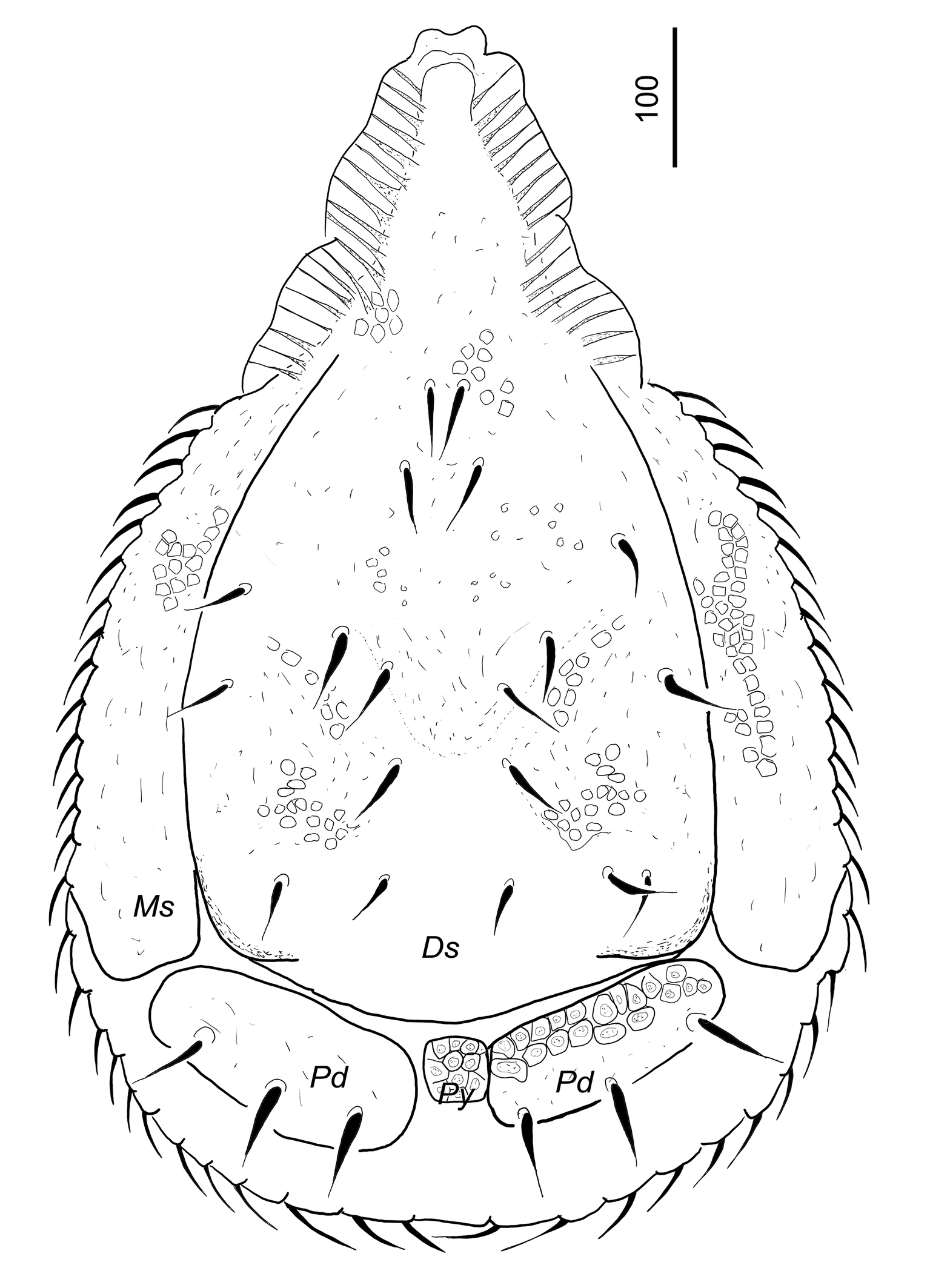
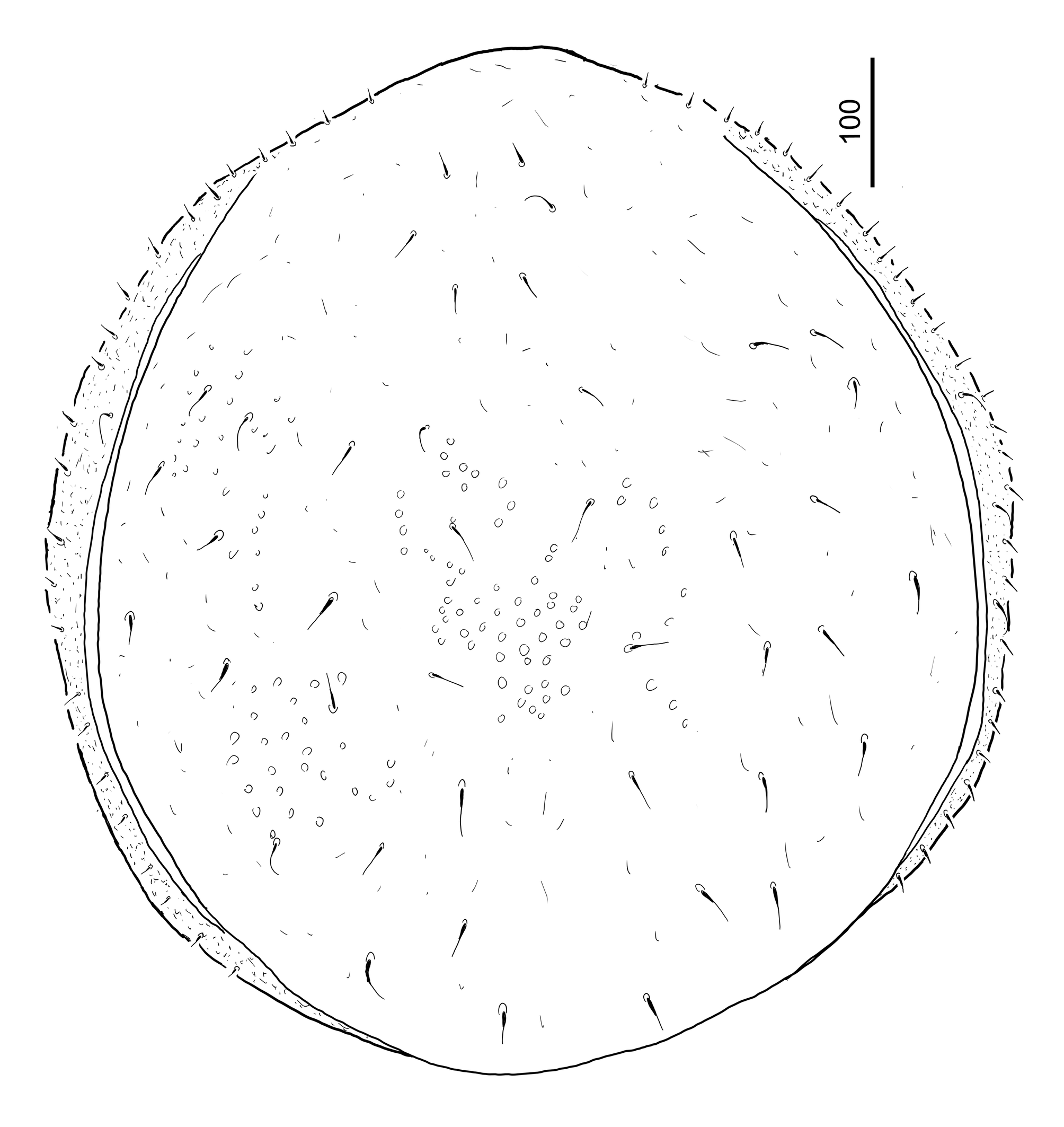
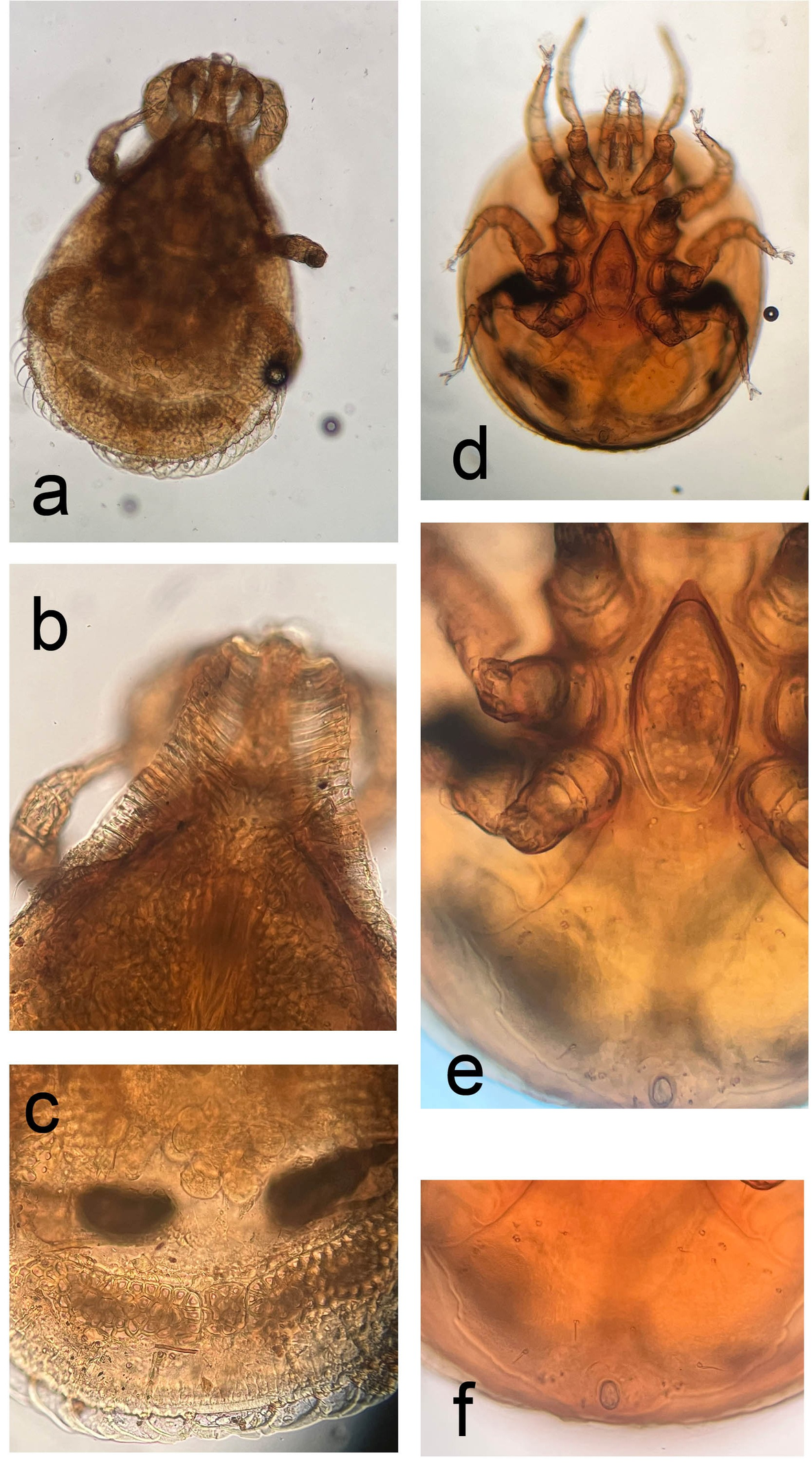
Dorsal idiosoma (Figures 1, 8a, b and c). Wide and ribbed lateral sections on vertex present. Marginal and dorsal shields completely separated. All setae on dorsal and postdorsal shields smooth, needle-like and ca 54–61 long. Setae on marginal shield, smooth, needle-like and ca 35–41 long. Surface of dorsal shield covered by deep, irregular pits (ca 6–11 long × 8–15 wide), posterior edges of dorsal shield with a strongly sclerotized elbow. Postdorsal shield divided. Separated postdorsal shields two times wider (ca 174–176) than long (ca 67–70 wide) and bears three pairs of setae. Pygidial shield small (ca 46 × 44) and quadrangular, placed among posterior margin of dorsal shield and lateral margin of postdorsal shields. Surface of pygidial and postdorsal shields with irregular pits which contain shallow, rounded central hollows. Sculptural pattern of marginal shields same as on dorsal shield.
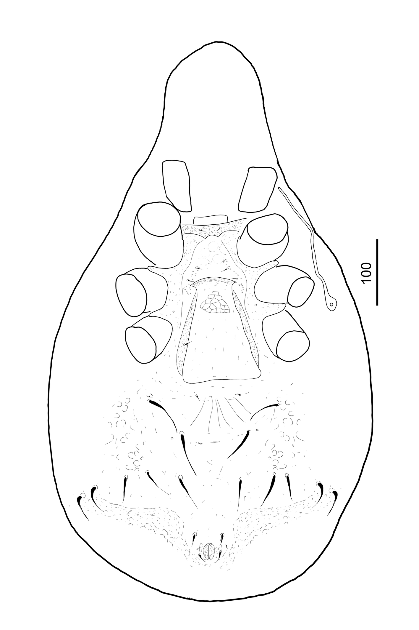
Ventral idiosoma (Figure 2). All sternal setae smooth, needle-like and ca 21–26 long. Setae st1 situated close to anterior margin of sternal shield, st2 and st3 near the anterior margin of genital shield, st4 at level of posterior margin of coxae III, st5 on adgenital platelets, st6 near basal margin of genital shield. Majority of surface of sternal shield smooth, only some very small oval pits situated around setae st2 and st3 (Figure 3a). Sternal-, inguinal- and ventral shields fused.
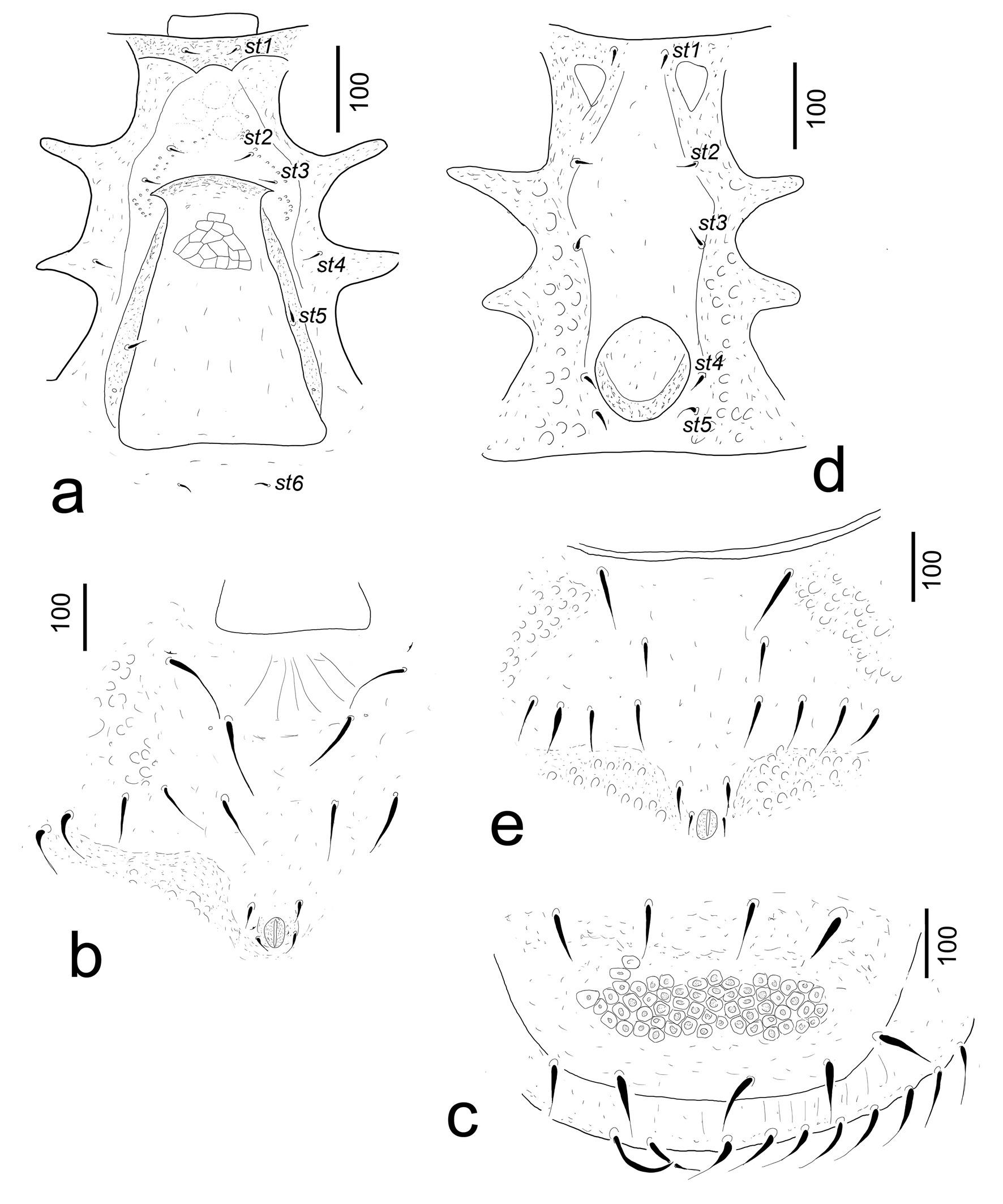
Inguinal area covered by irregular pits, remainder of the fused shield with smooth surface, except the posterior part of ventral shield where some oval pits visible. Fused shields bear seven pairs of smooth and needle-like setae (ca 69–110 long). Two pairs of adanal setae needle-like short (ca 29–30) and situated near anal opening (Figure 3b).
Genital shield axe-like (ca 306 long and ca 223 wide at its base), its surface covered by web-like sculptural pattern on anterior area. Adgenital platelets present without processes on their outer margins. Genital shield situated between coxae III and IV (Figure 3a). Peritremes long and straight, stigmata situated between coxae II and III. Tritosternum (Figure 8) with wide base, tritosternal laciniae bifurcated.
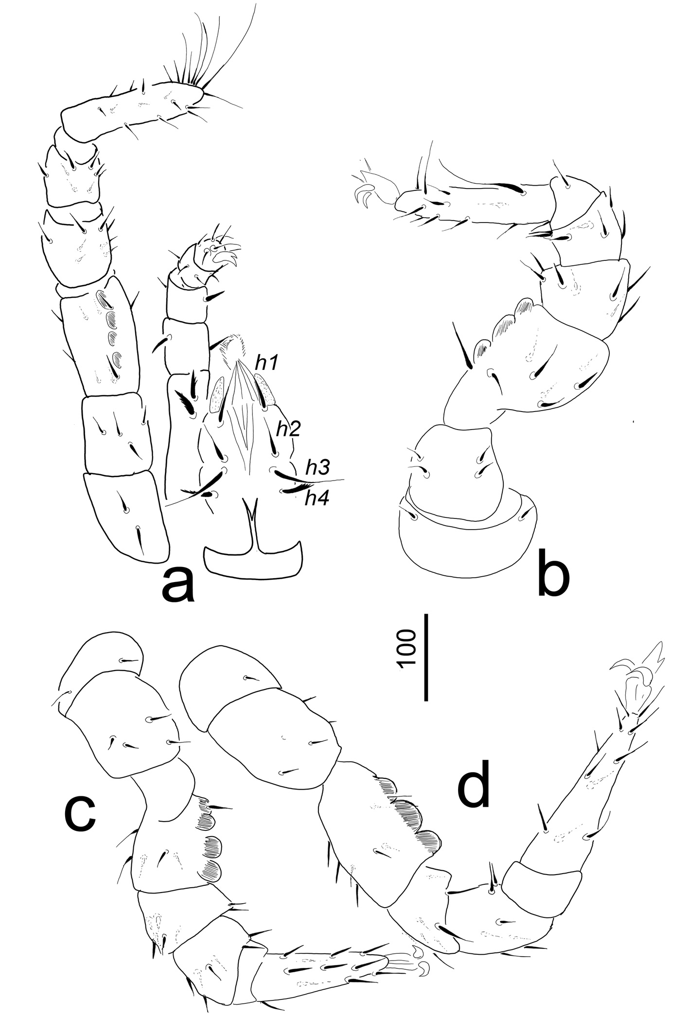
Gnathosoma (Figure 4a). Corniculi horn-like (ca 42–45), internal malae longer (ca 60–63) than corniculi and smooth. Hypostomal setae h1 long (ca 63–64) and smooth, h2 shorter (ca 45–47) and smooth, h3 similar to h1 in shape, but ca 74–76 long, h4 marginally serrate and ca 33–35 long. Chelicerae with long and pointed apical process on fixed digit, movable digit shorter than fixed digit. Epistome marginally serrate. Palp ca 377–380 long and with two serrate ventral setae (ca 30–42) on trochanter, other setae on palp smooth (Figure 4a).
Legs (Figures 4a–d). Leg I without ambulacral claws, all legs with smooth and needle-like setae; femora of legs I–IV with three-four rounded ventral processes. Leg I 547–549, leg II 699–702, leg III 579-580, leg IV 745–746. Leg setation Polyapsis-type after Evans (1972).
Male Length of idiosoma 610–630, width 390–405 (n=7).
Dorsal idiosoma. Shape and dorsal aspect of idiosoma as in female, except the caudal area of dorsal idiosoma: pygidial shield absent, postdorsal shield fused with dorsal shield and covered by irregular pits which contain shallow, rounded central hollows (Figure 3c).
Ventral idiosoma. Surface of sternal shield with some oval pits on marginal areas. Five pairs of smooth, short (ca 17–21 µm) and needle-like sternal setae present. Setae st1 situated close to anterior margin of sternal shield, st2 at level of posterior margin of coxae II, st3 at level of midcoxae III, st4 and st5 close to posterolateral margin of genital opening. Genital shield circular, without sculptural pattern and without eugenital setae, ca 100–102 long and ca 110–115 wide. Shield situated between coxae IV (Figure 3d). Position and shape of ventral setae and ornamentation of ventral shield as in female (Figure 3e).
Nymphs and larvae are unknown
Etymology
We dedicate the new species to the memory of the famous Hungarian writer Mór Jókai (1825–1904), who was born 200 years ago and who wrote a plant protection book and several important and significant novels.
Remarks
So far only eleven Trachytes species were presented from North America (Kontschán & Starý 2012, Kontschán 2017), but only six species have a wide and ribbed vertex in the Nearctic realm: T. aegrota (C. L. Koch, 1841), T. kaliszewskii Błoszyk & Szymkowiak, 1996, T. canadiensis Huţu, 2000, T. aegrotasimilis Huţu, 2000, T. californica Kontschán & Starý, 2012 and T. virginiana Kontschán, 2017. The proposed new species can be distinguished from the aforementioned species by the following: T. canadiensis and T. californica has a linguliform female genital shield, contrary to the new species, which has an axe-like genital shield. The inguinal shield is free and does not fuse with the ventral shield in T. aegrota and T. kaliszewskii, but this shield is fused with a ventral shield in the new species. The pygidial shield is rhomboid in T. aegrotasimilis and oval in T. virginiana, but quadrangular in the new species.
Key to the North American Trachytes species (only for females)
1. Vertex with wide and ribbed lateral sections
...... 2
— Vertex without wide and ribbed lateral sections
...... 8
2. Inguinal shield free, not fused to ventral shield
...... 3
— Inguinal shield fused to ventral shield
...... 4
3. Surface of genital shield smooth, setae X4 placed on ventral shield
...... T. aegrota (C. L. Koch, 1841)
— Genital shield with reticulate sculptural pattern, setae X4 situated on small platelets in membranous cuticle
...... T. kaliszewskii Błoszyk & Szymkowiak, 1996
4. Genital shield linguliform
...... 7
— Genital shield axe-shaped
...... 5
5. Wide and ribbed lateral sections presented on whole margins of idiosoma
...... Trachytes virginiana Kontschán, 2017
— Wide and ribbed lateral sections presented only on vertex
...... 6
6. Inguinal shield fused to anal shield, sternal shield separated
...... T. canadiensis Huţu, 2000
— Inguinal shield fused to anal shield and sternal shield
...... T. jokaii n. sp.
7. Pygidial shield rhomboid; surface of genital shield smooth
...... T. aegrotasimilis Huţu, 2000
— Pygidial shield oval; surface of genital shield reticulate
...... T. californica Kontschán & Starý, 2012
8. Genital shield linguliform, anterolateral angles of genital shield rounded
...... 9
— Genital shield axe-shaped, anterolateral angles of genital shield pointed
...... 10
9. Ventral shield separated from inguinal and sternal shields; surface of genital shield ornamented by irregular pits
...... T. balazyi Wiśniewski & Hirschmann, 1994
— Ventral shield fused with inguinal and sternal shields; surface of genital shield smooth
...... T. marilynae Huţu, 2000
10. Inguinal shield fused to ventral shield; apical part of genital shield relatively wide (apical: basal = 1:1.3–2)
...... 11
— Inguinal shield separated from ventral shield; apical part of genital shield narrow (apical: basal = 1: 3)
...... T. axe Kontschán & Starý, 2012
11. Sternal shield fused to inguinal and ventral shields; ventral and dorsal idiosoma ornamented by irregular pits
...... T. minima Trägårdh, 1910
— Sternal shield separated from inguinal and ventral shield; surface of ventral and dorsal idiosoma without irregular pits
...... T. nortoni Huţu, 2000
Uropodidae Kramer, 1881
Jedediella hoffmani Kontschán, 2017
Jedediella hoffmani Kontschán, 2017: 346–350.
New records — USA, Virginia, Henry county, 1 miles SW of Figsboro, Fagus litter, 15.XI.1981, leg. Hoffmann.
Notes — Only three Jedediella species are known from the North America. The distinguishing characters are presented in Kontschán (2017: 350).
Klebensbergia n. gen.
ZOOBANK: 8D68328E-5A8A-495D-9CE0-A2247709C521 ![]()
Diagnosis
Idiosoma oval, dorsal and marginal shield fused on anterior area. Dorsal shield with oval pits and needle-like setae. Marginal shield without sculptural patterns and bears several short and needle-like setae and some longer, slight curved smooth seta. Ventral shield reduced in caudal part. Genital shield of female scutiform, but apically a little peaked. Peritreme straight. Base of tritosternum wider than long. Gnathosomal setae h1 long and needle-like, internal malae longer than horn-like corniculi and apically pilose. The first ventral setae on palp trochanter situated on small protuberance. Chelicerae without internal sclerotized node.
Type species
Klebensbergia virginiana n. sp. Monotypic. Genus based on adult female material representing one newly described species.
Etymology
We dedicate the new genus to Kuno Klebensberg (1875–1932), who was born 150 years ago. He was the most important reformer of the Hungarian education system in primary school and in university levels.
Gender. Feminine.
Remarks
Based on long setae h1, internal sclerotized node absent on chelicerae, apically smooth corniculi and first ventral setae of palp trochanter situated on small protuberance we can place the new genus in the family Uropodidae; however all taxa within this family merit revision. But the general shape of idiosoma with the shape of the female genital shield resembles the uropodid genus Penicillaturopoda Hirschmann, 1979. The most important differences are the following between these two genera: the ventral shield of Penicillaturopoda is complete and reduced in the new genus, and the peritreme of Penicillaturopoda is 3-shaped and it is straight in the new genus.
The base of the tritosternum of the new genus is wide. This phenomenon was mentioned only in the ''lower Uropodina» (see Lindquist et al. 2009), but this character is not also unusual within the Uropodidae, it was described earlier in some taxa (see Hirschmann 1993: 364, as catch-all genus Uropoda).
Klebensbergia virginiana n. sp.
ZOOBANK: 49434C6E-0058-4650-A5CA-6A9F036A9915 ![]()
(Figures 5–7)
Diagnosis
As for the genus.
Material examined
Holotype. Female. USA. Virginia. Tazawell county, Burkes Garden, east slope of Beartown Mountains, 1500 m. a.s.l., loose surface litter in Acer-Aesculus-Fagus forest. 19 May 1981, Hoffmann coll. Paratypes. Two females, with the same collection data as those for the holotype.
Description
Female (n=3). Length of idiosoma 840–850, width at level of coxae IV 660–675, color reddish-brown. Shape of idiosoma oval.
Dorsal idiosoma (Figure 5). Marginal and dorsal shields fused anteriorly. Dorsal shield covered by oval pits (ca 5–7×4–8). All dorsal setae (23–25 pairs) (ca 20–37) long and needle-like. Surface of marginal shield smooth and bears 27–30 pairs short (ca 10–12) and 3–4 longer and little curved (ca 14–16) setae.
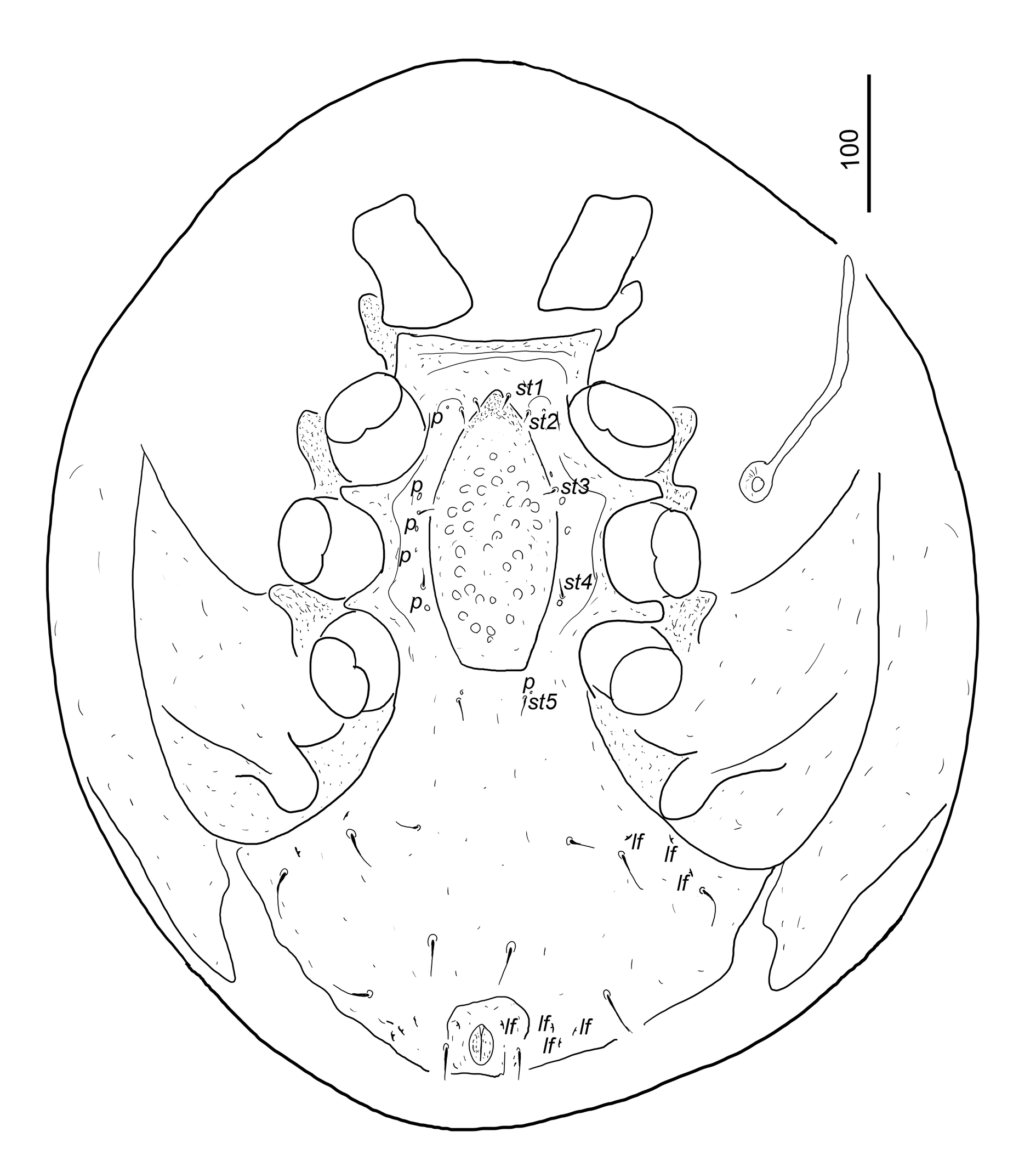
Ventral idiosoma (Figures 6, 8d, e and f). Five pairs of sternal setae short (ca 11–14), smooth and needle-like. Setae st1 inserted close to anterior margin of genital shield, st2 at level of anterior margin of coxae II, st3 at level of posterior margin of coxae II, st4 at level of posterior margin of coxae III, st5 close to basal edges of genital shield. Sternal shield smooth, six pairs of poroid-like structures situated close to st2, st3, between st3 and st4 and close to st4 and st5. Ventral shield reduced caudally; one pair of deep incision situated posterior to pedofossae IV. Surface of ventral shield without sculptural pattern. Five pairs of ventral setae smooth and needle-like, first pair shorter (ca 18–20), others longer (ca 29–36). Several lyriform fissures situated lateral to anal opening and close to metapodal suture. Anal opening oval (ca 27–29 long and ca 18–20 wide), anal valves smooth, without euanal setae. One pair of smooth and needle-like (ca 22–23) adanal setae inserted lateral to anal opening
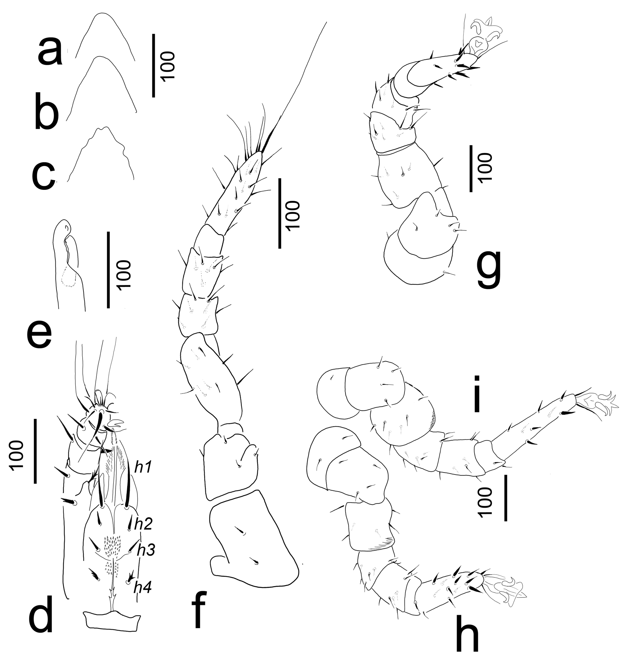
Genital shield scutiform, length 202–208, width 90–95 at level of coxae III, situated between coxae II and IV; anterior margin of female genital shield peaked, but very variable (Figures 7a–c), surface covered by oval pits (ca 7–8 × 8–9). Stigmata situated between coxae II and III. Peritremes with straight prestigmatid part, poststigmatid part absent. Pedofossae deep, their surface smooth, without separate furrow for tarsi IV. Tritosternum with wide base, tritosternal laciniae long and with two pairs of lateral spines (Figure 7d).
Gnathosoma (Figure 7d). Corniculi small (ca 50–52), smooth and horn-like, internal malae long (ca 87–90) and pilose, two times as long as corniculi. Hypostomal setae h1, h2 and h3 smooth and needle-like, h4 with serrate margins. Setae h1 long (ca 100–103), h2, h3 and h4 short (ca 19–25). Deutosternal groove narrow, deutosternum covered with numerous denticles between h2 and h4. Chelicerae without internal sclerotized nodes (Figure 7e). Fixed digit of chelicerae without teeth, longer (ca 83–85) than movable digit (ca 65–66); movable digit bearing a small central tooth. Palp ca 365–370 long, palp trochanter setae serrate, v1 longer (ca 50–52) and situated on small protuberance, v2 shorter (ca 24–26). Other setae on palp segments smooth. Palp apotele bifurcated. Epistome marginally pilose.
Legs (Figures 7f–h). Length of legs (from base of coxae to apex of tarsi): I 580–592, II 620–634, III 567–571, IV 625–632. Leg I without ambulacral claws; majority of setae on all legs needle-like, but some setae serrate on tarsi II–IV. Leg setation Uropoda-type after Evans (1972).
Male and developmental stages are unknown
Etymology
The name of the new species refers to the state where it was collected.
Acknowledgements
We are very grateful to Dr. Peter Schwendinger (MHNG) for his kind hospitality during the first author's visit to Geneva
References
- Błoszyk J., Szymkowiak P. 1996. Trachytes kaliszewskii, n. sp. (Acari: Uropodina) from the Great Basin (Utah, USA), with remarks on the habitats and distribution of the members of the genus Trachytes. Great Basin Nat. 56: 59-72. https://doi.org/10.5962/bhl.part.4108
- Evans G.O. 1972. Leg chaetotaxy and the classification of the Uropodina (Acari: Mesostigmata). J. Zool., London. 167: 193-206. https://doi.org/10.1111/j.1469-7998.1972.tb01729.x
- Farrier M.H., Hennesey M.K. 1996. Soil-Inhabiting and Free-living Mesostigmata (Acari-Parasitiformes) from North- America. An Annotated Checklist with Bibliography and Index. Technical Bulletin 302. North Carolina: North Carolina Agricultural Service, North Carolina State University Raleigh. pp. 408
- Hirschmann W. 1979. Stadiensystematik der Parasitiformes Teil 1. Stadienfamilien und Stadiengattungen der Atrichopygidiina, erstellt im Vergleich zum Gangsystem Hirschmann, 1979. Acarologie. 26: 57-70.
- Hirschmann W. 1993. Gangsystematik der Parasitiformes Teil 550. Bestimmungstabellen der Uropodiden der Erde, Atlas der Ganggattungen der Atrichopygidiina. Acarologie. 40: 292-370.
- Huţu M. 2000. Neue Trachytes-Arten (Anactinotrichida: Uropodina: Trachytidae) aus Canada. Acarologia. 41: 5-24.
- Koch CL. 1841. Deutschlands Crustaceen, Myriapoden und Arachniden. Heft. 32. Regensburg: F. Pustet. pp. 222.
- Kontschán J. 2017. New species and new records of Uropodina from Virginia, USA (Acari: Mesostigmata). Zootaxa. 4347(2): 346-360. https://doi.org/10.11646/zootaxa.4347.2.9
- Kontschán J., Starý J. 2012. New Uropodina (Acari: Mesostigmata) from California, USA. Zootaxa. 3210: 26-38. https://doi.org/10.11646/zootaxa.3210.1.2
- Kramer P. 1881. Über die Prinzipien der Classification bei den Gamasiden. Zeitschrift für ges. Naturschft. 54: 638-642.
- Lindquist E.E., Krantz G.W., Walter D.E. 2009. Order Mesostigmata. In: Krantz G.W., Walter D.E. (Eds). A Manual of Acarology. Third edition. Lubbock: Texas Tech University Press. p. 124-232. https://doi.org/10.1111/j.1365-2818.1894.tb00029.x
- Trägårdh I. 1938. Further contributions towards the comparative morphology and classification of the Mesostigmata. Entomol. Tidskr. 59: 123-158.
- Wiśniewski J. 1993. Gangsystematik der Parasitiformes. Teil 549. Die Uropodiden der Erde nach zoogeographischen Regionen und Subregionen geordnet (mit Angabe der Lande). Acarologie. 40: 221-291.
- Wiśniewski J., Hirschmann W. 1994. Neue Uropodina-Arten (Acarina) aus USA. Acarologia. 35: 83-96.


2025-05-16
Date accepted:
2025-10-02
Date published:
2025-10-03
Edited by:
Faraji, Farid

This work is licensed under a Creative Commons Attribution 4.0 International License
2025 Kontschán, Jenő and Ermilov, Sergey G.
 Download article
Download the citation
RIS with abstract
(Zotero, Endnote, Reference Manager, ProCite, RefWorks, Mendeley)
RIS without abstract
BIB
(Zotero, BibTeX)
TXT
(PubMed, Txt)