


Further mechanistic insights into the trophic design of free-living mites
Bowman, Clive E.  1
1
1✉ Mathematical Institute, University of Oxford, Oxford, OX2 6GG, United Kingdom
2025 - Volume: 65 Issue: 3 pages: 677-707
https://doi.org/10.24349/sah5-bh2dPresented at X EurAAc Symposium 2–6 September 2024 Athens
Keywords
Abstract
Introduction
Heterotrophic animals must feed and within any one community will therefore potentially directly compete against each other. Their comparative design therefore matters. De Lillo et al. (1994, 2001) provide recent detailed micro-anatomical and histological accounts of various mite chelicerae building upon foundational work of acarologists stretching back to the 1800s. Adaptations for grasping, piercing/perforating, salivary injection and juice sieving/suction involved in prey and host processing are outlined.
This paper references unusual material for an acarological topic. Acarine examples will be linked to vertebrate ecology, the mechanical construction of 19th century objects to handle rainfall, human weaponry and even dinosaur palaeobiology. As Mark Purnell said on NBC News in 2009 (https://www.nbcnews.com/science/cosmic-log/how-dinosaurs-chewed-flna6c10404397  ):
):
''The more we understand the ecosystems of the past, and how they were affected by global events like climate change, the better we can understand how changes now are going to pan out in the future.''
Mites are useful'bellweather' species in times of global change. Potential insights gained from acarine ecomorphological analyses are enhanced when the morphological variables measured can be interpreted in a clear functional context (Wainwright and Ricard 1995). It is essential to test form to postulated function relationships quantitatively (Lautenschlager et al. 2016). Mathematics and matching designs to physical macro-scale tools can help here for free-living mites (Bowman 2020, 2021, 2023a, 2023b, 2024a, 2024b). However, sometimes there may not be as a strong a relationship as first thought (e.g., beak shape and trophic function in birds, Navalón et al. 2019). Nevertheless, in selecting variables with clear functional consequences, interpretations of acarine morphology-diet relationships must be couched in terms of the behavioural capabilities of the animal concerned. After all any particular tool can be used in different ways according to the task in hand (although it just may not be very efficient at all of them).
Indeed, the morphology of the whole structures involved and not just only part of them needs to be considered to prove any posed functionality. How oral structures move as a whole, their integration and relationships with the other mouthparts needs careful evaluation especially if the posed movements of the mouthparts are the result of anatomic structures like the muscles which must assume certain arrangements. All aspects must not be forgotten in ascribing functions. This paper is a'call to arms' for acarologists to pursue possibilities and seek proof of natural selection pressures.
Aim
Mites are an increasing focus of modern life, especially those controlling pests of human food production. So, accordingly, the opening question of this paper is:
''How do phytoseiid use tools to eat things?''
Exemplifying that they do this by
- Piercing and slashing things
- Sensing, cracking and tearing things
- Prising things open
- Opening things like a can
- Cutting curves in'tough' things
- Snipping a hole in'metal-like' things
- and, not'dribbling'
Then, this paper widens matters to other acarines and asks:
''How can other mites attack things with other tools?''
Exemplifying that they do this by
- Levering things up and sawing
- Un-peeling things
- 'Trap-jaw snapping' on things and'rearing up'
- and, locking onto things
Then, this paper asks:
''What about handling fluids?''
Exemplifying that this is done by
- Controlling the flow
- Feeding in fluids
- and,'mopping up'
Before concluding with the question:
''What other tool types are there left to find in mites?''
Highlighting macro-scale tools for acarologists to search for and match to mite chelicerae such as:
- Tools for cutting into bodies
- Tools for cutting off legs
- Tools for cutting bodies up
- and, tools for grabbing, scratching and holding
To those ends, certain recently developed mite related topics will be discussed.
Results and discussion
''How do phytoseiid use tools to eat things?''
Piercing and slashing things
One might ask, to what extent are phytoseiid chelicerae designed like bladed weapons such as swords? Following Peter Saveliev's detailed explanation (see https://calculus123.com/wiki/What_shape_of_sword_is_best_for_cutting/%3F ), curved swords (e.g., the shamshir, Figure 1 left) cut better than straight swords. This is true even of tools like a scythe (Figure 1 right) where the cutting edge is opposite to that of a downward striking sword blade.
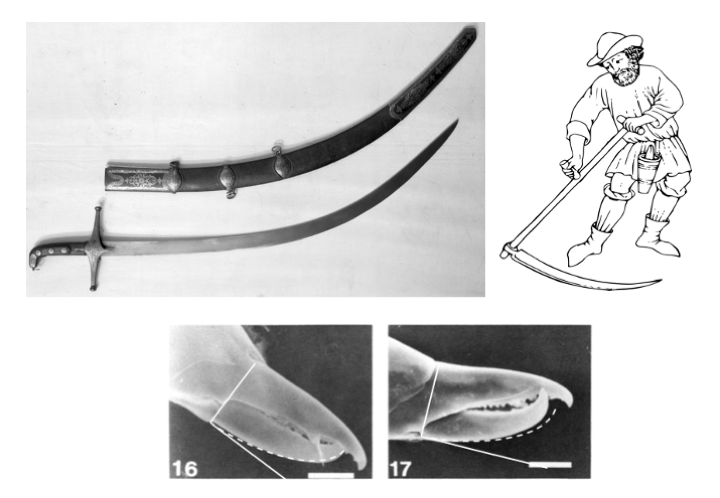
The best sword shape is one where the curvature is decreasing towards the tip, i.e., the involute sword. The mathematical equation that describes this based upon a circle is [x=r.(cos(t)+t.sin(t)), y=r.(sin(t)-t.cos(t))], t\textgreater0 and r=radius of circle. The path that this traverses is the same as an object connected to a string that wraps or unwraps around a circular post. In form it looks like an Archimedes spiral or a less expanding logarithmic spiral (found widespread in Nature).
Overall, do predatory mite moveable digits overall show such a form (i.e., a parallelism of blade location, with its'propagation' and with its'rotation' when occluding)? Certainly the Figures in Liu et al. (2017) suggest that phytoseiid mites probably do (but therein the quoted'morphometric angle 15' values for the various phytoseiid feeding groups at around 30o are much higher than 60o=tan-1\{y/((x-1))\}, @ t=1.57 for the involute equation, r=1). Global rotation of the reference axes by around 10-15o does produce visual congruence on the SEMs shown in Flechtmann and McMurtry (1992b) (e.g., Figure 1 lower, and so consequently generate consilient lower final morphometric angle values). More work, of course, is needed. For instance, perhaps just the region from the tip to the first tooth could be also modelled in this way and compared to the [`gape':`bite/throat'] ratios of fishing hooks (of known different function already) found in Allen (1996) and in Beverly (2010)?
The involute shape has some interesting properties.
- From a fully closed chela, as the moveable digit opens (rotating in its condyle), a particular food morsel at first resting proximal to that condyle on the ventral surface of the moveable digit, will be continually pushed vertically downwards as the chela uniformly opens, as long as the moveable digit ventral surface can flow (in a relative sense) past it. That is, on rotation, ''... the tangents to the involute having a given direction move in a uniform translation motion'' (see the animation by Robert Ferréol of a cam at https://mathcurve.com/courbes2d.gb/developpantedecercle/developpantedecercle.shtml ). So food material will be compacted, or an existing wound prised open, i.e., until the chela is fully open. Clearly, muscle arrangements and the relative power of adductors and abductors compared to any handling resistance of food material needs consideration here for this assertion to be definitive.
- Conversely, for a fully open chela (i.e., biting at a maximum gape), a particular food morsel at first resting on the dorsal surface of the moveable digit proximal to the tip, will be continually pushed vertically upwards as the chela uniformly closes, as long as the moveable digit dorsal surface can flow (in a relative sense) past it (until the chela is fully shut). A consequent slicing action is ensured for a narrow blade given high inertia in the food material. Forward facing teeth would of course prevent this smooth relative sliding of the digit surface against the food. Again, muscle arrangements and the relative power of adductors and abductors compared to any handling resistance of food material needs consideration here for this assertion to be definitive
Sensing, cracking and tearing things
In a later section, the design for pollenophagy is discussed in detail. For this (and other gripping tools), it would be advantageous to directly sense when the pollen grain (or food morsel) is successfully held on the inner side of the chelicera. The pilis dentilis, e.g., in Amblyseius similioides (Figure 2), as a sensory seta on the outside flank of the moveable digit is such a mechanism. Nuzzaci et al. (1992) and de Lillo et al. (1997) detail various other mouthpart sensilla in economically important mesostigmatids. Note that many melicharids which include several pollen specialists have their long pili dentili enlarged into membranous hyaline flang for fluid control or support (Lindquist and Hunter 1965, Masan 2022) They may also pick up other small objects like fungi which could be similarly sensed. Many other predatory mites also have the distal'raptor' hook suitable for tearing at food (Figure 2). A SEM follow-up study is indicated.
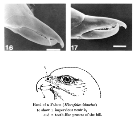
Moreover the tubercle found here in phytoseiids may function like the tomial tooth of a peregrine falcon. This'tooth' is also present in most celaneopsoid mites, (Seeman 2023, Seeman and Miranda 2024), where it is always the largest tooth.
The raptor-like hooked tip of chelal digits e.g., Amblyseius similioides, may facilitate tearing at prey flesh.
Prising things open
The crossed-over digit tips in some of the phytoseiids illustrated by other workers (e.g., Figure 2 top right) are reminiscent of the beak of crossbill birds (Jerry Krantz pers.comm.). These finches' unusual bill shape (see https://en.wikipedia.org/wiki/Red_crossbill ) is an adaptation which enables them to extract seeds from pine cones by twisting their mandibles (see https://ebird.org/species/redcro9 ). For sure, if predatory mite digits were closed shut, this form would effectively widen the wound in any prey on cheliceral retraction (Bowman 2020).
For asymmetric-shaped prey like the snails attacked by the limpkin (https://en.wikipedia.org/wiki/Limpkin ), beak shape needs to match the handedness of shell coiling to be maximally efficient. As each chelicera is a mirror image of the other so that both moveable digits swing'inwards' i.e., there are not'left-handed' mites and'right-handed' mites only left-hand and right-hand chelicerae (within an individual). Mites by virtue of having two mirror image chelicerae (e.g. Figure 13 in Bowman 2020) thus can match either prey handedness. However, it is not clear how often predatory mites initially grab prey by'shooting' both chelicerae out together in a tandem'snap' versus a single chelicera being used in that first attack.
Opening things like a can
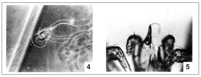
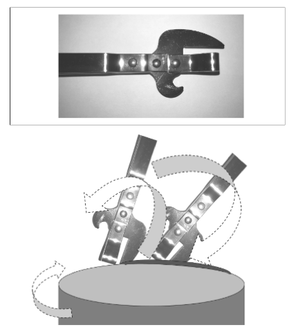
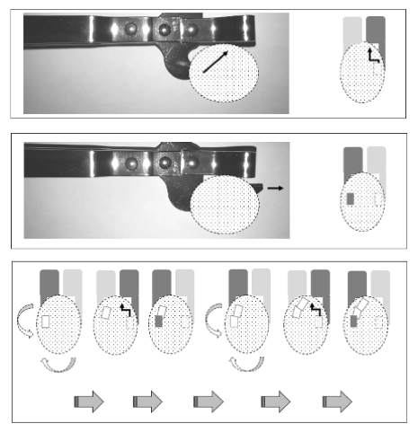
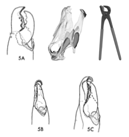
Flechtmann and McMurtry (1992a) describes a'side-bite' mechanism by which a pollen grain is snatched and held by one chela and then ruptured by the other, all above the hypostome (Figure 3). How firmly the pollen grain is held during any fluid suction needs more clarity. However, if one focusses upon the stabbing moveable digit for such'plant' feeders, the mode of action of a certain human kitchen utensil comes to mind here, i.e., the'stab can-opener' (Figure 4) with an alternating rotating and piercing mechanism like in Figure 5. Indeed, Goleva and Zebitz (2013) show shrunken pollen with'feeding holes' after phytoseiid attack. Of course each chelicera can swop their role interchangeably as they go along. The end result of the'side-swipe' holding plus stabbing action of such a tool is that the pollen grain can be repeatedly punctured and sliced open held up over the hypostome fully releasing its contents for imbibition.
Cutting curves in'tough' things
Is there a design that is consilient with other phytoseiids feeding where they are known to puncture prey legs and drain their body fluids (Flechtmann and McMurtry 1992a)? Their cheliceral form (Flechtmann and McMurtry 1992b) is more like Figure 6, suitable for cutting curves in tough material.
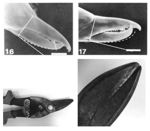
There is an extreme design found in'hawkbill cutters' used in podiatry and chiropody (Figure 7). This curved cutting style is necessary because metal (and keratin in nail) is stiff and heavy and does not move out of the way readily when cutting around a curve. Chitin is a laminated material like melamine, both of which can be stiff like metal. The respective styles of these pliers move such material out of the way when cutting in the direction that they are designed for. Mirror-image chelicerae with moveable digits formed accordingly would allow cutting in either direction.
Interestingly, estimating the moveable digit claw or hook angle (γ) and claw-length equivalent measures (see Bowman 2024a) for the phytoseiids illustrated in Adar et al. (2012) gives the following means per feeding style group: Group I-II: 126o, 13.4 μm, Group III: 127o, 13.8 μm, Group IV: 136o, 13.1 μm. This shows little differentiation between generalist and specialist predators and pollenophages for their ability to cut bent material without buckling it.
Snipping a hole in'metal-like' things
`Aviator snips' which come in left-cutting and right-cutting versions are used for cutting tight curve holes in pipes. The shape of the blades allow for sharp turns without buckling flat sheet metal that they are also used for. Arthropod cuticle is tough. Again the mirror-image form of acarine chelicerae allow cutting curves in either direction (i.e., clockwise or anti-clockwise). Aviator snips blades are usually serrated to prevent material slippage, just like phytoseiid digits. Note that teeth serrations apparently do not affect stress dissipation during feeding (at least not in herbivorous dinosaurs, Reichel 2010). Blunt curved pliers as opposed to such cutters are also known being usually called curved jaw or bent nose pliers.
And.. not'dribbling'
Many vertebrate animals have cheeks. Could mites have anything similar (recall the hyaline flange of Proctolaelaps and some other melicharids), and are there unusual actions related to them?
Pergamasid mesostigmatids have been observed to drink from droplets (despite the challenges of viscosity at that scale, Bowman 2023b). Humming bird flower-inhabiting mites handle nectar not just pollen, depleting flower stocks significantly if in large population numbers. Various subsidiary structures are described to facilitate fluid feeding in some phytoseiids (Flechtmann et al. 1994). How do these function? Some clues as to unusual fixed digit actions in some phytoseiid mites are the location of apparent cuticular extensions to the sensory dorsal proprioreceptive lyrifissure on some species (e.g., in euseiids, Flechtmann and McMurtry 1992b). These augmentations extend laterally around the digit shaft. The lyrifissure functions as a strain gauge for fixed digit cuticular flexing under moveable digit occlusion against foodstuffs in mites. These extra cuticular folds suggest a degree of movement outwards (i.e., obliquely sidewards) on chewing as the moveable digit slides against the upper fixed chelal teeth. This is consilient with the side-bite action discussed above. It also is much like the action (Weishampel 1984) arising from the unique'hinge' between the upper jaws and the rest of the skull in ornithopods (which compensates for the lack of a flexible lower jaw joint prevalent in modern day mammalian herbivores) and also in the beaks of crossbills.'Cheeks' would hide this action during chewing (Williams and Purnell 2008).
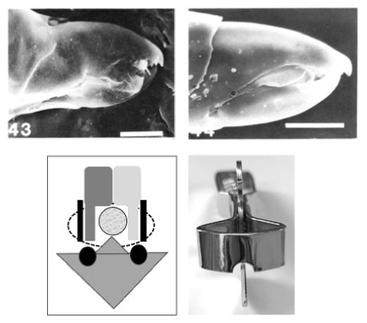
Hamsters are of course well known for sequestering foodstuff in their cheek pockets. Indeed, fluids released from pollen by some phytoseiid mites can be channelled (much like how a straw functions) between the chelicerae by the exterior lobes on the chelae (Figure 8). Such lobes are functioning like how the duck-billed hadrosaur cheeks (Galton 1973) did in keeping food within the oral area. Of course, any lengthening of the distance liquids have to flow before entering the pre-oral food channel and being imbibed by mites may affect feeding efficiency. However, prevention of loss by stopping overflow'spilling' may be a selective advantage. Just as a cheek-delimited'pocket' retaining material for the continual intensive mastication and further extraction of fluids would too. Such lobes appear in other taxa (e.g. trigynaspids) – so perhaps Proctolaelaps solved this problem by modifying the pilus dentitlis, while others modified other structures like the membranes where the moveable digit connects with the fixed digit.
''How can other mites attack things with other tools?''
Levering things up and sawing
Underbite in a chelicera occurs when the length of the moveable digit is more than that of the fixed digit i.e., VPmd\textgreater DPfd (Adar et al. 2012). In contrast, overbite occurs when the length of the moveable digit is less than that of the fixed digit i.e., VPmd> DPfd. Both such malocclusions can be adaptive. What biomechanical consequences might they have?
An example of underbite is the chelicera of certain mesostigmatids (Figure 9). Here the long serrate moveable digit may be used to lever up between the joints of millipedes scales (Seeman 2022) lifting them up so that the mite can access its host's body fluids after sawing into the underlying tough cuticle. Note that the'tomial' tooth seems to be secondarily lost in Terrogynium weatherwaxae unsurprisingly (Figure 9).
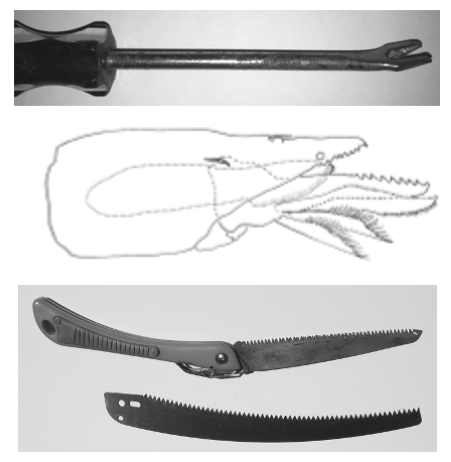
Overbite is a feature of many uropodoid mites (e.g., Cilliba cassidea). Here the length of the fixed digit is visually approximately 125% that of the moveable digit (Athias-Binche 1977). Such'small-headed' mites may still have a powerful chelal crunch force as the adductive muscles can be stored well back in the larger cheliceral base segments within the gnathosoma. The third baso-basal segment in some species may increase such muscle power even more, making them designed'track nuts'.
In the gamasines, Adar et al. (2012) found that plant feeding phytoseiids had \(\frac{D P}{V P}>1.0\) compared to non-plant feeders at \(\frac{D P}{V P}<1.0\), i.e., there is a degree of overbite in the (facultative) herbivores (or conversely a degree of underbite in the predators). Liu et al. (2017) found that pollen feeding phytoseiids had fixed digit dorsal profile perimeters greater than their moveable digit ventral perimeters (i.e., \(\frac{D P}{V P}>1.0\)) compared to generalist and specialist predators (who showed the opposite i.e., \(\frac{D P}{V P}<1.0\)). All phytoseiids illustrated by Flechtmann et al. (1994) and Flechtmann and McMurtry (1992b) appear to have overhanging fixed digits in lateral SEM pictures of the chelicerae. Overbite can be useful for a'can-opening' action (see above). For sure, to the author's knowledge there is no evidence for a supernumerary process on mite moveable digits (like the predentary bone in ornithischian jaws (Nabavizadeh and Weishampel 2016)) to compensate for any overbite.
Un-peeling things
Are certain chelicerae designed for browsing or designed for grazing? A question that can be often asked of herbivorous and saprophagous mites. There is already the same long-standing debate regarding such in different hadrosaurs amongst palaeontologists. So, what are the key design features of each modality?
For essentially tubular animals, metazoans accessing terrestrial material has its challenges (Wassenbergh et al. 2006). Mites with their anterior gnathosoma and oral opening above a hypostome need to get food up and into their gut. Grazers (like vertebrate sheep or cows) feed on vegetative material close to the ground and are morphologically adapted accordingly (i.e., by bending their necks down and cropping food). Browsers (like vertebrate deer or giraffes) consume higher growing material (such as leaves and twigs) and are designed differently. Indeed what are the micro-scale equivalents of trees, bushes, twigs and leaves for mites? Some chelae are designed not just to crop but also to unpeel material (Figure 10).
For sure, the former near-ground grazer habit requires a klinorhynchid pose or an ability to access material essentially underneath the main axis of a mite's idiosoma. Uropodoids certainly have adaptations for this (with effectively teeth on the end of a long snout like in weevils). Just what might be the micro-equivalent of grasslands like the Serengeti for mites? Browsing is facilitated by a long flexible neck on an airorhynchid pose or the ability of an animal to rear up (especially onto rear legs like for instance Brontosaurus). Are there such mite analogues? More detailed observations of certain free-living astigmatan and mesostigmatid feeding in the wild are needed to see if either behavioural modality is observed to match their cheliceral design.
For sure, a rasping type of overbite (Figure 10) is found in the form of the blattisociid Hoploseius cometa fixed chela (Lindquist 1963). This species shows a blunt apical end to the fixed digit with a curved row of small teeth along the apical margin, plus a sharp'normal' moveable digit. In general form this looks like a version of a mini-top cutter, however, where one'digit' surface remains curved while the other is flattened out, like Stubai standing seam opening pliers. The latter are used to open up seams on metal roofs (see https://www.stortz.com/product/standing-seam-opener-364-a/ ). This serrate broad snout matches the upper mandible of Diplodocus longus, itself like the shape of a horse's head. This dinosaur is posed to be a branch stripper, smoothly gleaning foliage from such stems or cropping soft water plants as the animal's head moved backwards. Other camel-like gleaning hadrosaurs like Edmontosaurus share the same distal design. Hoploseius is associated with shelf and bracket fungi (and drosophilids therein, Lindquist 1963). Its ecology is not clear. However, these multiple notches on the mite's digit of course would also engage with material for the moveable digit to act like a can-opener blade - another example of a polyfunctional composite tool.
In bee-hive living astigmatans, gleaning was associated with chelae like'pliers' (Bowman 2024b) and browsing with saw-like moveable digits (Bowman 2024a), the latter design also having a greater occlusive chelal force against foodstuffs (Bowman 2021). Amongst herbivorous dinosaurs, Stegosaurus had a more powerful bite than its peg-like teeth would suggest (although this is a lot less than what its five ton body weight might suggest, Lautenschlager et al. 2016). Is this true also of mites in general? Indeed why are there seemingly no powerful jawed herbivorous mesostigmatids (or even spiders for that matter) designed like a Giant Panda (Bowman 2020)? Indeed, why have no non-acarine chelicerates figured out how to stab plants with their fangs and feed upon phloem (like aphids do)? Why are there no large uropodoids (in the micro'rainforest' environment of temperate soils) matching the equivalent damp niche of extinct land-based giant turtles like Carbonemys (Cadena et al. 2012) with its relatively massive jaw? Is it that such trophic opportunities are dominated by oribatids which effectively exclude other acarine competitors? What is it about Acariformes that make them successful herbivores? Where are the predatory mite equivalents of snake-necked turtles (like Chelodina)? Is it that tracheal respiration effectively restricts activity in flood-able environments so that active predators need significant plastrons (like in mesostigmatids)? Can oribatids actually ever be truly predatory in design (as opposed to be just opportunistic or facultative zoophages)? Many questions remain.
Indeed, is it that a very long chelicera (as in uropodoids) is an energy efficient adaptation for these mites to browse across wide swathes of potential food without moving much as posed for sauropods (and they are not indeed'crevice feeders')? Could such uropodoids live in semi-sedentary, slow-moving herds (or flocks)? Could the trophic niche of uropodoids simply be equivalent to that of dry habitat ostriches (i.e., extant ratites) or even ornithomimosaurians (Barrett 2005)? But then, why are there seemingly no high speed moving and kicking uropodoids? For sure, the uropodoid strategy seems to be based around limpet-like protection (their pedofossae are significant, letting them pull their legs under their body and then they'hunker down', Evans 1972). Some uropodoids are also cryptic, covering themselves with grit etc., so running and kicking probably needs a big change in life strategy. Indeed, kicking itself is not commonly reported in mites (usually it is males kicking other males while engaged in precopula).
Much previous work in oribatids examined the degree to which different feeding styles are reflected in gnathosomal morphology (e.g., Perdomo et al. 2012). This can, like in dinosaur assemblages (Mallon and Anderson 2014), explain co-existent species in communities. However, Bowman (2024a) makes the point that at least in some astigmatans, chelae are composite (i.e., not of a single function) tools. Indeed Bowman (2024b) illustrates how for Carpoglyphus lactis the moveable digit may be simultaneously both a picker, a fluid slicer and a hyphal cutter/crusher (much like'long nose' cutting pliers). Similarly, Tyrophagus putrescentiae may both grip and crush fungal and nematode food as well as strip (like'slip joint pliers') conidia and spores off of the hyphal stalks. Is this an analogue of how some Serengeti ungulates perform? As the conclusion of Klunk et al. (2023) shows for insect mandibles, it is the active use of morphology, not just its static form, that matters.
`Trap-jaw snapping' on things and'rearing up'
Bowman (2020) offered a variety of functions for the'Rollplatte' in the chelicerae of uropodoid mites. Such mites have elongate chelicerae acting almost like an elephant's trunk. The unguintractor in the leg pre-tarsi of other arthropods may help in understanding which of the posed functions is more likely.
Gorb (1966) discusses the functional design of the insect leg pre-tarsus (a strongly modified structure originating from the crustacean dactylopodite with only a flexor muscle). The pre-tarsus connects to the terminal segment of the tarsus by a sclerotised plate called the unguitractor (or unguitrador plate). This attachment structure (Ditsche and Summers 2014) has long been used as a taxonomic feature in insects (e.g., Ruiter 2004). The unguitractor (plate) is an insertion point of the claw flexor internal muscle. This system is interpreted as an energy-saving unit that fixes the claws in the grasping position and can vary ecomophologically (Matsumura et al. 2022). In Diptera, Coleoptera, Hemiptera (Rebora et al. 2021) and Hymenoptera (Asperges et al. 2017) the roughened surface plate is semi-external to the body to facilitate insect attachment by adhesion.
If the seemingly internalised Rollplatte in uropodoids acted with the same ratchet-like function (except here the van der Waals forces-mediated adhesion is not to the external substrate surface as in insect claws, but to the internals of what was the ancestral tarsal shaft), then Figure 11 indicates its likely role. This would mean that the adductor muscle would not need to resist the over-opening forces of an open chela being moved forward plough-like into the foodstuff (thus saving muscle energy in maintaining the orientation of its moveable digit mastication surface, teeth and blades with respect to foodstuffs). This fits with idea of the chela being used like a mechanical shovel-bucket digger or bulldozer (Bowman 2020).
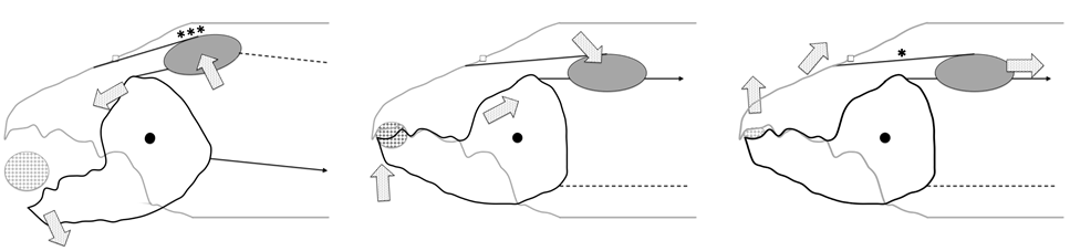
The idea of an internalised plate is not so fanciful. In the claw of the midge Chironomus riparius, the unguitractor is recessed so deeply into the cuticular pocket of the tarso-pre-tarsal joint membrane that even when the pre-tarsus is extended it does not reach the walking surface (Seifert and Heinzeller 1989). Follow-up acarological work is needed to look for scaly (Gladun 2008) or micro-trichial roughed surfaces on Rollplatten (and their corresponding cheliceral base engagement surfaces like in Plecoptera, Nelson 2009) using scanning electron microscopy of freeze-dried fractured chelicerae. However, it can be difficult to demonstrate physical contact between such surfaces (Gorb et al. 2019).
A similar internal mechanism is found in the tendon locking mechanism (TLM) of falcon (and parrot) claws (Einoder and Richardson 2006). There, a ventrally located tubercle pad on the tendon interacts with a stationary plicated sheath (and the phalangeal bone) to keep the claw closed with less continual muscle effort. Of course, the exact movement of the acarine fixed digit in this scheme will depend upon the geometry of the articulating condyle (or equivalent flexible cheliceral sheath) as well as the exact location of the tendon junctions with the fixed digit (see Bowman 2024a for a discussion on coronoid process design margins).
More morphological and embryological work is needed to regularise what parts of mite chelicerae might be related to the arci, arolia (Federle et al. 2001), auxilliae, empodia, manubria, planta and pulvilli found in arthropod claws (https://en.wikipedia.org/wiki/Arthropod/_leg ) and the precise way that they interact (e.g., see Gladun 2008). For sure there are known phylogenetic, body size and ecological correlates of the TLM in birds (Einoder and Richardson 2006). Could the sizes of Rollplatten be informative too?
Instead, could some uropodoids be considered as'arthrodires' (https://en.wikipedia.org/wiki/Arthrodira )? That is, if the fixed digit of the uropodoid chela is rapidly pulled up and backwards at the same time as the moveable digit opens (sabre-tooth like as Bowman 2020 suggested), then it is possible at a very local micro-scale to produce a tiny suction effect (much like at the macro-scale the extinct apex predator Dunkleosteus terrelli skull functioned). Such would bring material into the chelal occlusion area. Is this sudden dorsal flexure what the jointed basal third cheliceral segment might be able to achieve for the chelal head in uropodoids? It is worth pointing out that suction feeding in the analogous aquatic very long-necked hunting placoderm Dinocephalosaurus (Spiekman et al. 2024) has not been universally accepted (https://en.wikipedia.org/wiki/Dinocephalosaurus ).
Moreover, this ''jointed-neck» placoderm had another possible commonality with these mites - the armoured fish lacked distinctive teeth, using instead strengthened edges of a bony plate on their bony jawbone as a biting surface. The exposed upper and lower jaw plates came together to form a scissor-like cutting edge that would also self-sharpen every time the fish jaws opened and closed. Uropodoid moveable digits simply by virtue of their smallness must consiliently have rather small thickness especially distally (thus approximating such bony'edges'). Indeed, the left inferognathal anterior in the largest currently known individual (CMNH 5936) of D.terrelli (Engelman 2023) looks very much like the typical moveable digit dentition of some soil living oribatids (e.g., Steganacarus magnus).
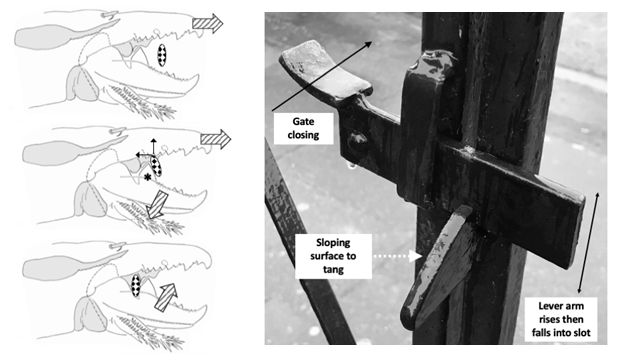
Finally, if there were special condylar modifications that would allow not just pivoting, but also'slip joints' or'cocking slip joints' (Kaji et al. 2018) in the chela, then chela in uropodoids with Rollplatten could function as ultrafast'snapping claws' like in shrimps or have a trap-jaw snapping action as in some ants (https://en.wikipedia.org/wiki/Odontomachus ). Here, from an open jaw'cocked' position with the Rollplate'anchored' (Figure 11 Left), tension is accumulated in the adductive tendon. This continues, until suddenly the joint slips un-cocking the digit with an attendant release of the inner adhesion of the Rollplatte (Figure 11 Middle). The energy stored in the stretched spring-like tendon is released, slamming the digit shut and continuing into the upper flexing action of the chelal head (Figure 11 Right). In this way, the mite'ploughs' (deep into the substrate), and the moment sensory receptors on the fixed digit tip detect something - the mite instantly grabs it (before it can move even a tiny bit away) and tears the morsel up and out from its original location all in one scheme. Nano-computerised tomography and confocal microscopy is needed in follow-up work to check for such joint architectures.
Locking onto things
A further unusual action is the possible'latch' mechanism (Figure 12) in the diplogyniid Weiseronyssus mirus, an associate of scarabaeid beetle larvae (Zhang et al. 2024). In this species an object hitting the large tooth may be cut or sliced by it, but almost certainly drives the moveable digit open as it rises up the sloping surface until it transverses the zenith of the mastication surface and drops into the nadir where membranous excrescences seal around it. A novel idea is to suggest that the trapped material is the chitinous exoskeleton of the beetle larva and the various membranes are to channel host fluids into the oral area while the mite is stuck fast on its food source. Eventually courtesy of the short output moment arm, any held-fast material can be easily crushed. As a polyfunctional tool, the chela of course is also able to stab, grip and crush other foodstuffs (like acarid mites or nematodes) more distally in line with these mesofaunal predatory scavenging mites typical habits.
''What about handling fluids?''
Preventing'dribbling' has been already discussed above
Controlling the flow
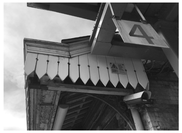
Daggerboards are a feature of historical UK railway station canopies (many having been built in the Victorian era -
https://roofingtoday.co.uk/the-distinctive-designs-of-railway-station-canopy-roofs/ .
Such crenelations facilitate rain handling directing the fluid downwards. Are there similar bladed structures in mites?
Efficient fluid handling whether of prey/host liquids or coxal gland debouched (Nuzzaci et al. 1999) material during feeding (Bowman 2014, 2017a, 2017b, 2019) is important in efficient mesostigmatid feeding (Bowman 2023b) not just via gnathosomal grooves and channels but also through using droplet'depots' and distributive'points'. Surface characteristics are the key (Figure 13). A good example of controlling fluids with'depots' at a macro-scale, are the gaps between Victorian vertical flat daggerboards (Figure 14) on the margins of old UK railway station roofs. Since fluids try to form a surface of minimal energy, surface tension will suck water films along the edges of adjacent daggerboards (near their base) and accumulate rain on the surfaces in the spherical depot areas, which when sufficiently full will collapse (due to gravity) and flow down towards the sharp drip point.
Indeed the constriction (even in an open channel like this) of flowing down the narrow slots will induce a Venturi effect by which there will be a reduction in fluid pressure and an increase in fluid speed. The cupped wing profile of diving peregrine falcons also increases air flow speed via a similar effect (Ponitz et al. 2014). This may be the reason why in places the mesostigmatid deutosternal groove in cross-section is concave. One would be most intrigued to see if dagger boards are more effective in shifting rainfall if the edges facing the previous/next board are routed out into a shallow central groove.
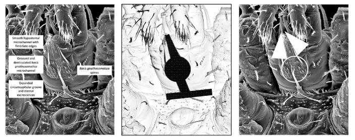
Daggerboard drip points distribute the rain away much like the various fimbriate excrescences and tips of internal male on a mesostigmatid mite's hypostome (albeit at a different scale and by an edge crawling mechanism). If the tips were (super)hydrophobic, subsequent droplet formation and loss by gravity would be facilitated. Some canopies can have a string of depots too (just like the localised deutosternal differentiations first categorised by Karg 1965).
Figure 15 illustrates the mapping between daggerboards and the forward flow of gnathosomal fluids on the ventral surface of the mesostigmatid hypostome. Further examples with the same topology can be seen in various Uropodina (e.g., Figs 3 and 11 of Bal 2006, Fig 3A of Bal and Özkan 2007, Fig 5 of Gwiazdowicz et al. (2023), Fig 4D of Kazemi and Klompen (2022), etc.,).
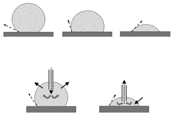
Depots can have another possible function. Addition of a surfactant or an oil into the depot can cause effective unidirectional movement driven by changes in surface tension. This is the mechanism that can propel a flat floating toy boat with soap, or a flat paper fish by a droplet of oil (see Dev Vries 1974) over a water surface. Sugar secretion will have the opposite effect removing water from surface depots. This is all mediated by the flat boat or fish surface sitting on the fluid and a change in wetting angle (Figure 13) which can have dramatic effects upon droplet shapes and flows. Looking for secretory pores and ducts leading to acarine fluid depots whether hypostomal or idiosomal is needed in follow-up morphological work.
Of course, on mite surfaces fluids move, advancing and receding within the gnathosoma, according to a scheme summarised in Bowman (2023b). Indeed, Jiang (2021) gives a nice recent review of how oscillatory mechanisms can move fluids in unexpected ways. The advancing (wetting) angle of a fluid is thought to be more sensitive to hydrophobic components of the solid surface. The receding (de-wetting) angle has been shown to correlate well with the adhesion force between the solid and the liquid. The difference between the two is called contact angle hysteresis (Eral et al. 2013). It is widely accepted that practically all real surfaces exhibit contact angle hysteresis arising from chemical and topological heterogeneities (e.g., roughness of the nanometer scale and chemical heterogeneity as small as 6-12 nm can contribute to this contact angle hysteresis). Narrow rectangular planes (like for example a deutosternal groove) alters advancing and wetting behaviour (Hong et al. 2013). Fluids can be pinned (and thus de-pinned) when advancing and receding (Chou et al. 2012), phenomena which may occur at the deutosternal ridges. Indeed, wettability is a complex topic. Dorrer and Rühe (2006) is a useful entry point to the literature.
Although not obviously crenelated, there are a variety of other flat membraneous structures within the gnathosoma of some mites other than the'cheeks' discussed above. All Celaenopsoidea and Megisthanoidea have two'membranous flanges' on the inner surface of the fixed digit, the base of which itself is a broad membranous area Owen Seeman pers.comm.). It is not yet clear exactly how these work when the chelicerae are working back and forth, but when together these membranous areas would certainly appress channelling fluids accordingly. They are not'pukka' cheeks as they are on the inner surface, true cheeks need to be on the outside. An SEM follow-up study is needed.
Feeding in fluids
Astigmatans exemplify different design solutions for feeding in fluids. For example, if carpoglyphids do not'skim' fluids (Bowman 2024a) for nutrition, how else might they feed?
Putting aside histiostomatids (and probably Hericia) for the moment, who access and filter essentially'planktonic' particulate material, the chelicerae of some carpoglyphids are designed in such a way as to be effective in skimming for food through fluids if necessary (Bowman 2024a). This is a limnetic style of feeding (McGee et al. 2013). However, this relies upon them (or at least their cheliceral bases or gnathosoma) being propelled forward through very wet food. Perhaps the mites are very motile and march their idiosoma forward continually with their gnathosoma tilted obliquely down when feeding? More detailed observations in the wild are needed.
If skimming does not occur,
- Is there any actual evidence that carpoglyphid mites collect or pick specific material from the fluid's benthic surface (McGee et al. 2013)? Biting and manipulating such material is often not needed (such for example does not occur in wrasses, Ferry-Graham et al. 2002). However, the ability to bend the oral apparatus down (like in the evolution of terrestrial vertebrates, Wassenbergh et al. 2006) is needed.
- Is there any actual evidence of these mites striking forward or'ramming' (Wasiljew et al. 2022) and attacking aquatic-based food material episodically (like an ice-spear, as posed in Bowman 2024a)? For sure there are a diversity of food capture styles, different behaviour and mechanical solutions to the problems of feeding in water shown in snakes (Alfaro 2002). Aquatic strikes are less successful (Vincent et al. 2005) presumably in part due to the resistive thickness of the medium compared to moving through air. Could fast moving aquatic micro-prey be efficiently stabbed by carpoglyphids during feeding (freshwater rotifers can be as small as 50 μm)?
- If there was a suction mechanism driven by some sort of rapid pharyngeal or gut expansion mechanism, could soft food specialist mites be striking straight-on into food with some sucking action like fish jaws to engulf food?
Manipulators (Higham 2011) need not necessarily move forward nor rely upon moving foodstuff by pressure changes. Indeed, can astigmatan bite size (estimated by Bowman 2023a) be validated by using scanning electron microscopy to measure the actual surface area removed from say (micro-sized) standardised gelatine blocks (John et al. 2020) at various hydration levels?
Fish can be categorised as'grabbers' versus'engulfers' (Mihalitsis and Bellwood 2021). Are stenophagous carpoglyphids therefore engulfers compared to the euryphagous biting acarids and glycyphagids who may grab drier food and manipulate it? What does the gut contents of wild-collected carpoglyphids actually look like?
However astigmatan feeding in fluid is done, (most) mites are visually blind (although Carpoglyphus do have structures called'eyes'). Could carpoglyphids have pressure sensors in the tips of their cheliceral digits (much like those in Kiwis and those purported to have been found in pterosaurs Martill et al. 2021). For sure, there is no distal complicated sensory apparatus as in some uropodoids.
Returning to the derived histiostomatids whose feeding is very distinct (Wirth 2010). Can the equivalent filter feeding apparatus as that found in baleen whales or Mesozoic marine reptiles (Fang et al. 2023) be found in mites? More SEM studies are needed.
`Mopping up'
Paramegistids are medium to large (0.48 - 1.65 mm long), circular mites whose adult life stages have a penchant for long, thin slippery animals like millipedes or snakes (Baker and Seeman 2008). Their biology is poorly known but there are reports of them feeding on their hosts' external secretions, organic particulates and fluids. Lawrence (1939) took them to be spermatophagous. They have various derived characters like an enormous pilus dentilis and paddle-like ventral setae (Owen Seeman pers.comm.). Do such sequester fluids?
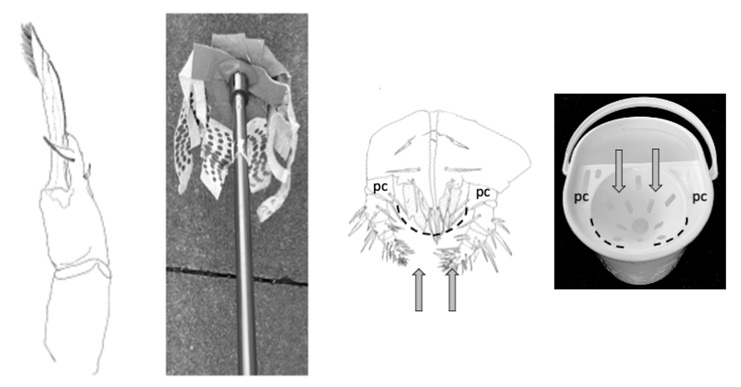
Neomegistus remus from an Australian millipede has an unusual chelicera and membraneous corniculi on its conical hypostome (Figure 16). This matches the design of a'cloth mop' and its'wringing out' apparatus. The very long moveable digit with its long membranous excrescence, finely toothed inner margin and many fine hairs distally on its external margin could'suck up' liquids when it is dipped in by surface tension onto itself. Such'mops' are also found in Fedrizzioidea where they are used to clean out the insides of dead arthropods (Seeman 2007) and in Parantennuloidea (Seeman 2025). In Micromegistus it is the cheliceral seta that is extraordinary rather than the pilus dentitlis. In both groups'beaded' filamentous excrescences help mop up fluid. Fluid feeding Diptera use a similar mechanism to lap-up nutrients. Indeed, Brazilian bearded capuchin monkeys use their long slender furry tails to dip into water stored deep in tree holes - the wet tail is then run through their mouths to extract fluids (Castro et al. 2017). Fluids in the mite excrescences can be similarly squeezed out as the chelicera is retracted and compressed between the gnathotectum, palp coxae and the fimbriate hypostomal floor. Fluids could be prevented from leakage by the membraneous corniculi sealing the gnathosomal'tube' and then the fluids being subsequently imbibed much like the strainer functions in a mop bucket when a liquid laden mop is pressed into it.
What other tool types are there left to find in mites?
Tools for cutting into bodies
At one level the moveable digits of Carpoglyphus lactis look like needle-nosed pliers (with the three small teeth acting like the pliers' transverse striations in order to facilitate grip Bowman 2024a). However, acarine exemplars of many other macro-scale tool types remain to be found (see Figures 17-19).
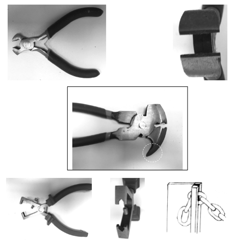
Are there gleaner-habit mites who preferentially use their chelal tips distally like wire-stripping pliers (Figure 17 bottom row) rather than proximal digit mastication surface notches (like Tyrophagus putrescentiae, Bowman 2024b) to strip material? Even a single digit with a distal notch would catch onto linear material (like a farm gate catch does, Figure 17 bottom row) and enable the stripping of say conidia from a hypha.
A possible analogy (in part) to the leverage of the fencing plier design (Figure 17 middle row) might be the unexplained function in Katydiseius (see Fain and Lukoschus 1983) and Berlesia chelae (Lindquist et al. 2020). The'jaws' are at the end of elongate chelicerae (weevil-like). However, here the exact internal arrangement of tendons and the apparent inverted position of the fixed digit compared to the bladed moveable digit awaits further detailed micro-investigation. At one level, the sharp robustly toothed moveable digit could saw and slice like a can-opener blade, but it seemingly has nothing to press the food material against in order to laterally grip such. Rather than a chelate fixed digit, there appears to be an offset pointed outgrowth of the distal cheliceral segment comprising it. This perhaps could stab, root into or lever material. A detailed discussive scheme for how it might work (comparing it to Varroa and ixodid ticks) is given in Lindquist et al. (2020). A SEM follow-up study is needed to better understand the chelal geometry exactly in these laelapids (which were previously in the Otopheidomenidae).
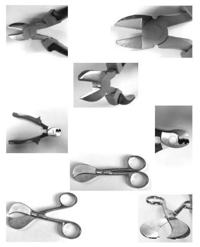
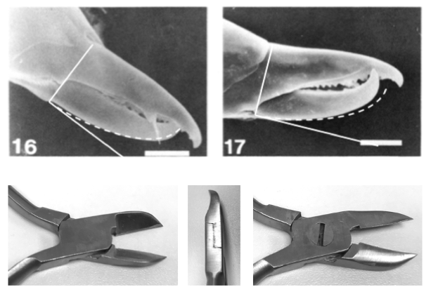
Rhagidiids like Robustocheles may have a fixed digit that works like a wire-stripper and a moveable digit acting like hawkbill pliers or diagonal/side-cutters (see Zacharda et al. 2012). Diagonal cutting or side-cutting pliers (Figure 19 top and second row) have heavy blades sharpened at their edge and excavated on one side. They are ideal for cutting medium and medium-hard wire close to the surface. Could this design (like plastic pipe cutters) be the optimal form for the chelae of nematode feeding mites or mites rupturing dipteran larvae, enchytraeids etc.?
Tools for cutting off legs
Steel electric cable wire cutters (Figure 19 third row) have specially shaped shearing surfaces designed to slice through very tough material. To my knowledge phytoseiids have not been observed cutting off legs. However, could this be the chelal design for other predatory mites to attack heavily armoured tube-like material like arthropod legs at the junction with their bodies?
Tools for cutting bodies up
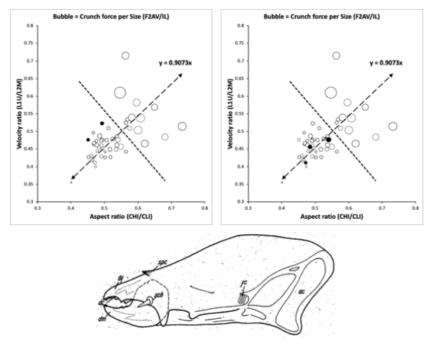
If deep digit blades were flattened and curved like scimitar-shaped plates one obtains the form of umbilical cord scissors (Figure 19 fourth and bottom row). These are smoothly rounded for safety when delicately inserted amongst tissues, and specifically designed to easily cut tubes (≡ nematodes for a mite). Are there any mite chelae like this? Lardoglyphus zacheri can be found in surface or shallow-buried dead bodies (where the species is active in the'bloated' and'dry' stages of decomposition)? Lardoglyphus zacheri has an extreme cheliceral design position amongst the microsaprophagous free-living astigmatans. Inspection of Figure 18 upper, shows it to be one of the most westerly and north-westerly located taxon in the lower group of the gnathosomal design space of Bowman (2021).
Indeed, lardoglyphids have a distinct flesh-slicing cheliceral form (Figure 18 lower). For the scale of its chelal velocity ratio (being like that of the more centrally located Acarus siro, A10b), the cheliceral base of L.zacheri has a relatively more elongate shape than expected. Or for that relatively elongate shape of its cheliceral base (like that of the more centrally located Tyrophagus putrescentiae, T13) its chela has a higher velocity ratio than expected. The former inference suggests lardoglyphids (like Lardoglyphus konoi categorised as a surface-living, potential crevice feeding/excavating specialist consuming only small and soft food morsels, Bowman 2021), feeds on more easily available resources than the'demolition-feeding' storage acarid also found in cadavers (which excavates into material Bowman 2021). The latter inference, suggests an ability to tackle relatively tougher surface material than the mycophagous Tyrophagus putrescentiae. However, either way it has a feeble chelal crunch force for its body size indicating a soft flesh slicing habit. Whether its moveable digit ascending ramus indicates that it could function like a'stitch unpicker' as in Carpoglyphus lactis (Bowman 2024b) awaits investigation.
This taken all together with the fact that lardoglyphids have few if any cheliceral teeth (e.g. Lardoglyphus konoi, Figure 18 lower), marks them out more like a weak-jawed extinct ankylosaur (Haas 1969) which had small leaf-shaped teeth to aid in stripping and pulping soft leaves from plants rather than breaking up large material or grinding such. This match to a non-selective low-browse cropper, suggests that lardoglyphids are likely to be consiliently consuming relatively soft non-abrasive material. This matches the conclusion of Bowman (2021) where they are archetypal feeble effort, small food material, little morsel biting, tiny mouthful feeders. For sure, L.zacheri neither shows clear heterodonty, nor the single kind of tooth for instance found in dolphins or porpoises that deal with grasping slippery or evasive prey (Das et al. 2023). Given that lardoglyphids are not armoured like ankylosaurs (or even uropodoid mites), this poses the question - what is their defence mechanism against predation from mesostigmatids also known in cadavers for instance? Is it that they'outbreed' their opponents (they are not high speed movers who could flee) or do they just rely upon tough skinned hypopal stages to'weather any storm' (Iverson et al 1996)? Do they produce noxious oily secretions? More work for forensic acarologists beckons.
Tools for grabbing, scratching and holding
Are there wide-mouthed mites designed like a white rhinoceros (https://en.wikipedia.org/wiki/White_rhinoceros ) or a hippopotamus (https://en.wikipedia.org/wiki/Hippopotamus ) that match end-cutter pliers (Figure 17 top row)? If the illustrated mini-top cutter design is blunted and made much larger one obtains the form of traditional carpenter's pincers, which are used to grab, grip and extract nails without damaging the surface of the substrate. Are there mites that pull their foodstuffs out like this?
Rather than an'unpeeling' design (Figure 10), if both the upper and lower cutting edges of a mini-top cutter design were flattened out (into smooth mastication surfaces), then one forms flat welding pliers, glass breaking pliers, canvas and sheet metal pliers. Do any mite chelae grab and grip such wide flat surfaces to snap material off?
Do mites use digit excrescences to lever up or grub up material or root about in the substrate so material could be levered out like how fencing pliers (Figure 17 middle row) are used? This is how extinct tusked deinotherids may have grabbed and fed even on trees and bushes (https://en.wikipedia.org/wiki/Deinotheriidae ). Indeed do saprophagous mites hoe? More live observations are needed.
`Dual chain catches' (Figure 19 bottom row) may perhaps be useful in phoretic species? The large proximal tooth in Weiseronyssus mirus (Figure 12) could function like the setscrew in the upper jaw of the wire-stripping pliers illustrated in Figure 17 (Jerry Krantz pers.comm.) as it would preclude complete digit closure unless socketed into the fixed digit (see Bowman 2024a for discussion on effective gape and tooth position).
Many trophic modalities need acarologists to search for mites designed appropriately.
Conclusion and Future challenges
Any one mite species might do one or all the feeding actions discussed above at once!
Applied acarologists are asked:
"Which of your species are designed as
- A specialist feeding tool ?
- A generalist feeding tool ?"
and,
''What might that mean for your success in biological control against different pests?''
The discussions above rely upon fixed structures within and between identified taxa (recall that the bizarre chelicerae and membranous or modified corniculi of several trigynaspid mites only develop in the adult stage indicating that the adults are doing something different to the juveniles). However, could there be acarine gnathosomal polymorphisms driven by living conditions? Round-worms can develop into a wide-mouthed predator or a narrow-mouthed bacteria eater (Ragsdale et al. 2013) controlled by their environment. When the animals were starved or when too many worms crowded the Petri dish, the researchers observed the increased development of the wide-mouthed variant. Can acarologists find similar mite examples?
What about'fasteners' and not just tools? Ixodid tick mouthparts with their jagged hypostome inserted into host epidermis mimic dry wall anchors, and with the palps splayed out during feeding bear a strong resemblance to plaster-board spring-loaded fasteners. Are there parasitic mites with structures like hollow-wall fasteners, cavity-wall or butterfly anchors, rag- or through- or'molly'- or toggle-bolts etc.? Many challenges await future acarologists - mite chelae or gnathosomal parts like all of these macro-devices might exist.
Finally, perhaps there are acarologists out there who can draw together the scattered diverse literature to compile for all mites the equivalent of the recent book on dinosaurs i.e., Nabavizadeh and Weishampel (2023)? However, even though the parallelism and comparison with other phyla is intriguing, doing this is not just to categorise different species but to crucially relate their evolutionary forms to feeding guilds unequivocally determined in the wild using isotopic ratios (as done in mesostigmatids and oribatids by Perdomo et al. (2012), Díaz-Aguilar and Quideau (2013) etc.,. That would be truly impressive.
Acknowledgements
Analysis and reporting of this work was self-funded. I dedicate this manuscript to the extraordinary Donald E Johnston (b.1934, d. 9th August 1994) of the Acarology Laboratory, Ohio State University for introducing me to the work of D'Arcy Wentworth Thompson and Robert McNeill Alexander. Thanks go especially to Evert Lindquist, Jerry Krantz and Owen Seeman for suggesting various example species, together with a midwife neighbour and the staff at my local ironmongery store for their interest and supply of various key tools. I thank a legion of intellectually generous scientists for their friendly help during my varied career. Owen Seeman (in particular) and an anonymous referee made many useful suggestions to improve this paper. Above all, I thank my dear wife Diane for supporting my involvement in this field over many years. The author is a Royal Society Industrial Fellow (IF110047) at the Mathematical Institute, University of Oxford.
Author contributions
All aspects were solely carried out by CEB.
Declarations
Conflicts of interest
The author declares that they have no known conflicts of interest. No competing claims are known.
Human or animal rights
This article does not contain any studies with human participants or vertebrate animals performed by any of the authors.
References
- Adar A, Inbar M, Gal S, Doron N, Zhang X-Q, Palevsky E (2012) Plant-feeding and non-plant feeding phytoseiids: differences in behavior and cheliceral morphology. Experimental and Applied Acarology. https://doi.org/10.1007/s10493-012-9589-y
- Akimov I A (1985) Biological foundations of harmfulness in acaroid mites. Naukova Dumka, Kiev, 160 pp (in Russian).
- Alfaro ME (2002) Forward attack modes of aquatic feeding garter snakes. Functional Ecology 16(2): 204-215. https://doi.org/10.1046/j.1365-2435.2002.00620.x
- Allen MS (1996) Style and function in East Polynesian fishhooks. Antiquity 70(267): 97-116. https://doi.org/10.1017/S0003598X00082922
- Asperges M. D'Haen J, Lambrichts I, Belleghem FV (2017) The pretarsus of the honeybee. Belgian Journal of Zoology 147(2): 87-103. https://doi.org/10.26496/bjz.2017.8
- Athias-Binche F (1977) Observations morphologiques au cours du dévloppement postembryonaire chez Cilliba cassidea (Hermann 1804) (Anacinotriches, Uropodides). Acarologia 18(4): 575-593.
- Baker MR, Seeman OD (2008) Mites and millipedes: a new Neomegistus (Acari: Mesostigmata: Paramegistidae) from Australia. Systematic & Applied Acarology 13: 204-213. https://doi.org/10.11158/saa.13.3.7
- Bal DA (2006) New species of mites in the genera Uropoda, Crinitodiscus and Uroobovella from Turkey (Acari: Mesostigmata: Uropodidae, Urodinychidae). Zootaxa 1368: 19-40. https://doi.org/10.11646/zootaxa.1368.1.2
- Bal DA, Özkan M (2007) Uropoda abantica n.sp., a new mesostigmatid mite (Acari: Uropodidae) from Turkey. International Journal of Acarology 33(1): 41-47. https://doi.org/10.1080/01647950708684499
- Barrett PM (2005) The diet of ostrich dinosaurs (Therapoda: Ornithomimosauria). Palaeontology 48(2): 347-358. https://doi.org/10.1111/j.1475-4983.2005.00448.x
- Beverly S (2010) Circle hooks in the Pacific. SPC Fisheries Newsletter 131: 22-25.
- Bowman CE (2014) Gut expansion and contraction in the predatory soil mite Pergamasus longicornis (Mesostigmata: Parasitidae) - a stiff system. Experimental & Applied Acarology 64(3): 337-359. https://doi.org/10.1007/s10493-014-9827-6
- Bowman CE (2017a) Modelling Malpighian tubule crystals within the predatory soil mite Pergamasus longicornis (Mesostigmata: Parasitidae) Experimental & Applied Acarology 72(1): 35-59. https://doi.org/10.1007/s10493-017-0137-7
- Bowman CE (2017b) Digestive half lives, gut contents and feeding state prediction in the soil predatory mite Pergamasus longicornis (Mesostigmata: Parasitidae) Experimental & Applied Acarology 73(1): 11-60. https://doi.org/10.1007/s10493-017-0174-2
- Bowman CE (2019) The gut epithelium from feeding to fasting in the predatory soil mite Pergamasus longicornis (Mesostigmata: Parasitidae) - one tissue, two roles. Experimental & Applied Acarology 77(3): 253-358. https://doi.org/10.1007/s10493-019-00356-6
- Bowman CE (2020) Feeding design in free-living mesostigmatid chelicerae (Acari: Anactinochitinosi). Experimental and Applied Acarology 84(1): 1-119. https://doi.org/10.1007/s10493-021-00612-8
- Bowman CE (2021) Cheliceral chelal design in astigmatid mites. Experimental and Applied Acarology 84(2): 271-363. https://doi.org/10.1007/s10493-021-00625-3
- Bowman CE (2023a) Variation in the trophic morphology of Astigmatid mites common in UK beehives. Acarologia 63(Suppl): 4-16. https://doi.org/10.24349/z9n6-u3t3
- Bowman CE (2023b) Looking for future biological control agents: the comparative function of the deutosternal groove in mesostigmatid mites. Experimental and Applied Acarology 91: 139-235. https://doi.org/10.1007/s10493-023-00832-0
- Bowman CE (2024a) Do astigmatid teeth matter: a tribological review of cheliceral chelae in co-occurring mites from UK beehives. Experimental and Applied Acarology 92(4): 567-686. https://doi.org/10.1007/s10493-023-00876-2
- Bowman CE (2024b) Transitional chelal digit patterns in saprophagous astigmatan mites. Experimental and Applied Acarology 92(4): 687-737. https://doi.org/10.1007/s10493-024-00907-6
- Button DJ, Rayfield EJ, Barrett PM (2014) Cranial biomechanics under-pins high sauropod diversity in resource-poor environments. Proceedings of Royal Society B 281: 20142114. https://doi.org/10.1098/rspb.2014.2114
- Cadena EA, Ksepka DT, Jaramillo CA, Bloch JI (2012) New pelomedusoid turtles from the late Palaeocene Cerrejón Formation of Colombia and their implications for phylogeny and body size evolution Journal of Systematic Palaeontology 10(2): 313-331. https://doi.org/10.1080/14772019.2011.569031
- Castro SCdN, Souto AdS, Schiel N, Biondi LM, Caselli CB (2017) Techniques Used by Bearded Capuchin Monkeys (Sapajus libidinosus) to Access Water in a Semi-Arid Environment of North-Eastern Brazil. Folia Primatologica 88: 267-273. https://doi.org/10.1159/000479106
- Chou T-H, Hong S-J, Sheng Y-J, Tsao H-K (2012) Drops Sitting on a Tilted Plate: Receding and Advancing Pinning. Langmuir 28(11): 5158-5166. https://doi.org/10.1021/la300257t
- Das K, Sköld H, Lorenz A, Parmentier E (2023) Who Are the Marine Mammals?. In: Brennecke D, Knickmeier K, Pawliczka I, Siebert U, Wahlberg M (eds) Marine Mammals. Springer, Cham. https://doi.org/10.1007/978-3-031-06836-2
- de Lillo E, Aldini P (1994) Contributo alla conoscenza delle parti boccali in femmine di Typhlodromus exhilaratus Ragusa (Acari: Phytoseiidae). Atti XVII Congresso nazionale italiano di Entomologia, Udine 13-18 giugno 1994: 287-294.
- de Lillo E, Nuzzaci G, Aldini P (1997) Fine morphology of the mouthpart sensilla in females of Typhlodromus exhilaratus Ragusa (Phytoseiidae). p287-295 In: Needham GR, Mitchell R, Horn DJ, Welbourn WC (eds) Acarology IX Proceedings. Ohio Biological Survey, 718pp.
- de Lillo E, di Palma A, Nuzzaci G (2001) Morphological adaptations of mite chelicerae to different trophic activities (Acari). Entomologica, Bari 35: 125-180.
- De Vries L (1974) The Book of Experiments. John Murray (Publishers), London 123pp.
- Díaz-Aguilar I, Quideau SA (2013) Trophic ecology of mesostigmatan and oribatid mites in harvested and control coniferous and deciduous stands of the boreal mixedwood forest determined using 15N stable isotopes. Soil Biology & Biochemistry 67: 147e154. https://doi.org/10.1016/j.soilbio.2013.08.019
- Ditsche P, Summers AP (2014) Aquatic versus terrestrial attachment: Water makes a difference. Beilstein Journal of Nanotechnology 5: 2424-2439. https://doi.org/10.3762/bjnano.5.252
- Dorrer C, Rühe J (2006) Advancing and Receding Motion of Droplets on Ultrahydrophobic Post Surfaces. Langmuir 22(18): 7652-7657. https://doi.org/10.1021/la061452d
- Einoder L, Richardson A (2006) An ecomorphological study of the raptorial digital tendon locking mechanism. Ibis 148,: 515-525. https://doi.org/10.1111/j.1474-919X.2006.00541.x
- Engelman RK (2023) A Devonian Fish Tale: A New Method of Body Length Estimation Suggests Much Smaller Sizes for Dunkleosteus terrelli (Placodermi: Arthrodira). Diversity 15(3): 318. https://doi.org/10.3390/d15030318
- Eral HB, Mannetje DJCM′t, Oh JM (2013) Contact Angle Hysteresis: a Review of Fundamentals and Applications. Colloid and Polymer Science 291(2): 247-260. https://doi.org/10.1007/s00396-012-2796-6
- Evans GO (1972) Leg chaetotaxy and the classification of the Uropodina (Acari: Mesostigmata). Journal of Zoology 167(2): 193-206. https://doi.org/10.1111/j.1469-7998.1972.tb01729.x
- Fain A, Lukoschus FS (1983) Katydiseius madchatram n.g, n.sp. (Acari, Otopheidomenidae) from the trachaea of a Malayasian katydid Chloracris brullei Pictet & Saussure, 1892 (orthoptera, pseudophyllldae). International Journal of Acarology 9(4):173-178. https://doi.org/10.1080/01647958308683333
- Fang Z-C, Li J-L, Chun-Bo Y, Zou Y-R, Tian L, Zhao B, Benton MJ, Cheng L, Lai X-L (2023) First filter feeding in the Early Triassic: cranial morphological convergence between Hupehsuchus and baleen whales. BMC Ecology and Evolution 23(36): 1-12. https://doi.org/10.1186/s12862-023-02143-9
- Federle W, Brainerd EL, McMahon TA, Hölldobler B (2001) Biomechanics of the movable pretarsal adhesive organ in ants and bees. Proceedings of the National Academy of Sciences of the United States of America 98(11): 6215-6220. https://doi.org/10.1073/pnas.111139298
- Ferry-Graham LA, Wainwright PC, Westneat MW, Bellwood DR (2002) Mechanisms of benthic prey capture in wrasses (Labridae). Marine Biology 141: 819-830. https://doi.org/10.1007/s00227-002-0882-x
- Flechtmann CHW, Evans GO, McMurtry JA (1994) Some noteworthy features of the chelicerae and subcapitulum of Phytoseiulus longipes Evans (Acari: Mesostigmata: Phytoseiidae), with observations on the preoral channel in the Phytoseiidae. Experimental & Applied Acarology 18: 293-299. https://doi.org/10.1007/BF00132318
- Flechtmann CHW, McMurtry JA (1992a) Studies on how phytoseiid mites feed on spider mites and pollen. International Journal of Acarology 18(3): 157-162. https://doi.org/10.1080/01647959208683946
- Flechtmann CHW, McMurtry JA (1992b) Studies of cheliceral and deutosternal morphology of some Phytoseiidae (Acari: Mesostigmata) by scanning electron microscopy. International Journal of Acarology 18(3): 163-169. https://doi.org/10.1080/01647959208683947
- Galton PM (1973) The cheeks of ornithischian dinosaurs. Lethaia. 6(1) 67-89. https://doi.org/10.1111/j.1502-3931.1973.tb00873.x
- Gladun DV (2008) Morphology of the pretarsus of the sawflies and horntails (Hymenoptera: 'Symphyta′). Arthropod Structure & Development 37: 13e28. https://doi.org/10.1016/j.asd.2007.04.002
- Goleva I, Zebitz CPW (2013) Suitability of different pollen as alternative food for the predatory mite Amblyseius swirskii (Acari, Phytoseiidae). Experimental & Applied Acarology 61: 259-283. https://doi.org/10.1007/s10493-013-9700-z
- Gorb SN (1996) Design of Insect Unguitractor Apparatus. Journal of Morphology 230: 219-230. https://doi.org/10.1002/(SICI)1097-4687(199611)230:2<219::AID-JMOR8>3.0.CO;2-B
- Gorb SN, Pullwitt T, Kleinteich T, Busshardt P (2019) The insect unguitractor plate in action: Force transmission and the micro CT visualizations of inner structures. Journal of Insect Physiology 117: 103908. https://doi.org/10.1016/j.jinsphys.2019.103908
- Gwiazdowicz DJ, Babaeian E, Ermilov SG (2023) A new species of Trigonuropoda (Mesostigmata: Uropodina) from Vietnam. Acta Zoologica Academiae Scientiarum Hungaricae 69(2): 117-126. https://doi.org/10.17109/AZH.69.2.117.2023
- Haas G (1969) On the jaw musculature of ankylosaurs. American Museum Novitates 2399: 1-11.
- Higham TE (2011) Feeding Mechanics. In: Farrell A.P., (ed.), Encyclopedia of Fish Physiology: From Genome to Environment, volume 1, pp.597-602 Academic Press, San Diego. https://doi.org/10.1016/B978-0-12-374553-8.00216-1
- Hong S-J, Chou T-H, Liu Y-Y, Sheng Y-J, Tsao H-K (2013) Advancing and receding wetting behavior of a droplet on a narrow rectangular plane. Colloid and Polymer Science 291: 347-353. https://doi.org/10.1007/s00396-012-2797-5
- Iverson K, OConnor BM, Ochoa R, Heckmann R (1996) Lardoglyphus zacheri (Acari: Lardoglyphidae), a Pest of Museum Dermestid Colonies, with Observations on Its Natural Ecology and Distribution. Annals of the Entomological Society of America 89(4): 544-549. https://doi.org/10.1093/aesa/89.4.544
- Jiang D, Liu J, Pan Y, Zhuang L, Wang P (2021) Surface acoustic waves (SAW) techniques in tissue engineering. Cell and Tissue Research 386: 215-226. https://doi.org/10.1007/s00441-020-03397-1
- John MESt, Holzman R, Martin CH (2020) Rapid adaptive evolution of scale-eating kinematics to a novel ecological niche. Journal of Experimental Biology 223:jeb217570. https://doi.org/10.1242/jeb.217570
- Kaji T, Anker A, Wirkner CS, Palmer AR (2018) Parallel Saltational Evolution of Ultrafast Movements in Snapping Shrimp Claws. Current Biology 28: 106-113. https://doi.org/10.1016/j.cub.2017.11.044
- Karg W (1965) Larvalsystematische und Phylogenetische Untersuchung sowie Revision des Systems der Gamasina Leach, 1915 (Acarina, Parasitiformes). Mitteilungen aus dem Zoologischen Museum, Berlin 41(2): 193-340. https://doi.org/10.1002/mmnz.19650410207
- Klunk CL, Argenta MA, Rosumek FB, Schmelzle S, van de Kamp T, Hammel JU, Pie MR, Heethoff M (2023) Simulated biomechanical performance of morphologically disparate ant mandibles under bite loading. Scientific Reports 13: 16833. https://doi.org/10.1038/s41598-023-43944-8
- Lautenschlager S, Brassey CA, Button DJ, Barrett PM (2016) Decoupled form and function in disparate herbivorous dinosaur clades. Scientific Reports 6: 26495. https://doi.org/10.1038/srep26495
- Lawrence RF (1939) Notes on the habits of two mites living on South African millipedes. Transactions of the Royal Society of South Africa, 27: 233-239. https://doi.org/10.1080/00359193909519793
- Lindquist EE (1963) A Taxonomic Review of the Genus Hoploseius Berlese (Acarina: Blattisocidae). Canadian Entomologist 95(11): 1175-1185. https://doi.org/10.4039/Ent951175-11
- Lindquist EE, OConnor BM, Shaw MD, Sidorchuk EA (2020) Review of the genera Berlesia Canestrini, 1884, and Katydiseius Fain & Lukoschus, 1983, the subfamily Katydiseiinae Fain & Lukoschus, 1983, and their family group relationships (Acari: Mesostigmata: Gamasina), with description of three new species parasitic on gryllacridid crickets (Orthoptera). Zootaxa 4857(1):005-070. https://doi.org/10.11646/zootaxa.4857.1.4
- Lindquist EE, Hunter PE (1965) Some mites of the genus Proctolaelaps Berlese (Acarina: Blattisociidae) associated with forest insect pests. Can. Ent. 97: 15-32. https://doi.org/10.4039/Ent9715-1
- Liu S, LV J, Wang E, Xu X (2017) Life-style classification of some Phytoseiidae (Acari: Mesostigmata) species based on gnathosoma morphometrics. Systematic & Applied Acarology 22(5): 629-639. https://doi.org/10.11158/saa.22.5.3
- Mallon JC, Anderson JS (2014) Implications of beak morphology for the evolutionary paleoecology of the megaherbivorous dinosaurs from the Dinosaur Park Formation (upper Campanian) of Alberta, Canada. https://doi.org/10.1016/j.palaeo.2013.11.014
- Palaeogeography, Palaeoclimatology, Palaeoecology 394: 29-41. https://doi.org/10.1016/j.palaeo.2013.11.014
- Martill DM, Smith RE, Longrich N, Brown J (2021) Evidence for tactile foraging in pterosaurs: a sensitive tip to the beak of Lonchodraco giganteus (Pterosauria, Lonchodectidae) from the Upper Cretaceous of southern England. Cretaceous Research 117: 104637. https://doi.org/10.1016/j.cretres.2020.104637
- Masan P (2022) The family Melicharidae (Acari, Mesostigmata) in Slovakia, with description of new species, annotated faunal synopsis and identification keys of species from Europe. Zootaxa 5172: 1-449. https://doi.org/10.11646/zootaxa.5172.1.1
- Matsumura Y, Lima SP, Rafael JA, Câmara JT, Beutel RG, Gorb SN (2022) Distal leg structures of Zoraptera - did the loss of adhesive devices curb the chance of diversification? Arthropod Structure & Development 68: 101164. https://doi.org/10.1016/j.asd.2022.101164
- McGee MD, Schluter D, Wainwright PC (2013) Functional basis of ecological divergence in sympatric stickleback. BMC Evolutionary Biology 13: 277. https://doi.org/10.1186/1471-2148-13-277
- Mihalitsis M, Bellwood DR (2021) Functional groups in piscivorous fishes. Ecology and Evolution 11: 12765-12778. https://doi.org/10.1002/ece3.8020
- Nabavizadeh A, Weishampel DB (2016) The Predentary Bone and Its Significance in the Evolution of Feeding Mechanisms in Ornithischian Dinosaurs. The Anatomical Record 299: 1358-1388. https://doi.org/10.1002/ar.23455
- Nabavizadeh A, Weishampel DB (2023) An Illustrated Guide to Dinosaur Feeding Biology. John Hopkins University Press 368 pp. https://doi.org/10.56021/9781421445878
- Navalón G, Bright JA, Marugán-Lobón J, Rayfield EJ(2019) The evolutionary relationship among beak shape, mechanical advantage, and feeding ecology in modern birds, Evolution 73(3): 422-435. https://doi.org/10.1111/evo.13655
- Nelson CH (2009) Surface ultrastructure and evolution of tarsal attachment structures in Plecoptera (Arthropoda: Hexapoda). Aquatic Insects 31(1): 523-545. https://doi.org/10.1080/01650420802598210
- Nuzzaci G, de Lillo E, Porcelli F (1992) Functional morphology of the mouthpart sensilla in females Varroa jacobsoni Oudemans (Acari: Varroidae). Entomologica, Bari 27: 41-67.
- Nuzzaci G, Di Palma A, de Lillo E, Aldini P (1999) Prosomal glands in Typhlodromus spp. females (Mesostigmata: Phytoseiidae). In: Bruin J, van der Geest LPS, Sabelis MW (eds), Ecology and Evolution of the Acari, p637-649, Kluwer Academic Publishers. https://doi.org/10.1007/978-94-017-1343-6_55
- Perdomo G, Evans A, Maraun M, Sunnucks P, Thompson R (2012) Mouthpart morphology and trophic position of microarthropods from soil and moss are strongly correlated. Soil Biology and Biochemistry 53: 56-63. https://doi.org/10.1016/j.soilbio.2012.05.002
- Ponitz B, Triep M, Brücker C (2014) Aerodynamics of the Cupped Wings during Peregrine Falcon's Diving Flight. Open Journal of Fluid Dynamics 4: 363-372. https://doi.org/10.4236/ojfd.2014.44027
- Ragsdale EJ, Müller MR, Rödelsperger C, Sommer RJ (2013) A Genetic Switch Coupled to Micro- and Macroevolution of a Developmental Plasticity Acts Through a Sulfatase. Cell 155(4): 922-33. https://doi.org/10.1016/j.cell.2013.09.054
- Rebora M, Salerno G, Piersanti S, Gorb EV, Gorb SV (2021) Attachment devices and the tarsal gland of the bug Coreus marginatus (Hemiptera: Coreidae). Zoomorphology 140: 85-102. https://doi.org/10.1007/s00435-020-00515-z
- Reichel M (2010) A model for the bite mechanics in the herbivorous dinosaur Stegosaurus (Ornithischia, Stegosauridae). Swiss Journal of Geoscience 103: 235-240. https://doi.org/10.1007/s00015-010-0025-1
- Ruiter D (2004) A review of the adult Anagapetus (Trichoptera: Glossomatidae). Western North American Naturalist 64(4): 454-464.
- Seeman OD (2007) Revision of the Fedrizziidae (Acari: Mesostigmata: Fedrizzioidea). Zootaxa 1480: 1-55. https://doi.org/10.11646/zootaxa.1480.1.1
- Seeman OD (2022) Three new genera of Schizogyniidae (Parasitiformes: Mesostigmata) from a New Caledonian millipede (Spirobolida). Zoosymposia 22: 195-222. In: Zhang Z-Q., Fan Q-H, Heath ACG, Minor MA. (Eds) (2022) Acarological Frontiers: Proceedings of the XVI International Congress of Acarology (1-5 Dec. 2022, Auckland, New Zealand). Magnolia Press, Auckland, 328 pp. https://doi.org/10.11646/zoosymposia.22.1.128
- Seeman OD (2023) Paraschizogynium Hedwig sp.n., Australia's first species of Schizogyniidae (Parasitiformes: Celaenopsoidea). Australian Entomologist 50, 221-236.
- Seeman OD (2025) Australia's first described species of Parantennulidae: Micromegistus thorpei sp. nov. (Acari: Mesostigmata: Antennophorina) from Trichosternus fax (Coleoptera: Carabidae). Systematic and Applied Acarology 30(4): 770–786. https://doi.org/10.11158/saa.30.4.9
- Seeman OD, Miranda RJ (2024) A new genus and species of Diplogyniidae (Acari: Mesostigmata: Celaenopsoidea) from the mammoth wasp Aelocampsomeris variegata (Fabricius) (Hymenoptera: Scoliidae) in Panama, International Journal of Acarology 51: 25-33. https://doi.org/10.1080/01647954.2024.2427672
- Seifert P, Heinzeller T (1989) Mechanical, sensory and glandular structures in the tarsal unguitractor apparatus of Chironomus riparius (Diptera, Chironomidae). Zoomorphology 109: 71-78. https://doi.org/10.24349/6gmw-wgod
- Shahrooz K, Klompen H (2022) Description of a new species of Protodinychus (Mesostigmata: Protodinychidae), and a key to deutonymphs of the genus. Acarologia 62(3): 585-596. https://doi.org/10.24349/6gmw-wgod
- Spiekman SNF, Wang W, Zhao L, Rieppel O, Fraser NC, Li C (2024) Dinocephalosaurus orientalis Li, 2003: a remarkable marine archosauromorph from the Middle Triassic of southwestern China. Earth and Environmental Science Transactions of the Royal Society of Edinburgh 1-33. https://doi.org/10.1017/S175569102400001X
- Vincent SE, Herrel A, Irschick DJ (2005) Comparisons of Aquatic Versus Terrestrial Predatory Strikes in the Pitviper, Agkistrodon piscivorus. Journal of Experimental Zoology 303A: 476-88. https://doi.org/10.1002/jez.a.179
- Wainwright PC, Ricard BA (1995) Predicting patterns of prey use from morphology of fishes. Environmental Biology of Fishes 44: 97-113. https://doi.org/10.1007/BF00005909
- Wasiljew BD, Pfaender J, WipflerB, Wantania LL, Herder F (2022) Functional morphology of prey capture in stream-dwelling sailfin silversides (Telmatherinidae) based on high-speed video recordings. Zoomorphology 141: 347-359. https://doi.org/10.1007/s00435-022-00570-8
- Wassenbergh SVan, Herrel A, Adriaens D, Huysentruyt F, Devaere S, Aerts P (2006) A catfish that can strike its prey on land. Nature 440: 881. https://doi.org/10.1038/440881a
- Weishampel DB (1984) Evolution of jaw mechanisms in ornithopod dinosaurs. Advances in Anatomy Embryology and Cell Biology 87: 1-109. https://doi.org/10.1007/978-3-642-69533-9_1
- Williams V, Purnell M (2008) Duck-billed dentistry: evidence for hadrosaur diet from tooth microwear. Transactions of the Leicester Literary & Philosophical Society 102: 58-59.
- Wirth S (2010) Food competition and feeding behavior and its implications for the phylogeny of the Histiostomatidae (Astigmata). pp 37-40 In: Sabelis MW and Bruin J (eds.), Trends in Acarology: Proceedings of the 12th International Congress, Springer Science+Business Media. https://doi.org/10.1007/978-90-481-9837-5
- Zacharda M, Ueckermann EA,,Rostami E, Abbasipour H, Khanjani M (2012) A survey of Rhagidiidae (Acari: Prostigmata) in fruit orchards in Iran, with description of a new species of Robustocheles. International Journal of Acarology 38(1): 30-34. https://doi.org/10.1080/01647954.2011.577454
- Zhang S, Seeman OD, Xie L, Yan Y (2024) Redescription of Weiseronyssus mirus Samsinák, 1962 (Mesostigmata: Diplogyniidae) from the larvae of Allomyrina dicohotoma (Coleoptera: Scarabaeids: Dynastinae). Systematic & Applied Acarology 29(2): 187-199. https://doi.org/10.11158/saa.29.2.1
to post any comment or correction.


2024-09-29
Date accepted:
2025-06-09
Date published:
2025-06-30
Edited by:
Tsagkarakou, Anastasia

This work is licensed under a Creative Commons Attribution 4.0 International License
2025 Bowman, Clive E.
 Download article
Download the citation
RIS with abstract
(Zotero, Endnote, Reference Manager, ProCite, RefWorks, Mendeley)
RIS without abstract
BIB
(Zotero, BibTeX)
TXT
(PubMed, Txt)