


Three new feather mites of the genus Proterothrix Gaud (Astigmata: Proctophyllodidae) from passerines (Aves: Passeriformes) in China
He, Shu-Xiang1
; Sun, Li-Hua2
; Liu, Huai3
; Yuan, Yu-Chuan4
and Wang, Zi-Ying  5
5
1College of Plant Protection, Southwest University, No. 2, Tiansheng Road, Beibei district, Chongqing, China.
2College of Plant Protection, Southwest University, No. 2, Tiansheng Road, Beibei district, Chongqing, China.
3College of Plant Protection, Southwest University, No. 2, Tiansheng Road, Beibei district, Chongqing, China.
4Nanjian administration and Protection Bureau of Yunnan Wuliangshan National Nature Reserve, No. 56, Boai Road, Nanjian district, Yunnan, China.
5✉ College of Plant Protection, Southwest University, No. 2, Tiansheng Road, Beibei district, Chongqing, China.
2024 - Volume: 64 Issue: 3 pages: 843-864
https://doi.org/10.24349/y1xh-2eq2ZooBank LSID: A882B50D-A456-45DE-A0F1-EB77DD46BECE
Original research
Keywords
Abstract
Introduction
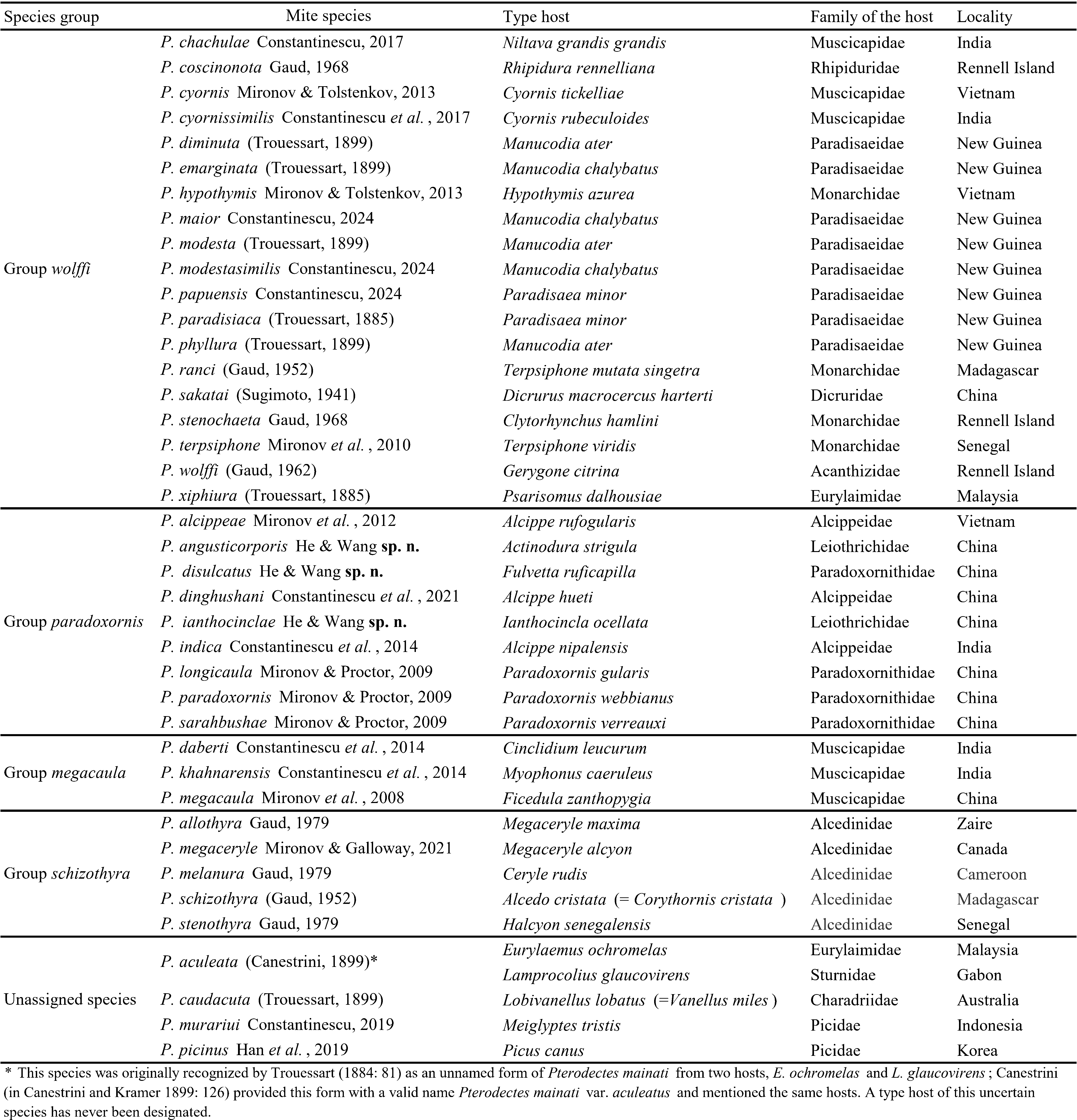
Feather mites of the genus Proterothrix Gaud, 1968 (Astigmata: Proctophyllodidae: Pterodectinae) have been found, to date, on birds of the orders Passeriformes, Coraciiformes, and Piciformes. According to the current taxonomic concept, the subfamily Pterodectinae is divided into the tribes Ramphocaulini and Pterodectini; in turn, the tribe Pterodectini is divided into the generic groups Pterodectes and Proterothrix (Mironov, 2009; Hernandes and Valim 2014). The genus Proterothrix belongs to the latter group and, is characterized by setae ps3 situated anteromesal to the adanal suckers in males and seta wa situated anterior to setae ra and la on tarsi Ⅰ and Ⅱ in both sexes (Mironov 2009; Mironov and Proctor 2009; Mironov and OConnor 2017; Constantinescu et al. 2017b, 2019). To date, the genus Proterothrix has included 37 species; of these, 33 species were arranged in four species groups: wolffi (19 species), paradoxornis (6 species), schizothyra (5 species) and megacaula (3 species) (Table 1). Representatives of the schizothyra group are restricted to kingfishers (Coraciiformes: Alcedinidae). Species of the wolffi group are associated with various passerine families (Passerifomes), while those of the megacaula group are only known from the passerine family Muscicapidae. (Gaud 1952, 1962, 1968, 1979; Park and Atyeo 1971; Mironov 2009; Mironov and Proctor 2009, 2023; Mironov et al. 2008, 2010, 2012, Mironov and Tolstenkov 2013; Constantinescu et al.2014, 2017a, 2017b, 2018, 2019, 2021, 2024; Han et al. 2019; Mironov and Galloway 2021). Mites of the schizothyra group have coxal fields III in males open, the terminal cleft in females with distinctly divergent margins (V-shaped); those of the megalcaula group are characterized by the absence of dorsal setae c1 in both sexes and an extremely long aedeagus in males (Constantinescu et al., 2014, 2021). The wolffi group is characterized by the coxal fields Ⅲ in males closed or with a very narrow gap in the anteromesal part and the terminal cleft in females usually parallel-sided (Mironov et al. 2008). In the wolffi group, Mironov and Proctor (2009) recognized the paradoxornis species complex, which is characterized by lanceolate seta e on tarsus Ⅰ in males. As recently proposed by Mironov and Galloway (2021), it is reasonable to treat this complex as a fourth species group in Proterothrix.

In this paper, we describe three new Proterothrix species belonging to the paradoxornis species group collected in China.
Materials and methods
The materials used in the present work were collected by Xiao-Ling Li in Yunnan Province, China, in April 2019 and by Shu-Xiang He and Li-Hua Sun in Sichuan Province, China, in September 2021. Avian hosts were captured by mist nets, identified and visually checked for the presence of mites. If mites were detected on feathers, they were brushed down by a small brush and put into a tube with 99% ethanol. Captured birds were released to the wild after the processing. In the laboratory, mite specimens were mounted on microscope slides in Hoyer's medium (Krantz and Walter 2009). Photos of mites were taken with a digital camera attached to the Olympus BX51 microscope. Drawings (line art) were made by importing photos into an APP called ''Paintwork» on ipad, creating a new layer on the photos, and using an Apple Pencil to trace the shape of mites and their morphological structures. General morphological terms and the legs chaetotaxy follow Gaud and Atyeo (1996), the idiosomal chaetotaxy is that of Griffiths et al. (1990) with modifications to coxal setae by Norton (1998). Measuring techniques of particular structures follow the descriptive method presented in previous papers on the genus Proterothrix (Mironov and Proctor 2009; Mironov and Galloway 2021). All measurements are in micrometers (µm). The taxonomic system and scientific names of birds mentioned in the paper follow Clements et al. (2022). Type materials of new species are deposited in the Chinese Academy of Sciences, Beijing, China (IOZ) and the College of Plant Protection, Southwest University, Chongqing, China (IESWU).
Taxonomy
Family Proctophyllodidae Trouessart & Mégnin, 1884
Subfamily Pterodectinae Park & Atyeo, 1971
Genus Proterothrix Gaud, 1968
Proterothrix angusticorporis He & Wang sp. n.
ZOOBANK: E1FAD665-A89A-41A6-BCCD-390A72BD1347 ![]()
(Figs. 1–4)
Type Material
Holotype — Male, male and 5 female paratypes from Chestnut-tailed Minla Actinodura strigula (Hodgson, 1837) (Leiothrichidae), China, Yunnan Province, Wuliangshan Nature Reserve, 24°89΄N, 100°32΄E, April 2019, collector Xiao-Ling Li.
Depository — Holotype and one paratype — Chinese Academy of Sciences; other paratypes — College of Plant Protection, Southwest University.
Description
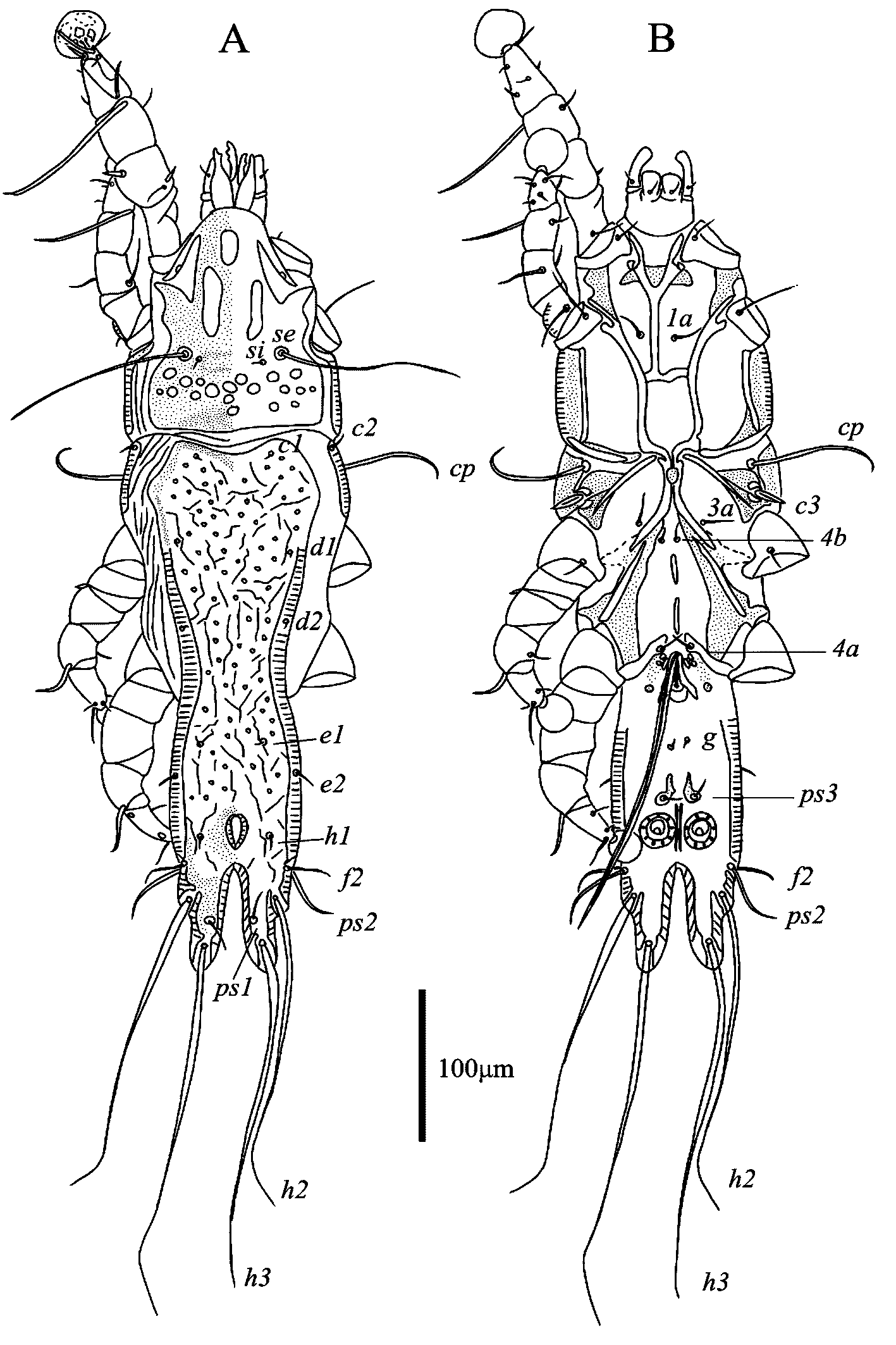
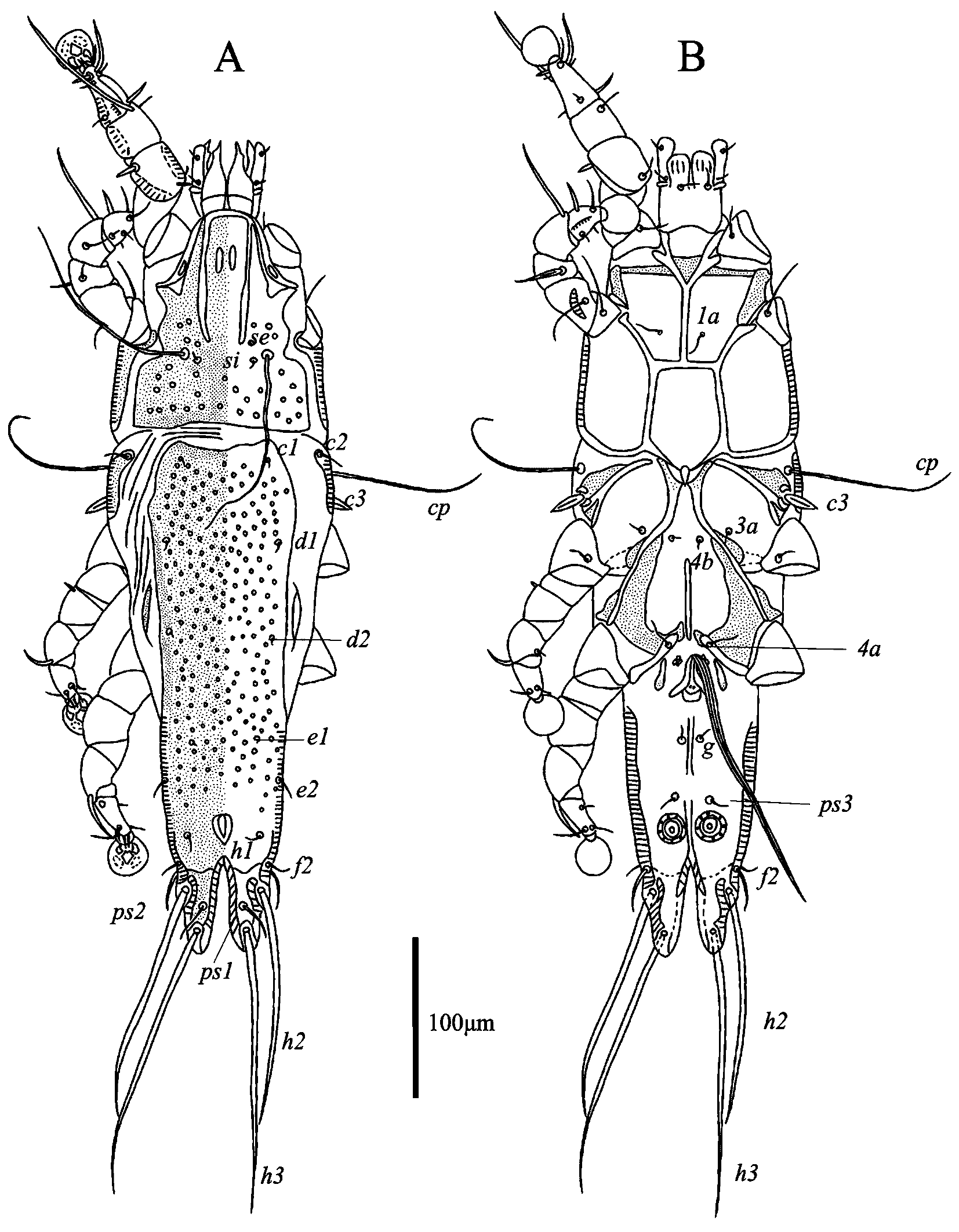
Male — (holotype, range for 4 paratypes in parentheses) (Figs. 1, 2). Length of idiosoma 518 (519–529), width 149 (148–155), length of hysterosoma 358 (355–368). Prodorsal shield: entire, anterolateral extensions acute, lateral margin slightly concave, posterior margin straight, length 152 (142–151), width 107 (110–123). Shield ornamentation: anterior half with large roughly ovate median lacuna and a pair of large strongly elongate lateral lacunae; posterior part with numerous circular and ovate lacunae. Distance between scapular setae se 63(63–68). Scapular shields narrow. Humeral shields narrow, fused with epimerites Ⅲ. Setae cp situated on soft tegument near ventral margin of humeral shield. Setae c2 situated on anterior margins of humeral shields. Subhumeral setae c3 lanceolate, 22 (22–23) ×8 (8–9). Hysteronotal shield: anterior margin slightly concave and sclerotized stronger than median part of the shield; surface with sparsely distributed small circular lacunae and numerous wavy striae of different length; greatest length from anterior margin to the base of setae h3 351 (333–377), width in anterior part 111 (105–109). Distance between prodorsal shield and hysteronotal shield 15 (16–22). Lateral hysteronotal shield absent. Opisthosomal lobes elongate, without membranes, lobar apices semi-ovate, lateral margins of lobes with short rounded extensions bearing setae h2, setae h3 situated near lobar apices. Terminal cleft narrow U–shaped, length 69 (72–74), width at the level of setae h3 11 (10–12). Supranal concavity clearly outlined, shaped as inverted teardrop, separated from terminal cleft. Setae h1 situated at midlevel of supranal concavity, closer to level of setae f2 than of setae e2. Setae h3 represented by macrosetae, length 264 (201–263), greatest width 5 (4–6). Setae h2 shorter than h3, length 246 (152–229), width 7 (5–8). Setae ps1 filiform, 29 (23–33) long, situated at midlevel between setae h2 and h3, slightly closer to inner margin of opisthosomal lobes than to outer margin. Setae ps2 setiform, 44 (30–39) long, setae f2 situated at level of setae ps2 (Fig. 1A). Dorsal measurements: c2–d2 122 (199–124), d2–e2 107 (105–112), e2–h3 113 (111–117), d1–d2 56 (59–65), e1–e2 22 (25–32), h1–ps2 19 (18–23), h2–h2 59 (53–62), h3–h3 40 (31–44), ps2–ps2 72 (66–74).
Epimerites Ⅰ fused into a Y, posterior end of sternum connected with middle parts of epimerites Ⅱ by transverse sclerotized bands. Epimerites Ⅱ long, their posterior ends almost touching each other and fused with corresponding anterior ends of epimerites Ⅲa forming a pentagonal structure in median area of propodosoma (Fig. 1B). Coxal fields Ⅰ closed, coxal fields Ⅱ, Ⅲ almost closed, with narrow gap between tips of corresponding epimerites Ⅱ, Ⅱa and epimerites ⅡI, Ⅲa. Anterior parts of epimerites Ⅲa fused with posterior ends of epimerites II encircle small drop-shaped sclerotized area. Epimerites Ⅳa well developed, seate 4a situated on their inner tips. Coxal fields Ⅳ with triangular sclerotized areas connecting epimerites IV and bases of epimerites Ⅳa. Pregenital sclerite stick-shaped and usually divided into two parts, posterior part not connected with inner tips of epimerites Ⅳa, distance between anterior and posterior ends of pregenital sclerite 55 (55–60). Genital arch of moderate size, length 25 (23–24), width 25 (24–28). Paragenital sclerites present; their poorly sclerotized anterior areas connected with epimerites IVa. Aedeagus long whip-shaped, directed anterior from genital arch, immediately bent backward and extends to midlength between setae h2 and h3, length from bend to apex 206 (208–217). Genital papillae anterolateral to genital arch, their bases contiguous. Adanal suckers 20 (21–23) in diameter, corolla with 8–9 denticles. Setae ps3 situated on comma-shaped adanal shields. (Fig. 1B). Ventral measurements: 3a–4b 10 (7–9), 4b–4a 73 (70–76), 4a–g 66 (62–73), g–ps3 36 (36–41), ps3–ps3 20 (19–23), ps3–h3 98 (98–100).
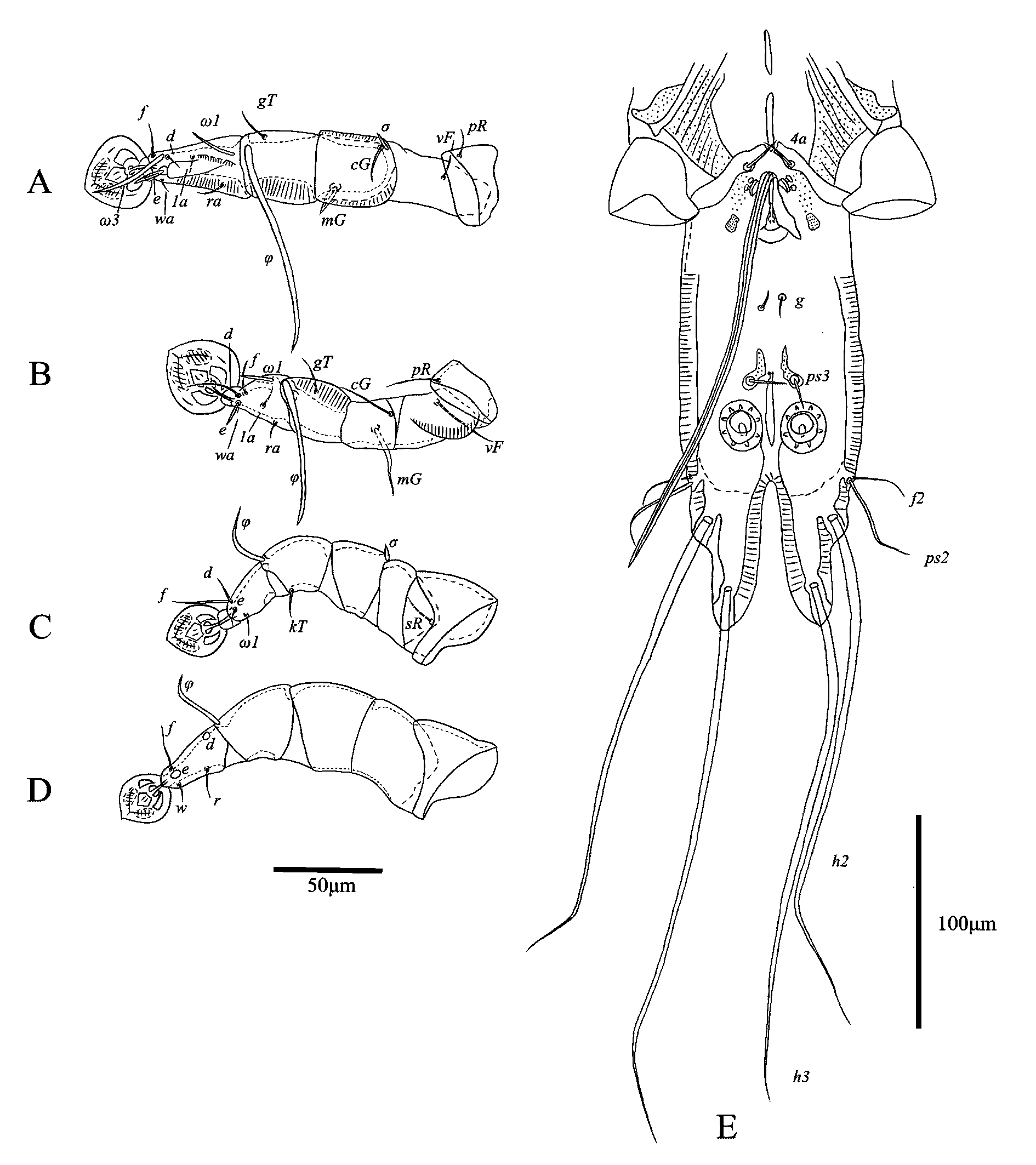
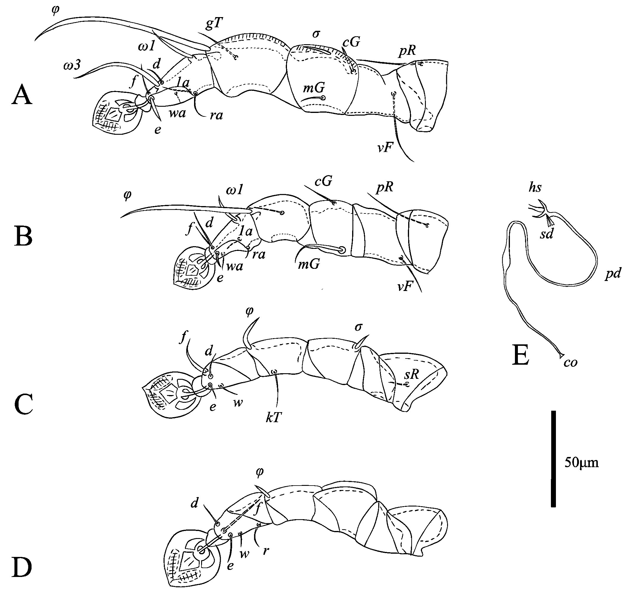
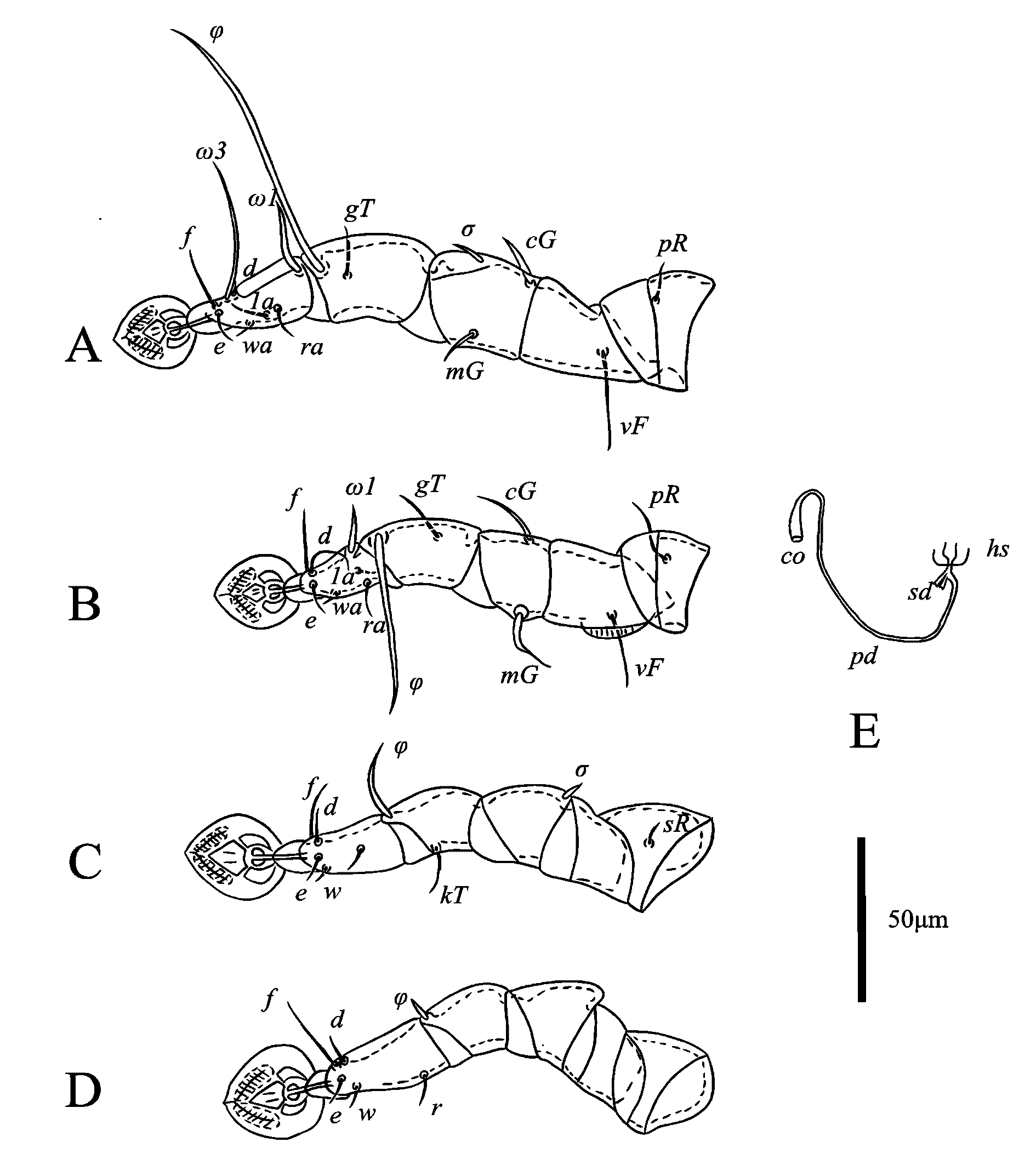
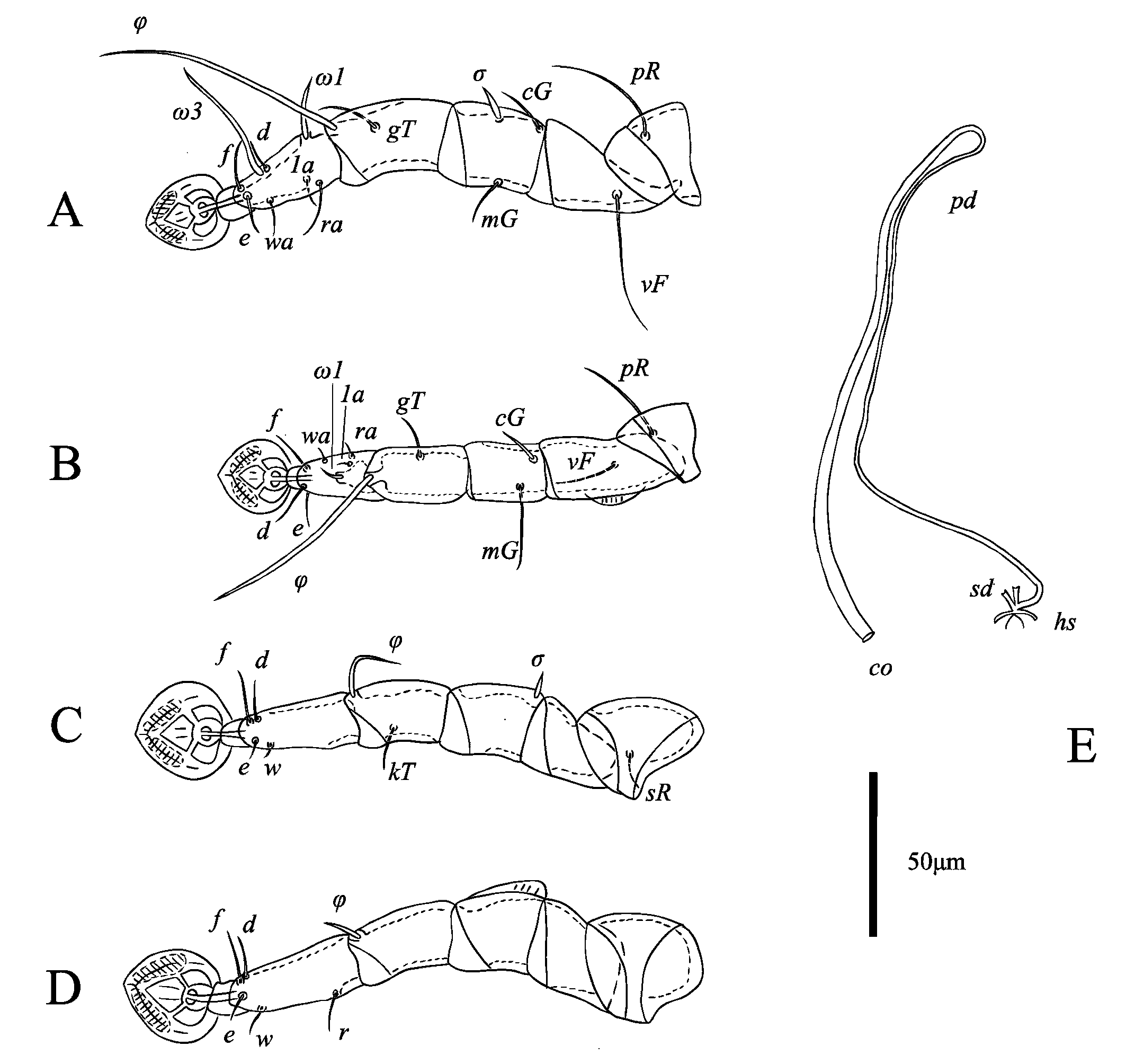
Legs Ⅰ longer and thicker than legs Ⅱ. Femur Ⅱ with ventral crest. Genu Ⅰ with dorsal and lateral longitudinal crests. Tarsus and tibia Ⅰ with lateral longitudinal crest. Tarsus Ⅱ with lateral longitudinal crest. Tarsus Ⅰ with thumb-like dorsal process. Solenidion σ of genu Ⅰ stick-like, 8 (7–11) long, situated at base of this segment. Setae cG Ⅰ, cG Ⅱ setiform, setae cG Ⅱ much longer than setae cG Ⅰ. Setea mG Ⅰ spiniform, 14 (12–15) long; setae mG Ⅱ thickened, with filiform apex 34 (32–34) long. Setae e of tarsus Ⅰ lanceolate, 15 (12–18) long. Setae d, f of tarsus Ⅱ subequal in length (Fig. 2A, B). Seta f of tarsus Ⅲ approximately 2.5 times longer than seta d. Leg Ⅲ, Ⅳ similar in size. Tarsus Ⅳ 37 (36–38) long, without claw-like apical process (except one of paratypes with apical claw); setae d, e button-like, situated in basal and apical parts of this segement, respectively (Fig. 2C, D). Lengths of solenidia: ω1Ⅰ 21 (15–18), ω1Ⅱ 13 (15–25), φⅠ 101 (101–111), φⅡ 66 (58–68), φⅢ 29 (23–29), φⅣ 29 (30–31).
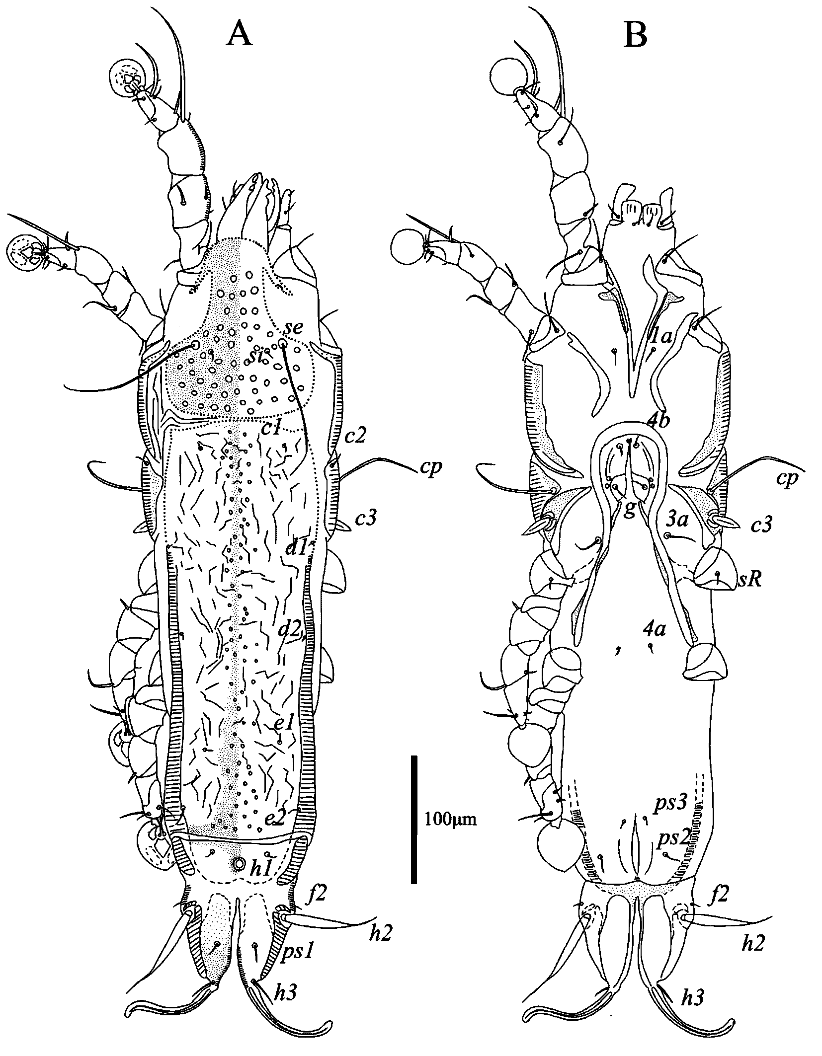
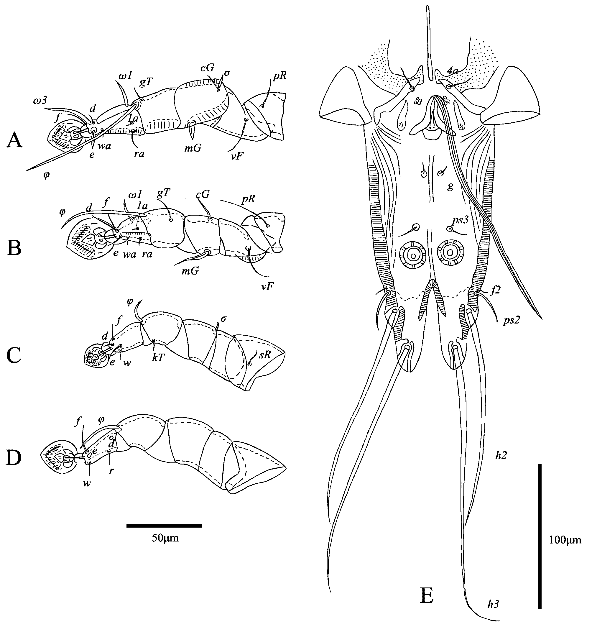
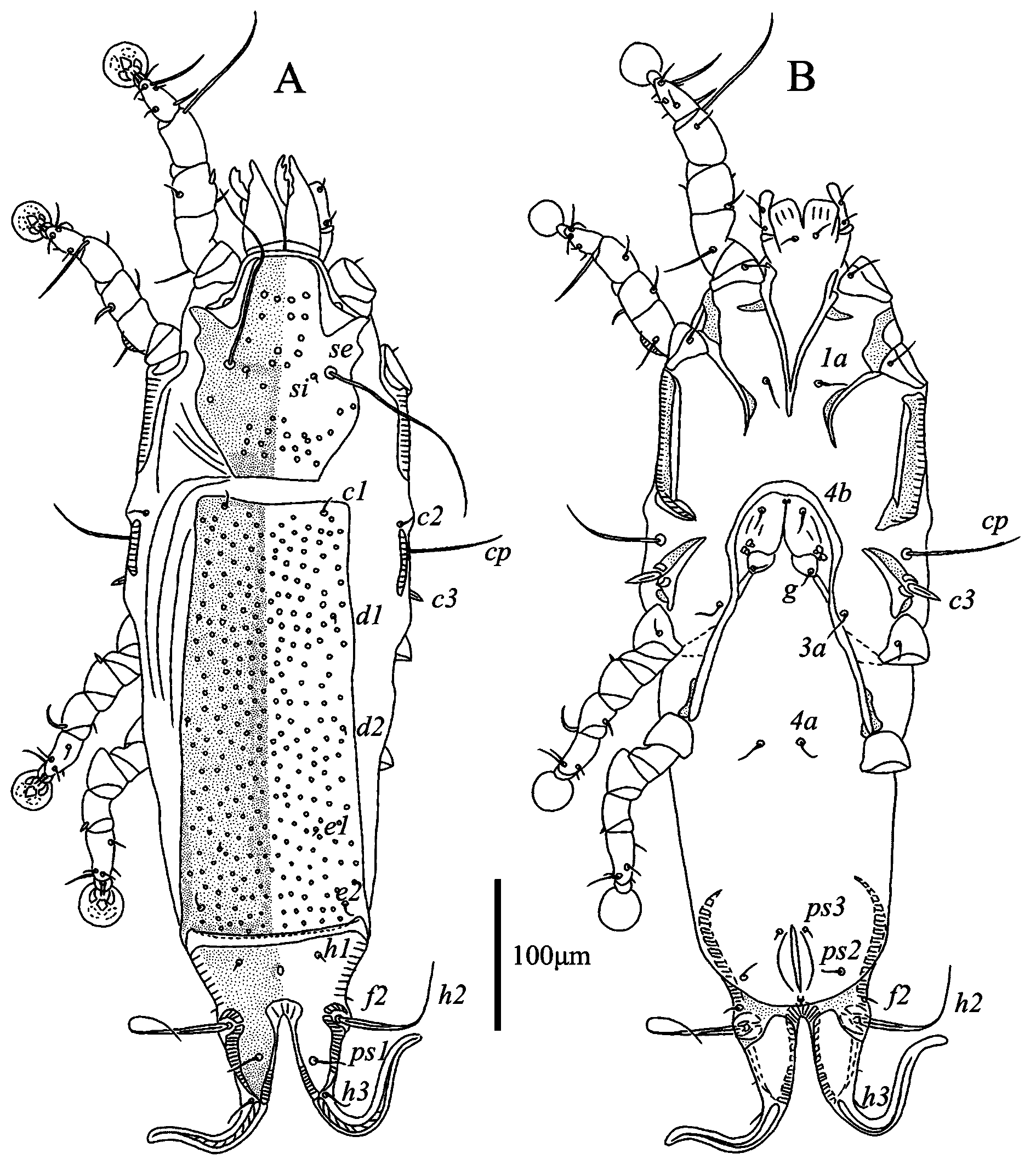
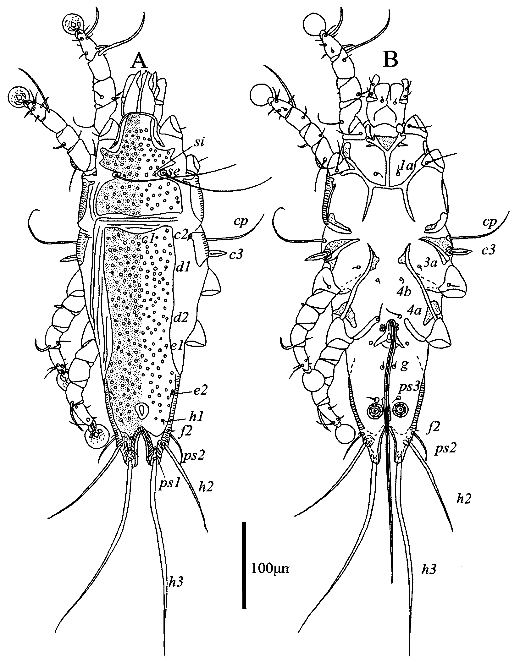
Female — (range for 6 paratypes) (Figs. 3, 4). Idiosoma length 539–588, width 140–176, hysterosoma length 371–421. Prodosal shield: entire, without clearly outlined border, without antrolateral extensions; most surface except anterior end with numerous circular and ovate lacunae; length146–163, width 105–131. Scapular setae se separated by 66–79. Scapular shield narrow. Humeral shields narrow, connected by posterior ends to epimerites Ⅲ. Setae cp situated on ventral margin of humeral shield, setae c2 situated on anterior end of humeral shield. Setae c3 lanceolate, 23–24 × 8–9. Distance between prodorsal and hysteronotal shields 1–4. Hysteronotal shield separated into anterior and lobar parts by narrow transverse band of soft tegument. Anterior hysteronotal shield: anterior margin and lateral margins up to level of trochanters III poorly outlined, narrow median area strongly sclerotized and with numerous small circular lacunae, remaining surface with irregular sparsely disposed striae, lateral areas are poorly sclerotized like soft tegument, length 305–103, width at anterior margin 146–163. Length of lobar region 104–117, width at level of setae h2 84–88. Terminal cleft shaped as a narrowly V, length 62–74, and greatest width at level of setae h3 12–20. Supranal concavity circular, clearly outlined. Setae h1 situated on lobar shield anterior to supranal concavity. Setae h2 spindle-shaped, with short terminal filaments, 92–114 × 8–10. Setae ps1 close to inner margin of opisthosomal lobes, approximately equidistant from levels of setae h2 and h3. Setae h3 19–26 long, about 1/3 the total length of terminal appendages (Fig. 3A). Dorsal measurements: c2–d2 129–149, d2–e2 111–133, e2–h2 78–83, h2–h3 45–55, d1–d2 53–78, e1–e2 46–53, h1–h2 43–50, h2–ps1 17–23, h1–h1 40–44, h2–h2 65–70.
Epimerites Ⅰ fused as a short-stemmed Y. Coxal fields Ⅰ, Ⅱ without heavily sclerotized areas. Epimerites Ⅳa absent. Translobar apodemes of opisthosomal lobes present, fused to each other anterior to terminal cleft. Epigynum horseshoe-shaped, greatest wide 58–66 (Fig. 3B). Primary spermaduct with spindle-shaped enlargement in distal one third; secondary spermaducts short (Fig. 4E). Distances between pseudanal setae: ps2–ps2 47–64, ps3–ps3 16–21, ps2–ps3 19–28.
Legs Ⅰ thicker and longer than legs II. Genu, tibia Ⅰ with lateral longitudinal crest. Setae cGⅠ spiculiform, setae mGⅡ filiform with thickened basal part; setae cG Ⅱ and setae mG Ⅰ filiform, former longer than latter. Setae d of tarsus Ⅱ half the length of corresponding setae f (Fig. 4A, B). Setae f of tarsi Ⅲ, IV are 2 times longer than setae d. Genu Ⅳ slightly inflated dorsally, with narrow longitudinal crest (Fig. 4C, D). Solenidion φ of tibia Ⅳ much shorter than on tibia Ⅲ. Lengths of solenidia: ω1Ⅰ 13–21, ω1Ⅱ 10–16, φⅠ 65–96, φⅡ 63–72, φⅢ 19–29, φⅣ 6–8.
Etymology
The specific name is a contraction of angusus (L., narrow) and corporis (L., body) and refers to the narrow idiosoma in both sexes.
Differential diagnosis
The new species Proterothrix angusticorporis sp. n. belongs to the paradoxornis species group in having, in males, seta e of tarsus Ⅰ lanceolate. Among six previously known species of this group, the new species is most similar to P. sarabushae Mironov & Proctor, 2009 described from the Golden Parrotbill Suthora verreauxi (Sharpe, 1883) (Paradoxornithidae) in having the following characteristics in males: the scapular shields have hook-like anteromesal extension, the aedeagus almost extends to the level of lobar apices; tarsus Ⅰ has a thumb-like dorsal process. Proterothrix angusticorporis sp. n. differs from P. sarabushae by the following features: in males, the anterior half of prodorsal shield has one ovate median lacunae and a pair of long slit-like lacunae, the posterior ends of epimerites Ⅱ are not fused with the anterior tips of epimerites Ⅲa, the posterior end of pregenital sclerite is not connected with the inner tips of epimerites Ⅳa, and one pair of adanal shields is present; in females, the idiosoma length is 539–588; the anterior margin of the anterior hysteronotal shield in convex, the median area of this shield is heavily sclerotized and bears small circular lacunae, the lateral areas are poorly sclerotized like the soft tegument, and the posterior margin of the anterior hysteronotal shield is almost straight. In males of P. sarabushae, the prodorsal shield has numerous small circular lacunae, which are arranged in transverse rows in the anterior part of this shield; the posterior ends of epimerites Ⅱ are fused with anterior tips of epimerites Ⅲa, the posterior end of pregenital sclerite is connected with the inner tips of epimerites Ⅳa; and two pairs of adanal shields are present; in females, the idiosoma length is 422–482; the anterior margin of the anterior hysteronotal shield is straight, the entire surface of this shield is uniformly sclerotized and bears numerous small circular lacunae; the posterior margin has a pair of incisions extending beyond the level of setae e2 (Mironov and Proctor, 2009).
Proterothrix disulcatus He & Wang sp. n.
ZOOBANK: 41F7CE5A-1C5C-4D6E-A16F-9073341D732F ![]()
(Figs. 5–8)
Type Material
Holotype — Male , 4 male and 5 female paratypes from the Spectacled Fulvetta, Fulvetta ruficapilla (Verreaux, J, 1871) (Paradoxornithidae), China, Sichuan Province, Tangjiahe Nature Reserve, 32°60΄N, 104°69΄E, 25 September 2021, collector Shu-Xiang He and Li-Hua Sun.
Depository — Holotype and one paratypes — Chinese Academy of Sciences, other paratypes — College of Plant Protection, Southwest University.
Description
Male — (holotype, range for 3 paratypes in parentheses) (Figs. 5, 6). Length of idiosoma 472 (470–478), width 149 (150–156), length of hysterosoma 326 (322–336). Prodorsal shield: entire, anterolateral extensions acute, lateral margin shallowly concave, posterior margin slightly concave, length 137 (139–144), width 115 (117–121); anterior half with a pair of long and narrowly lateral lacunae and with a pair or one small longitudinal median lacuna, posterior half with numerous small circular lacunae. Distance between scapular setae se 55 (54–60). Scapular shield narrow, with hook-like anteromesal extensions. Humeral shield narrow, separated from epimerites Ⅲ. Setae c2 situated on anterior ends of humeral shields. Setae cp situated on soft tegument near ventral margins of humeral shields. Subhumeral setae c3 lanceolate, 23 (21–22) × 7 (6–7). Hysteronotal shield with anterior margin slightly concave, surface with numerous small circular lacunae, greatest length 326 (322–336), greatest width 98 (98–99). Distance between prodorsal shield and hysteronotal shield 16 (10–17). Lateral hysteronotal sclerites present, situated silghly anterior to level of trochanter Ⅳ. Opishosomal lobes elongated (approximately two times longer than wide), without membranes, lobar apices rounded and poorly sclerotized; lateral margins of lobes with convexities bearing setae h2, setae h3 situated near lobar apices. Terminal cleft V-shaped, length 57 (59–62), width at level of setae h3 10 (9–11). Supranal concavity clearly outlined, shaped as inverted teardrop. Setae h1 at level of supranal concavity, closer to level of setae f2 than of setae e2. Setae h3 represented by macrosetae, length 207 (188–207), width 4 (3–4). Setae h2 wider and shorter than setae h3, length178 (178–181), width 5 (5–6). Setae ps1 spiculiform, length 21 (20–23), situated at midlevel between setae h2 and h3, slightly closer to inner margin than to outer margin of opisthosomal lobes. Setae ps2 setiform, length 31 (29–31) (Fig. 5A). Dorsal measurement: c2–d2 114 (107–119), d2–e2 90 (89–101), e2–h3 95 (97–102), d1–d2 61 (56–61), e1–e2 24 (24–27), h1–ps2 19 (18–20), h2–h2 49 (49–54), h3–h3 32 (33–35), ps2–ps2 59 (59–65).
Epimerites Ⅰ fused into a Y with long stem, posterior end of stem connected with middle part of epimerites Ⅱ by transverse sclerotized bands. Epimerites Ⅱ stronly elongated, their posterior tips almost fused to each other forming a closed pentagonal structure in median area of propodosoma; posterior ends of epimetrites II fused with corresponding epimerites IIa and almost touching inner tips of epimerites Ⅲ and Ⅲa (Fig. 5B). Coxal fields I, II closed, coxal fields III with small gap between tips of epimerites III and IIIa. Epimerites Ⅳa well developed, setae 4a situated on their inner tips. Lateral areas of coxal fields Ⅳ between corresponding epimerites IV and IVa heavily sclerotized. Pregenital sclerite present, long stick-like, not connected with inner tips of epimerites Ⅳa, length 53 (49–55), width 2 (2–3). Genital arch of moderate size, length 19 (16–22), width 29 (28–30). Aedeagus long whip-shaped, directed anteriorly from genital arch and immediately bent backward, not extending to apices of opisthosomal lobes, length from bend to apex 189 (177–186). Genital papillae anterolateral to genital arch, their bases contiguous. Paragenital apodemes shaped as small longitudinal sclerites lateral to genital arch. Adanal suckers 17 (17–18) in diameter; corolla with 7–8 denticles. Setae ps3 situated anterior to adanal suckers (Fig. 6E). Ventral measurements: 3a–4b 6 (7–5), 4b–4a 66 (66–76), 4a–g 59 (58–61), g–ps3 37 (38–41), ps3–ps3 23 (24–25), ps3–h3 82 (83–87).
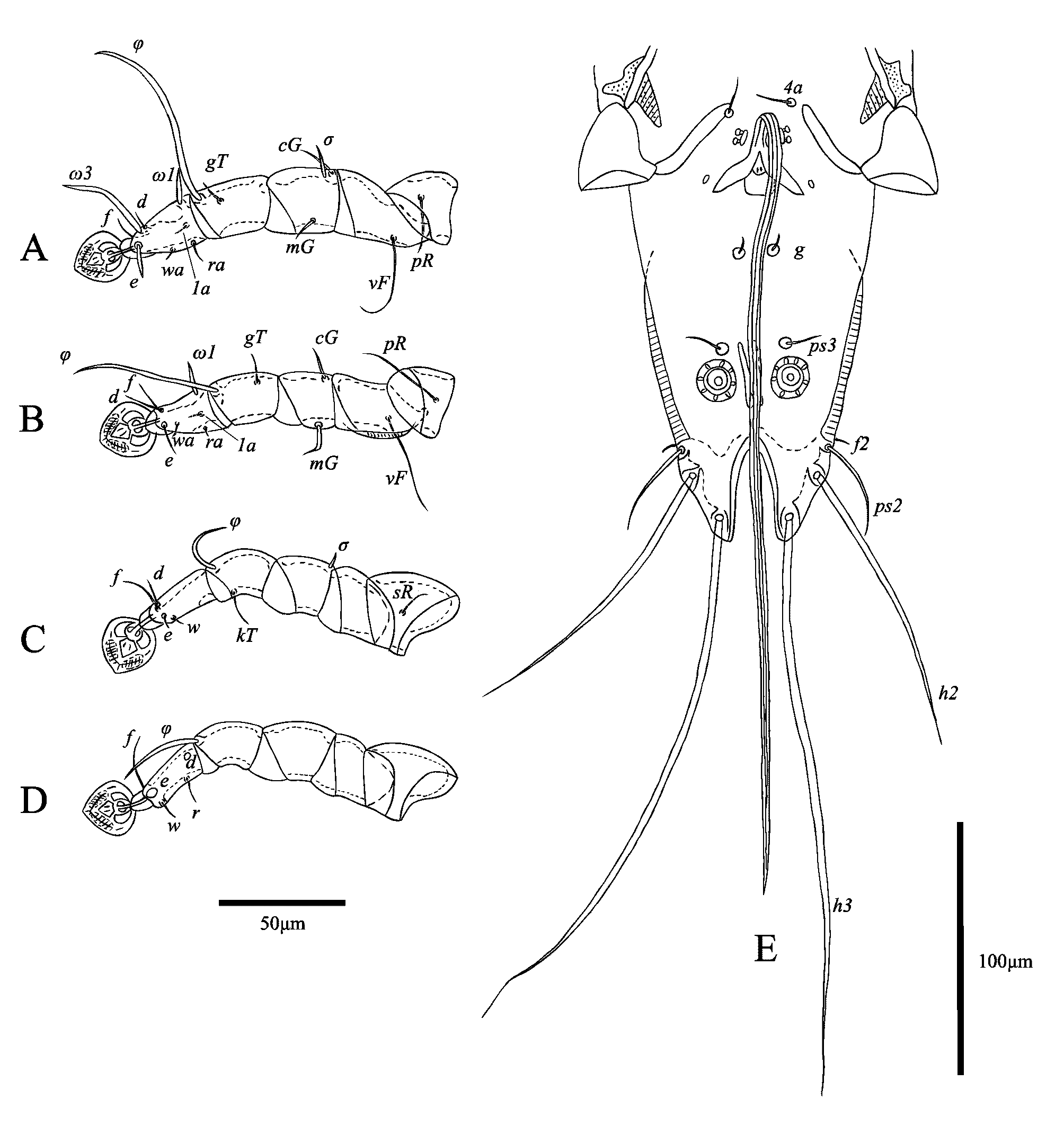
Legs Ⅰ longer and thicker than legs Ⅱ. Femur Ⅱ with ventral crest. Genu Ⅰ with dorsal and lateral longitudinal crest. Tarsus Ⅰ with thumb-like dorsal process. Tarsus Ⅱ with narrow lateral crest. Solenidion σ of genu Ⅰ stick-like, 9 (7–9) long, situated at base of segment. Setae cGⅠ, cGⅡ filiform, setae cGⅡ longer than setae cGⅠ. Setae mGⅠ lanceolate, 25 (21–30) long; setae mGⅡ thickened, with filiform terminal part, 53 (49–55) long. Setae e of tarsus Ⅰ lanceolate, 14 (12–17) long (Fig. 6A, B). Seta d of tarsus Ⅱ slightly longer than corresponding seta f. Sete d of tarsus Ⅲ shorter than corresponding seta f. Leg Ⅲ, Ⅳ similar in size. Tarsus Ⅳ long 28 (27–30), without apical claw, with small apicoventral extension bearing seta w. Setae d, e button-like, situated in basal and apical parts of this segment, respectively (Fig. 6D). Lengths of solenidia: ω1Ⅰ 16 (15–16), ω1Ⅱ 14 (11–13), φⅠ 89 (84–90), φⅡ 55 (50–56), φⅢ 16 (23–25), φⅣ 30 (22–29).
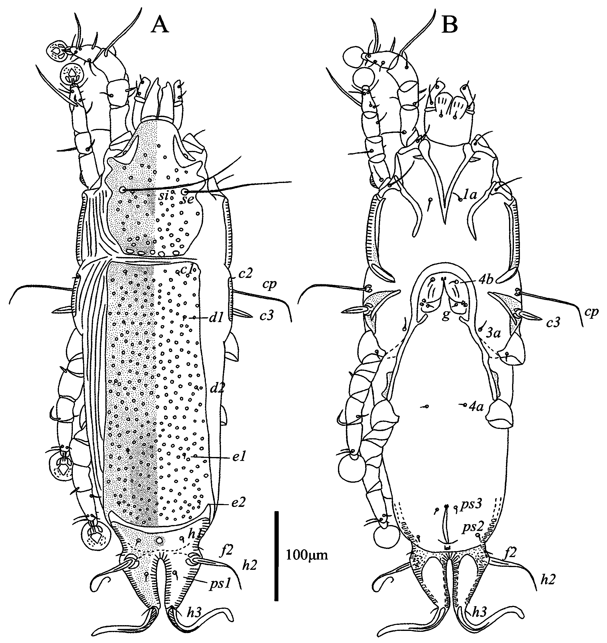
Female — (range for 5 paratypes) (Figs. 7, 8). Idiosoma length 540–557, width 176–188, hysterosoma length 377–385. Prodorsal shield: entire, anterolateral extensions acute, lateral margins without incisions, posterior half strongly attenuate posteriorly, posterior angles not expressed, posterior margin straight; anterior half with numerous medium-sized circular lacunae, posterior half with numerous small circular lacunae, length 140–150, width of posterior part 104–114. Distance between scapular setae se 63–73. Scapular shields narrow, with hook-like anteromesal extensions. Humeral shields well developed, separated from outer sclerotization of epimerites Ⅲ. Setae cp situated on soft tegument near ventral margins of humeral shields. Setae c2 situated dorsally on soft tegument near anterior ends of humeral shields. Subhumeral setae c3 lanceolate, 19–22 × 7–8. Distance between prodorsal and hysteronotal shields 13–18. Hysteronotal shield separated into anterior and lobar parts by narrow band of soft tegument. Anterior hysteronotal shield: anterior margin slightly concave, surface with small circular lacunae, median area with narrow heavily sclerotized band, length 282–291, width at anterior margin 100–103. Length of lobar region 104–107, width at level of setae h2 89–91. Terminal cleft V-shaped, anterior end rounded, length 54–61, greatest width at level of setae h3 23–29. Supranal concavity clearly outlined, circular. Setae h1 situated on lobar shield slightly anterior to level of supranal concavity. Setae h2 spindle-shaped, with terminal filaments, 82–95 × 6–8. Setae ps1 close to inner margin of opisthosomal lobes, approximately equidistant from levels of setae h2 and h3. Setae h3 18–20 long, about 1/6th the length of terminal appendages (Fig. 7A). Dorsal measurements: c2–d2 122–136, d2–e2 114–127, e2–h2 69–76, h2–h3 49–54, d1–d2 63–65, e1–e2 40–48,h1–h2 37–41, h2–ps1 21–24, h1–h1 50–51, h2–h2 70–72.
Epimerites Ⅰ fused as a short-stemmed Y. Coxal field Ⅰ, Ⅱ without heavily sclerotized areas. Epimerites Ⅳa absent. Translobar apodemes of opisthosomal lobes present, fused to each other anterior to terminal cleft. Epigynum horseshoe-shaped, greatest wide 72–78 (Fig. 8). Most part of primary spermaduct narrow, distal 1/7–1/8 of primary spermaduct enlarged forming bursa copulatrix; secondary spermaducts short; copulatory opening situated ventrally at anterior margin of fused translobar apodemes (Fig. 8E). Distances between pseudanal setae: ps2–ps2 59–71, ps3–ps3 18–21, ps2–ps3 28–34.
Legs Ⅰ slightly thicker and longer than legs Ⅱ. Femur Ⅱ with wide ventral crest. Solenidion σ of genu Ⅰ 7–10 long, situated closer to distal margin of this segment. Setae cGⅠ, cGⅡ, mGⅠ setiform; mGⅡ strongly thickened in basal part, 14–36 long. Seta e of tarsus Ⅰ short filiform (Fig. 8A, B). Setae d, f of tarsus II subequal in length. Setae d of tarsi Ⅲ, IV much shorter than corresponding setae f. Genu Ⅳ slightly inflated dorsally, with narrow longitudinal crest. Solenidion φ of tibia Ⅳ much shorter than on tibia Ⅲ (Fig. 8D). Lengths of solenidia: ω1Ⅰ 17–19, ω1Ⅱ 11–13, φⅠ 80–83, φⅡ 56–59, φⅢ 18–23, φⅣ 5–7.
Etymology
The specific name disulcatus is a contraction of dis (Gr., two, twice) and sulcus (L., groove) and refers to two long and narrow lacunae on the anterior half of the prodorsal shield in male.
Differential diagnosis
The new species, Proterothrix disulcatus sp. n. belongs to the paradoxornis species group due to having lanceolate seta e of tarsus Ⅰ in males. Among six previously known species of this group, the new species is most similar to P. sarabushae in having the following characteristics: in males, the scapular shields have hook-like anteromesal extensions, the lateral hysteronotal sclerites are present, the aedeagus almost extends to the level of lobar apices, tarsus Ⅰ has a thumb-like dorsal process, and setae mGⅠ are lanceolate; in females, setae mGⅡ are narrowly lanceolate. Proterothrix disulcatus sp. n. differs from P. sarabushae in having the following features: in males, the anterior half of the prodorsal shield has two long and narrow lateral lacunae and a pair or one short and narrow median lacuna, the pregenital sclerite is not connected with the inner tips of epimerites Ⅳa, the adanal shields are absent, and thickened setae mGⅡ have filiform apical part; in females, setae c2 are situated on soft tegument, the posterior margin of the anterior hysteronotal shield have very short incisions not extending to setae e2, and setae mGⅠ are filiform. In males of P. sarabushae, the anterior part of the prodorsal shield has large circular lacunae arranged in several transverse rows, the pregenital sclerite is connected with the inner tips of epimerites Ⅳa, two pairs of adanal shields are present, and setae mGⅡ are simple spiniform; in females, setae c2 are situated in the anteromesal angles of humeral shields, the posterior margin of the anterior hysteronotal shield has a pair of narrow incisions extending beyond the level of setae e2, and setae mGI are spiniform.
Proterothrix ianthocinclae sp. n.
ZOOBANK: 4DA2A498-3B4B-472D-B9CC-CD3B1BC7D4A9 ![]()
(Figs. 9–12)
Type Material
Holotype — Male holotype, 4 male and 5 female paratypes from Spotted Laughingthrush, Ianthocincla ocellata (Vigors, 1831) (Leiothrichidae), China, Sichuan Province, Tangjiahe Nature Reserve, 32°60΄N, 104°69΄E, 25 September 2021, collectors Shu-Xiang He and Li-Hua Sun.
Depository. Holotype and one paratypes — Chinese Academy of Sciences, other paratypes — College of Plant Protection, Southwest University.
Description
Male — (holotype, range for 4 paratypes in parentheses) (Figs. 9, 10). Length of idiosoma 406 (405–423), width 151 (141–146), length of hysterosoma 277 (272–288). Prodorsal shield: entire, anterolateral extensions acute, lateral margins with deep incisions encircling bases of setae se, posterior margin slightly convex medially, length 119 (119–129), width 102 (99–108), surface with numerous small circular and ovate lacunae. Distance between scapular setae se 52(48–53). Scapular shields narrow. Humeral shields narrow, separated from epimerites Ⅲ. Setae cp situated on ventral margin of humeral shields. Setae c2 situated on anterior end of humeral shields. Subhumeral setae c3 lanceolate, 23 (21–25) ×7 (7). Hysteronotal shield with anterior margin strongly concave, length from anterior margin to setae h3 on lobar apices 281 (276–290), greatest width 78 (80–88), surface with numerous small circular lacunae. Distance between prodorsal shield and hysteronotal shield 22 (14–19). Lateral hysteronotal shields absent. Opisthosomal lobes elongated, attenuate apically, lobar apices rounded, setae h3 situated near apices. Terminal cleft narrowly V-shaped, with membranous edges, length 43(39–45), width at the level of h3 17(12–19). Supranal concavity clearly outlined, shaped as inverted teardrop, not connect with terminal cleft. Setae h1 posterior to level of supranal concavity, much closer to level of setae f2 than of setae e2. Setae h3 represented by macrosetae, length 231 (215–240), width 3 (2–5). Setae h2 shorter than h3, length 126 (131–158), width 4 (3–4). Setae ps1 filiform, length 13 (8–10), situated closer to inner margins of opisthosomal lobes than outer ones, approximately equidistant from levels of setae h3 and h2. Setae ps2 setiform, close to setae f2, length 38 (32–44) (Fig. 9A). Dorsal measurements: c2–d2 97 (99–102), d2–e2 90 (90–97), e2–h3 69 (62–77), d1–d2 62 (50–61), e1–e2 26 (28–36), h1–ps2 19 (18–20), h2–h2 50 (47–52), h3–h3 28 (24–29), ps2–ps2 59 (58–65).
Epimerites Ⅰ fused into a Y with long stem, posterior end of stem connected with middle parts of epimerites Ⅱ by transverse sclerotized bands. Epimerites Ⅱ long, extending to sejugal area, posterior ends free. Epimerites Ⅲa elongated, extending to level of inner tips of epimerites Ⅲ. Coxal fields Ⅰ closed, coxal fields Ⅱ, Ⅲ open, without wide sclerotized areas. Rudimentary sclerites rEpⅡa absent. Epimerites Ⅳa well developed, setae 4a situated near or on their inner tips. Coxal fields Ⅳ with triangle-shaped sclerotized area at bases of trochanters Ⅳ (Fig. 9B). Pregenital sclerite absent. Genital arch of moderate size, length 24 (23–29), width 35 (32–34). Aedeagus long whip-shaped, directed anteriorly from genital arch, immediately bent backward and extend beyond lobar apices approximately by distal half, length from bend to apex 319 (318–324). Genital papillae anterolateral to genital arch, their bases contiguous. Adanal suckers 17 (15–17) in diameter, corolla with 7–8 denticles. Adanal shields absent, ps3 situated anterior to adanal suckers (Fig. 10E). Ventral measurements: 3a–4b 13 (7–15), 4b–4a 59 (60–61), 4a–g 56 (52–59), g–ps3 37 (36–38), ps3–ps3 24 (26–27), ps3–h3 68 (66–73).
Legs Ⅰ longer and thicker than legs Ⅱ; femur Ⅱ with ventral crest, Solenidion σ of genu Ⅰ stick-like, 10 (7–8) long, situated at base of segment. Setae cG Ⅰ, cG Ⅱ setiform; seta mGⅡ thickened with filiform apex, 14 (14–18) long, seta mG Ⅰ spiculiform, 11 (10–12) long; seta cG Ⅱ longer than seta cG Ⅰ. Seta e of tarsus Ⅰ thick spiniform, 15 (9–17) long. Setae d, f of tarsus Ⅱ subequal in length (Fig. 10A, B). Setae f of tarsus Ⅲ approximately 2 times longer than setae d. Leg Ⅲ, Ⅳ similar in size. Tarsus Ⅳ 28 (26–30) long, without apical claw, with a small apicoventral extension bearing seta w; setae d, e button-like, situated in basal and apical parts of this segment, respectively (Fig. 10D). Lengths of solenidia: ω1Ⅰ 13 (10–12), ω1Ⅱ 10 (7–11), φⅠ 79 (72–81), φⅡ 57 (54–59), φⅢ 22 (21–27), φⅣ 30 (24–28).
Female — (range for 5 paratypes) (Figs. 11, 12). Idiosoma length 535–580, width176–189, hysterosoma length 368–409. Prodorsal shield: entire, anterolateral extensions acute, lateral margin without incisions, posterior half slightly narrowed posteriorly, posterior angles not express, posterior margin straight; surface with numerous small circular lacunae and with a row of larger ovate lacunae at posterior margin; length149–157, width 104–114. Distance between scapular setae se 66–73. Scapular shields narrow. Humeral shields narrow, connected with epimerites Ⅲ. Setae cp situated on soft tegument near ventral margin of humeral shield. Setae c2 situated dorsally on anterior ends of humeral shields. Subhumeral setae c3 lanceolate, 21–26 × 7–8. Distance between dorsal and hysteronotal shield 8–11. Hysteronotal shield split into anterior and lobar parts by narrow band of soft tegument. Anterior hysteronotal shield: anterior margin slightly concave, entire surface with small circular and ovate lacunae, length 291–311, width 102–110. Length of lobar region 102–110, width at level of setae h2 90–102. Terminal cleft narrow U-shaped, length 52–61, greatest width at level of setae h3 8–14. Supranal concavity clearly outlined, circular. Setae h1 situated on lobar shield at level of supranal concavity. Setae h2 spindle-shaped, with terminal filaments, 67–91 × 7–9. Setae ps1 close to inner margin of opisthosomal lobes, approximately equidistant from levels of setae h2 and h3. Setae h3 24–42 long, about 1/3 the length of terminal appendages (Fig. 11A). Dorsal measurements: c2–d2 121–134, d2–e2 123–138, e2–h2 64–78, h2–h3 51–58, d1–d2 61–72, e1–e2 49–58, h1–h2 25–34, h2–ps1 25–28, h1–h1 47–57, h2–h2 67–81.
Epimerites Ⅰ fused in a short-stemmed Y. Coxal field Ⅰ, Ⅱ without heavily sclerotized areas. Epimerites Ⅳa absent. Translobar apodemes of opisthosomal lobes present, fused to each other anterior to terminal cleft. Epigynum horseshoe-shaped, greatest wide 72–79 (Fig. 11B). Proximal half of primary spermaduct narrow, distal half approximately 3 times wider than proximal half; secondary spermaducts short; copulatory opening ventral, situated at anterior margin of translobar apodeme (Fig. 12E). Distances between pseudanal setae: ps2–ps2 63–67, ps3–ps3 25–26, ps2–ps3 24–28.
Legs Ⅰ slightly thicker and longer than legs Ⅱ. Femur Ⅱ with wide ventral crest. Solenidion σ of genu Ⅰ long 10–13, situated slightly closer to distal margin of this segment. Genual setae cGⅠ, mGⅡ, mGⅠ filiform, cGⅡ spiculiform. Seta e of tarsus Ⅰ filiform. Setae d, f of tarsus Ⅱ subequal in length; setae d of tarsi Ⅲ, IV slightly shorter than corresponding setae f. Genu Ⅳ slightly inflated dorsally, with narrow longitudinal crest. Solenidion φ of tibia Ⅳ much shorter than of tibia Ⅲ (Fig. 12A-D). Lengths of solenidia: ω1Ⅰ 12–17, ω1Ⅱ 6–13, φⅠ 84–89, φⅡ 56–65, φⅢ 25–29, φⅣ 6–11.
Specific name
The specific name ianthocinclae is derived from the generic name of the type host and is a noun in the genitive case.
Differential diagnosis
Among six previously known species of the paradoxornis group, the new species is most similar to P. longicaula Mironov & Proctor, 2009 described from the Grey-headed Parrotbill Psittiparus gularis (Gray, GR, 1845) (Paradoxornithidae) in having the following characteristics in males: the surface of prodorsal shield has numerous small circular and ovate lacunae, coxal fields Ⅱ, Ⅲ are open, the aedeagus extends far beyond the apices of opisthosomal lobes, and femur Ⅱ has ventral crest. Proterothrix ianthocinclae sp. n. differs from P. longicaula in the following features: in males of P. ianthocinclae, the lateral margins of the prodorsal shield have angular incisions encompassing bases of setae se, the posterior end of stem formed by fused epimerites I is connected with the middle parts of epimerites Ⅱ by transverse sclerotized bands, coxal fields Ⅰ are closed; in females, setae h2 have terminal filaments, and the terminal cleft is parallel-sided. In males of P. longicaula, the lateral margins of prodorsal shield are shallowly concave, the stem formed by fused epimerites I is not connected with epimerites II and coxal fields Ⅰ are open; in females, setae h2 are spindle-like without terminal filaments, and the lateral margins of the terminal cleft are divergent posteriorly.
Acknowledgements
We are grateful to for Nanjian administration and Protection Bureau of Yunnan Wuliangshan National Nature Reserve, Yunnan Province and Administration Office of Tangjiahe National Nature Reserve, Sichuan Province allowing us to participate in the bird banding work. Special thanks are due to the staffs at Nanjian Fenghuangshan bird banding station (Dali, Yunnan), Tangjiahe bird banding station (Guangyuan, Sichuan) and Dr. Pinjia Que (Chengdu Research Base of Giant Panda Breeding) for their great help in collecting mites from the hosts. Finally, we sincerely thank the reviewers for their valuable suggestions on the revision of the article.
References
- Canestrini G., Kramer P. 1899. Demodicidae und Sarcoptidae. Das Tierreich, 7: 1-193. https://doi.org/10.5962/bhl.title.1226
- Clements J.F., Schulenberg T.S., Iliff M.J., Fredericks T.A., Gerbracht J.A., Lepage D., Billerman S.M., Sullivan B.L., Wood C.L. 2022. The eBird/Clements checklist of Birds of the World: v2022. Downloaded from https://www.birds.cornell.edu/clementschecklist/download/
- Constantinescu I.C., Chişamera G.B., Adam C. 2018. Redescription of six feather mite species of the genus Proterothrix Gaud, 1968 (Analgoidea: Proctophyllodidae: Pterodectinae) from the "Édouard Louis Trouessart» Collection. Zootaxa, 4486: 451-479. https://doi.org/10.11646/zootaxa.4486.4.3
- Constantinescu I.C., Chişamera G.B., Motoc R., Adam C. 2024. Three new feather mite species of the genus Proterothrix Gaud, 1968 (Analgoidea: Proctophyllodidae: Pterodectinae) from birds of paradise (Passeriformes: Paradisaeidae). Acarologia. 64 (2): 661-682. https://doi.org/10.24349/bx89-qaob
- Constantinescu I.C., Chişamera G.B., Motoc R., Gustafsson D.R., Zou F.S., Chu X.Z., Adam C. 2021.Two new species of feather mites (Acarina: Psoroptidia) from the Huet's fulvetta, Alcippe hueti (Passeriformes: Leiothrichidae), in China. Syst. Appl. Acarol., 26 (1): 146-165. https://doi.org/10.11158/saa.26.1.9
- Constantinescu I.C., Chişamera G.B., Mukhim D.K.B., Adam C. 2014. Three new species of feather mite of the genus Proterothrix Gaud, 1968 (Analgoidea: Proctophyllodidae: Pterodectinae) from passerines in Meghalaya, North East India. Syst. Parasitol., 89: 45-58. https://doi.org/10.1007/s11230-014-9508-1
- Constantinescu I.C., Chişamera G.B., Petrescu A., Adam C. 2019. Two new species of feather mites of the subfamily Pterodectinae (Analgoidea: Proctophyllodidae) from Indonesia. Acarologia. 59: 196-210. https://doi.org/10.24349/acarologia/20194324
- Constantinescu I.C., Cobzaru I., Geamana N.A., Mukhim D.K.B., Adam C. 2017a. Two new species of feather mites (Acarina: Psoroptidia) from the blue-throated blue flycatcher, Cyornis rubeculoides (Passeriformes: Muscicapidae). J. Nat. Hist., 51: 277-297. https://doi.org/10.1080/00222933.2017.1280194
- Constantinescu I.C., Popa O.P., Popa L.O., Cobzaru I., Mukhim D.K.B., Adam C. 2017b. A new feather mite species of the genus Proterothrix Gaud, 1968 (Acarina, Proctophyllodidae) from the Large Niltava, Niltava grandis (Passeriformes, Muscicapidae) - an integrative description. ZooKeys, 661: 1-14. https://doi.org/10.3897/zookeys.661.11793
- Gaud J. 1952. Sarcoptides plumicoles des oiseaux de Madagascar. Mém. de lʼInstit. Scien. Madagascar, 7: 81-107.
- Gaud J. 1962. Sarcoptiformes plumicoles (Analgesoidea) parasites dʼoiseaux de IʼIle Rennell. The Nat. Hist. Rennell Isl., Brit. Solomon Is., 4: 31-51.
- Gaud J. 1968. Sarcoptiformes plumicoles (Analgoidea) parasites dʼoiseaux de IʼIle Rennell. The Nat. Hist. Rennell Isl., Brit. Solomon Is., 5: 121-151.
- Gaud J. 1979. Sarcoptiformes plumicoles des oiseaux Coraciiformes dʼAfrique. Ⅱ. Parasites des Alcedinidae. Rev. Zool. Afr., 93: 245-266.
- Gaud J., Atyeo W.T. 1996. Feather mites of the world (Acarina, Astigmata): The supraspecific taxa. Ann. Mus. roy. Afr. centr., 277:1-193 (Part I, text), 1-436 (Part II, illustrations).
- Griffiths D.A., Atyeo W.T., Norton R.A., Lynch C.A. 1990. The idiosomal chaetotaxy of astigmatid mites. J. Zool., 220: 1-32. https://doi.org/10.1111/j.1469-7998.1990.tb04291.x
- Han Y.D., Mironov S.V., Min G.S. 2019. Two new feather mites (Acari: Analgoidea) isolated from the grey-headed woodpecker, Picus canus (Piciformes: Picidae) in Korea. Syst. Appl. Acarol. 24: 2167-2183. https://doi.org/10.11158/saa.24.11.9
- Hernandes F.A., Valim M.P. 2014. On the identity of two species of Proctophyllodidae (Acari: Astigmata: Analgoidea) described by Herbert F. Berla in Brazil, with a description of Lamellodectes gen. nov. and a new species. Zootaxa, 3794: 179-200. https://doi.org/10.11646/zootaxa.3794.1.8
- Krantz G.W., Walter D.E. 2009. A Manual of Acarology. 3rd ed. Texas Tech University Press, Lubbock, 807 pp.
- Mironov S.V. 2009. Phylogeny of feather mites of the subfamily Pterodectinae (Astigmata: Proctophyllodidae) and their host associations with passerines (Aves: Passeriformes). Proc. Zool. Inst. Russ. Acad. Sci., 313: 97-118. https://doi.org/10.31610/trudyzin/2009.313.2.97
- Mironov S.V., Diao W., Zhang Y., Zhang C., Yan Z. 2008. A new feather mite species of the genus Proterothrix Gaud (Astigmata, Proctophyllodidae) from Ficedula zanthopygia (Hay) (Passeriformes: Muscicapidae) in China. Acarina, 16: 31-38.
- Mironov S.V., Galloway T.D. 2021. Feather mites of the subfamily Pterodectinae (Acariformes: Proctophyllodidae) from passerines and kingfishers in Canada. Zootaxa, 5016: 1-55. https://doi.org/10.11646/zootaxa.5016.1.1
- Mironov S.V., Literak I., Čapek M., Koubek P. 2010. New species of the feather mite subfamily Pterodectinae (Astigmata, Proctophyllodidae) from passerines in Senegal. Acta Parasitol., 55: 399-413. https://doi.org/10.2478/s11686-010-0051-1
- Mironov S.V., Literak I., Hung M.N., Čapek M. 2012. New feather mites of the subfamily Pterodectinae (Acari: Proctophyllodidae) from passerines and woodpeckers (Aves: Passeriformes and Piciformes) in Vietnam. Zootaxa, 3440: 1-49. https://doi.org/10.11646/zootaxa.3440.1.1
- Mironov S.V., OConnor B.M. 2017. A new feather mite of the genus Neodectes Park and Atyeo 1971 (Acari: Proctophyllodidae) from New Zealand wrens (Passeriformes: Acanthisittidae). Acta Parasitol., 62: 171-177. https://doi.org/10.1515/ap-2017-0020
- Mironov S.V., Proctor H.C. 2009. Feather mites of the genus Proterothrix Gaud (Astigmata: Proctophyllodidae) from parrotbills (Passeriformes: Paradoxornithidae) in China. J. Parasitol., 95: 1093-1107. https://doi.org/10.1645/GE-1961.1
- Mironov S.V., Proctor H.C. 2023. New feather mites of the genus Neodectes (Acariformes: Proctophyllodidae) from honeyeaters (Passeriformes: Meliphagidae) in Australia. Zootaxa, 5330: 349-374. https://doi.org/10.11646/zootaxa.5330.3.2
- Mironov S.V., Tolstenkov O.O. 2013. Three new feather mites of the subfamily Pterodectinae (Acari: Proctophyllodidae) from passerines (Aves: Passeriformes) in Vietnam. Proc. Zool. Inst. Russ. Acad. Sci., 317: 11-29. https://doi.org/10.31610/trudyzin/2013.317.1.11
- Norton A.R. 1998. Morphological evidence for the evolutionary origin of Astigmata (Acari: Acariformes). Exp. Appl. Acarol., 22: 559-594. https://doi.org/10.1023/A:1006135509248
- Park C.K., Atyeo W.T. 1971. A generic revision of the Pterodectinae, a new subfamily of feather mites (Sarcoptiformes: Analgoidea). Bull. Univ. Nebr. State. Mus., 9: 39-88.
- Trouessart E.L., (1884) 1885. Note sur le classification des Analgésiens et diagnoses d'espèces et de genres nouveaux. Bull. Soc. Etud. Scient. Angers, 14: 46-89.


2023-09-28
Date accepted:
2024-06-05
Date published:
2024-06-24
Edited by:
Akashi Hernandes, Fabio

This work is licensed under a Creative Commons Attribution 4.0 International License
2024 He, Shu-Xiang; Sun, Li-Hua; Liu, Huai; Yuan, Yu-Chuan and Wang, Zi-Ying
 Download article
Download the citation
RIS with abstract
(Zotero, Endnote, Reference Manager, ProCite, RefWorks, Mendeley)
RIS without abstract
BIB
(Zotero, BibTeX)
TXT
(PubMed, Txt)