


Contribution to the life history and morphology of the water mite Panisellus thienemanni (Acari, Hydrachnidia: Hydryphantidae)
Wiggers, Rink1
and Boonstra, Harry  2
2
1Bureau Biota, Leningradweg 14, 9723 TP Groningen, the Netherlands.
2Wetterskip Fryslân, Fryslânplein 3, 8914 AZ Leeuwarden, the Netherlands.
2022 - Volume: 62 Issue: 3 pages: 608-620
https://doi.org/10.24349/qfy0-j67vOriginal research
Keywords
Abstract
Introduction
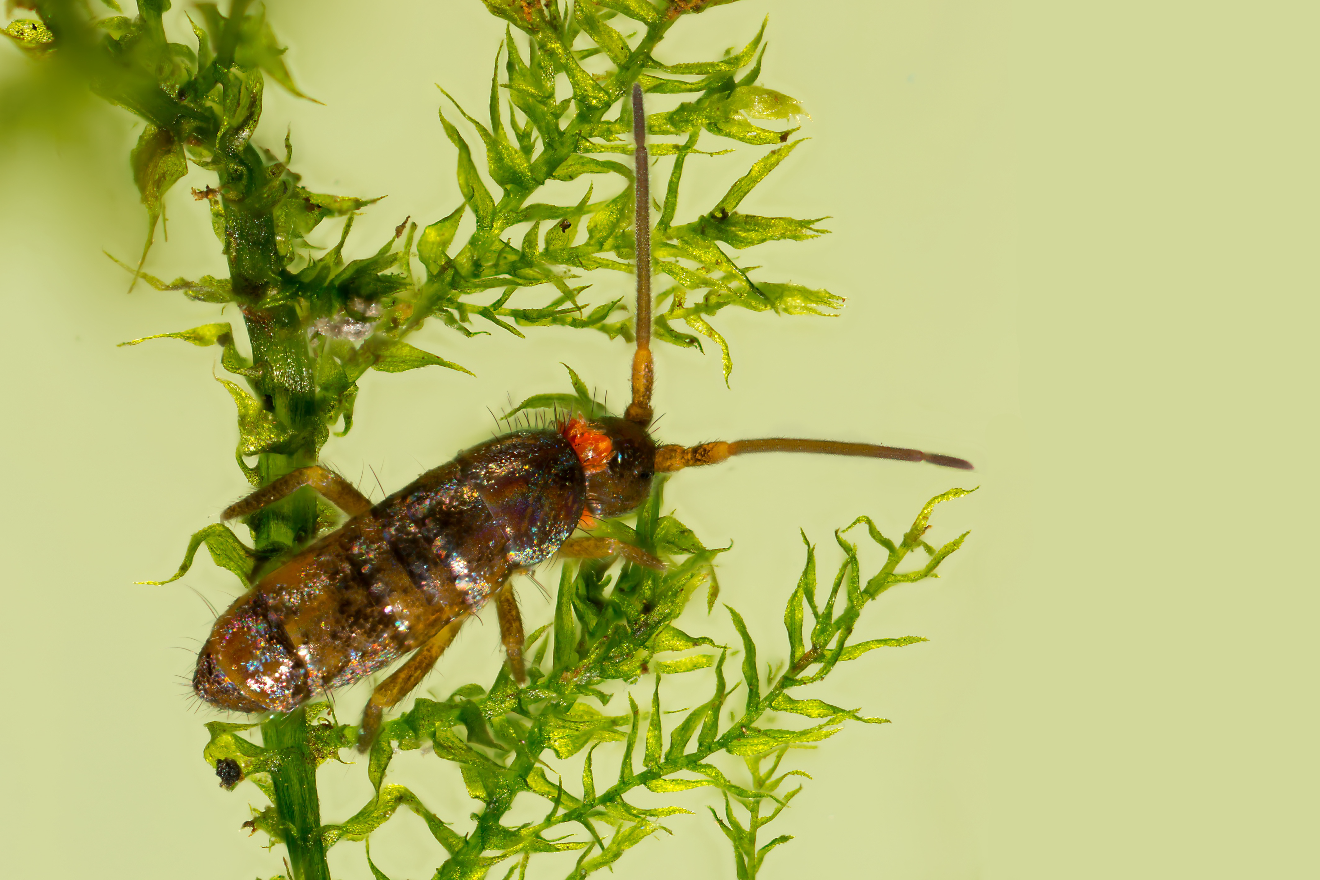
Panisellus thienemanni (K. Viets, 1920) is a spring-dwelling water mite with an interesting life cycle, using springtails (Collembola) as their host (Boehle 1996). With legs lacking swimming setae, P. thienemanni adults and deutonymphs have a crawling mode of locomotion. The species has a characteristic arrangement of dorsal and postero-ventral sclerites (Figure 1).

By using adult and juvenile stages of Collembola as hosts (Boehle 1996), P. thienemanni can be considered as one of the few exceptions to the rule by not resorting to flying insects.
Springtails infected by larvae of P. thienemanni (Figure 2) can be found from April-June (Boehle 1996; Wohltmann in Martin and Stur 2006).
Panisellus thienemanni has a scattered distribution throughout Europe, probably due to rare studies on spring habitats. The current state of knowledge of the distribution of P. thienemanni in the Netherlands is presented in Figure 3. Up till recently, P. thienemanni was only known from a single site in the Netherlands, a helocrene spring on a shaded, sloping bank of a headwater stream in the very east of the country (Mosbeek, N 52˚26.741′ E 6˚51.760′, Province of Overijssel) (Smit et al. 2012). In 2019 the first author also encountered the species in an area with seepage outflow on the shaded bankside of a stream in the southern part of the Netherlands (Tongelreep, Province of Noord-Brabant, N 51°20.449′ E 5˚29.060′, 22 Mar. 2019, 1 ♂, 2 ♀♀, 1 deutonymph).

In 2011 and 2012 we actively searched for P. thienemanni adults, deutonymphs and parasitized springtails in the helocrene springs adjacent to the Mosbeek. This paper describes the results of the survey, with special emphasis on the life cycle of the mite, selected hosts, and morphology.
Material and methods
Description of the sampling site
The sampling site is a helocrene spring, situated on the face of the stream bank of the Mosbeek, a small headwater stream near the hamlet of Hezingen (Province of Overijssel). The site is shaded by black alder (Alnus glutinosa), with an undergrowth of plants like wood club-rush (Scirpus sylvaticus), large bittercress (Cardamine amara), marsh marigold (Caltha palustris), water horsetail (Equisetum fluviatile), stinging nettle (Urtica dioica), and several moss species. The bankside is partially saturated with groundwater, with seepage outflow forming some trickles (Figure 4). Locally precipitation of iron oxide takes place.

Sampling
Sampling of water mites was carried out in the spring and autumn of 2011. A small shovel sampler as described in Bijkerk (2010) was used. A sample consisted of several shovel samples from the site. Sampling of springtails was carried out in the spring of 2012. Fifteen pitfall traps (Figure 5) containing some fixative (formaline, 6%) and a drip of detergent for breaking the surface tension, were placed at regular intervals on the slope of the bankside and collected again after twelve days (in place from 5 Apr. 2012 to 17 Apr. 2012). When collecting the pitfall traps, an additional shovel sample adjacent to every pitfall trap was taken for sampling water mites.

Abiotic parameters
To measure conductivity, acidity, and temperature of the groundwater discharged to the surface, groundwater monitoring wells were placed next to every pitfall trap (Figure 5). After twelve days, measurements were carried out in the monitoring wells. The electric conductivity, pH, oxygen saturation and concentration, temperature, and oxidation-reduction potential were determined.
Processing of samples, identification and measurements
All samples were taken back to the laboratory. In order not to miss the small mites while sorting, all shovel samples were divided into different fractions by gently washing the samples through a stack of sieves with different mesh sizes (mesh width bottom sieve: 300 µm). The fractions of the washed samples were sorted and water mites were stored in Koenike-fluid. When encountered, also infected springtails were collected and stored in ethanol (70%). Springtails from the pitfall traps were sorted and preserved in ethanol (70%).
Identification of mites and springtails was executed using a stereo microscope and a compound microscope. Measurements on adults and nymphs of P. thienemanni were executed using a calibrated compound microscope with graticule.
Results
Abiotic factors
The electric conductivity, pH, oxygen saturation and concentration, temperature, and oxidation-reduction potential of the groundwater measured in the 15 groundwater monitoring wells are shown in Table 1. The groundwater coming to the surface at the site can be described as weakly acidic. Groundwater temperature measured in the monitoring wells was very constant. Electric conductivity ranged from 203-450 µS/cm, with higher measurements at higher altitudes on the face of the stream bank.


Invertebrate assemblage
A complete list of other spring dwelling water mite species found at the sampling site is presented in Table 2. The assemblage of other invertebrates found at the site is also typical for helocrenes. Examples of encountered species are the beetles Agabus guttatus (Paykull, 1798) and Hydroporus discretus Fairmaire & Brisout, 1859, the stoneflies Nemoura dubitans Morton, 1894 and Nemurella pictetii Klapálek, 1900, and the chironomids Krenopelopia binotata (Wiedemann, 1817) and Heterotanytarsus apicalis (Kieffer, 1921). Wiggers et al. (2019) provide a full list of macro-invertebrates found at the site.
Morphology
Measurements of the idiosoma length and width, and number of acetabula (Ac) anterior and posterior to the genital plates of males, females and deutonymphs are presented in Table 3. The designation of the gender of adult specimens is based on the number of Ac anterior to each genital plate (one in males, two in females), which seems to be the most reliable feature (Figure 6). We found two aberrant adult specimens with the number of Ac anterior to the left genital plate being dissimilar to the number anterior to the right plate. These specimens have not been included in the measurements.


Adults, deutonymphs and larvae
The number of collected specimens of adult males and females, deutonymphs, and the presence or absence of larvae for each sampling date is presented in Table 4. We found adult males and females as well as deutonymphs in April and September. Adults and deutonymphs were mainly collected from groundwater saturated patches of moss. In all samples, females were found in greater numbers than males, most convincingly in autumn due to a higher number of adults collected compared to in spring. The main part of the females caught in April were still ovigerous and carried 2-9 eggs (average 6; n=6).
The number of deutonymphs found was low. Larvae attached to collembolans were only observed in spring.
Sampling effort and method of sampling differed between the sampling dates. Therefore, statements on the difference in number of specimens caught between dates cannot be made.

Infected springtails, number of larvae and attachment site
The total number of Collembola species caught in the pitfall traps, as well as the number and percentage of specimens with larvae of P. thienemanni attached to their body are shown in Table 5. In the pitfall traps we found nine species of springtails, of which three could be observed hosting larvae of P. thienemanni. The main two species hosting larvae were Tomocerus flavescens and T. minor (Tomoceridae). Of T. flavescens (n = 82) 6.1% of the specimens were infected, of T. minor (n = 56) 3.6%. Only a single specimen of Isotomurus fucicola (Isotomidae) was caught, and also hosted a larva. Of the remaining six species caught, no specimens could be observed hosting larvae, even not in the most abundant species, L. lignorum and I. plumosus.

The number of P. thienemanni larvae found attached to specimens of encountered host species caught in the pitfall traps and shovel samples are presented in Table 6. The number of larvae attached to an individual springtail ranged from 1-7 (n of springtails = 23, mean: 1.9). Larvae were exclusively found attached to the soft, reduced dorsal section of the prothorax. Occasionally we observed unengorged larvae clutched to the ventral side of the thorax and abdomen. However, these larvae were not fixed to the host's integument.

Discussion
Habitat
Both Dutch sites are shaded faces of stream banks with seepage outflow. At our study site, P. thienemanni could only be observed in samples of groundwater-soaked mosses on the bankside of the stream, and not in the main headwater itself. Also the known German sites are all shaded, weakly seeping helocrenes, which never dry out completely (personal communication Reinhard Gerecke). Andreas Wohltmann (personal communication) found P. thienemanni exclusively in wet mosses, which corresponds with our observations. The occurrence in shaded places only, implies that P. thienemanni depends on a stable thermal regime and can be considered a stenothermic species, which was classified in the past as ''cold-stenothermic″, a term which is not really fitting to a habitat which is relatively warm in the winter.
Sexual dimorphism
To distinguish adult males morphologically from females the number of Ac anterior to each genital plate can be used best. This feature mainly worked out, but we found two aberrant specimens with the number of Ac anterior to one genital plate being dissimilar to the number anterior to the other plate (1 versus 0 and 1 versus 2). Idiosoma length and width of both specimens would categorize them as males.
The position of the pregenital plate as well as the position of the setae on the genital plate as features to separate male and female as mentioned in Viets (1936) appeared unreliable in our specimens.
The number of Ac posterior to each genital plate we observed in adults varies and differs slightly from the number Di Sabatino et al. (2010) are reporting. They mention 4-6 posterior Ac, whereas we found 3-6 in males and 4-8 in females.
In general, females are larger than males but ranges of idiosoma length and width overlap.
Life cycle and phenology
Panisellus thienemanni is likely to be univoltine. Oviposition takes place in early spring, possibly in March-April. Oviposition and the subsequent hatching of larvae clearly takes place over a period of time. The main part of adult females caught at the study site on 17 Apr. 2012 could still be observed with eggs, whereas larvae were also present.
Hatching of larvae possibly starts as early as the end of March, since infected springtails were already observed on 10 Apr. 2011. It may well carry on until the end of spring, but this needs further investigation. Before attaching to a host, the bright red and terrestrial larvae are able to crawl freely in the moss (own observation in collected sample).
The larvae we found attached to springtails in our April samples, were not or not fully engorged. Combined with the presence of ovigerous females, this indicates that the time of sampling was at the beginning of the period host-seeking larvae were present. Boehle (1996) found infected hosts from the beginning of April until the middle of June. The length of the idiosoma of the larvae he observed increased from 0.2 mm to at most 0.7 mm when fully engorged. The larvae left their host after approximately three weeks and subsequently metamorphosed into deutonymphs in 20 days. This period of time seems temperature related. For the protonymphal stage, Wohltmann (unpublished data) observed a duration of an average of 50 days at a temperature of 15 °C before the active deutonymphs emerged. At 20 °C the protonymphal stage lasted on average 27 days. Considering the restricted seasonal presence of larvae, we assume only a single peak of emergence takes place. From these observations, we conclude that the development from larva to deutonymph takes place in the same year. From the scant data available, it is unclear when deutonymphs metamorphose into adults. At our study site, deutonymphs were at least still present on 28 Sep. 2011. In the temperate zone, deutonymphs of water mites typically feed and grow throughout the summer, and adults appear in late summer or early autumn and mate almost immediately (Proctor et al. 2015). However, since we also found a deutonymph the following spring (17 Apr. 2012), it can be inferred that not all deutonymphs metamorphose into adults before the winter. Part of the population seems to pass the winter in this stage. Another three records of deutonymphs found in March, April and May in the Netherlands, Luxembourg, and the Czech Republic (this publication; Martin 2006; personal communication Ian Smith; see below) are in line with this observation. This phenomenon of overwintering deutonymphs is not uncommon and has also been reported for other members of Hydryphantidae (Böttger and Völkl 1987; own observation). Those deutonymphs of P. thienemanni overwintering, might enter a reproductive diapause after turning into adults and partake in the following reproduction period.
Feeding behaviour of the deutonymphs is not known. Wohltmann (in Martin 1998) found that without nourishment deutonymphs were able to survive for four months.
As documented for other species within the Hydryphantidae and other early derivative groups (Witte 1991; Proctor 1992), sperm transfer in P. thienemanni is likely to take place without bodily contact (dissociated sperm transfer). Males deposit spermatophores on a substrate, while uptake of sperm packets by females takes place afterwards. In the temperate zone inseminated females of water mites typically overwinter and lay fertilized eggs the following spring (Martin 2010; Proctor et al. 2015), which possibly also applies to P. thienemanni.
Both adult males and females were found in both spring and autumn samples. This observation contradicts with the supposition of Martin and Rückert (2011) that P. thienemanni is an early spring species. Also Gerecke et al. (2009) mention records from Italy in samples from spring habitats taken in August, September, and October. The female-biased sex ratio observed might be explained by males having a shorter lifespan (Gerecke 2006), a phenomenon also observed in other species (Ullrich 1976).
Martin (1998, 2004) managed to feed a kept adult male with chironomids, which could be the main food source of adults and deutonymphs.
Parasite-host relationship
Panisellus thienemanni is probably highly host-specific to a limited number of springtail species. The dorsal side of the prothorax is the attachment site exclusively used by the larvae. Belonging to an early derivative family (Hydryphantidae), the species' specific site selection is striking. In many representatives of early derivative families, attachment sites are selected accidentally. Highly specific attachment sites are found to be more common in species of higher evolved families (Martin 2004). In many of these species, larvae are preparasitically attending their host awaiting the process of host ecdysis, during which they attach for feeding. This behaviour often correlates with aggregation of mite larvae at particular regions of the host's body. Such preparasitic attendance has also been observed in terrestrial Parasitengona (Johnstonianidae, Calyptostomidae, see e.g. Wohltmann 2000) and Hydryphantoidea (Lanciani 1971), again correlated with attachment site specificity. Further research is needed to confirm if larval attachment of P. thienemanni is likewise preceded by preparasitic attendance.
Springtail adults and nymphs of the genus Tomocerus are mainly selected (Boehle 1996; personal communication Andreas Wohltmann; this study). The three parasitized species we observed, T. flavescens, T. minor and I. fucicola, all belong to the Entomobryomorpha. Species within this large order are all exhibiting a reduced, not fully sclerotized prothorax. This feature enables the larva to attach itself onto the soft, membranous integument. Moreover, this obvious site specificity might be stimulated due to the site being out of reach by brushing movements of the host's legs (Wohltmann 2000). In line with Boehle (1996), the infected springtails we found were parasitized by a single larva. Occasionally higher numbers were found, with a maximum of seven. Wohltmann (personal communication) found 1-6 larvae on a single host. Boehle (1996) reported a maximum of four, a number found to be lethal to the host. As he observed, attached larvae are positioned in a fashion that their swelling idiosoma is supported by their host's head.
The range of host species selected by larvae of P. thienemanni seems limited. This narrow host range may be determined by different factors. Panisellus thienemanni is likely to exhibit a narrow niche breadth within its heterogeneous macrohabitat. Positive co-occurrence of P. thienemanni and a suitable host species due to overlap in habitat requirement therefore may be restricted. Of the three parasitized host species, Tomocerus flavescens and T. minor are common, drought sensitive, surface-dwelling species. In damp forests they can be abundantly found in leaf litter and mosses. In spring habitats their niche is rather broad and evidently overlaps with that of P. thienemanni larvae. Therefore, exposure of these species to mite larvae is comparatively high. Also Isotomurus fucicola, a much rarer species, apparently prefers very wet conditions along the shore of different waterbodies (Fjellberg 2007) and in spring habitats may tend to prefer similar sites as P. thienemanni.
Isotomurus plumosus is also a species found in wet habitats. Interestingly, no parasitized specimens of this species were found, although caught in substantial numbers at the study site. This apparent lack of susceptibility may be influenced by specific morphological, physiological, or behavioural host traits preventing successful settlement of larvae. Isotomurus plumosus is clearly a smaller species compared to the encountered host species and might therefore be an unsuitable host for development of P. thienemanni larvae.
Some of the unparasitized springtail species caught, exhibit a different habitat preference compared to P. thienemanni. For example, Entomobrya corticalis and E. nivalis can be mainly found on trees, where they live underneath bark or on mosses and lichens (Berg and Aptroot 2003). A host-parasite association between these species and P. thienemanni therefore seems highly unlikely.
The relatively high number of unengorged larvae encountered, as well as the presence of ovigerous females, implies our sampling was carried out at the beginning of the hatching period. The percentage of parasitized springtails determined by us reflects the situation at that period and is likely to underestimate the percentage expected at a later time in spring.
Smith and Oliver (1986) observed a larva, which they ascribed to the genus Thyas (currently Parathyas), attached to the thorax of Tomocerus sp. The record stems from a site in the Czech Republic, 1972. At the time of their research, larvae of P. thienemanni as well as their host species were not yet known. A request to reexamine the material resulted in a rectification, the Czech larvae found at the time on T. flavescens in fact belong to P. thienemanni (personal communication Ian Smith). No other cases of water mite species using springtails have been observed, making this host-parasite relationship of P. thienemanni and springtails unique among this group.
Low interspecific competition might be an advantage for P. thienemanni, since no other water mites utilize springtails. Furthermore, springtails are very abundant in spring habitats. However, there are some cases of terrestrial mites, with larvae known to parasitize on springtails (e.g. Greenslade & Southcott 1980; De Oliveira et al. 2016; Felska et al. 2018).
Dispersal
The main part of water mite species select flying insects as hosts. Besides providing the larvae with haemolymph for growth and development, the host facilitates dispersal of the mite. Selecting a flying insect therefore seems advantageous for dispersal.
Whereas springtails are not capable of flying, they have other means to reach a new site. Although springtails are mobile, dispersal by active locomotion is of minor importance, and they mainly rely on passive dispersal. However, successful aerial dispersal of wind-borne springtails has been proven (Hawes et al. 2007). Considering the predominantly sheltered biotopes of P. thienemanni, we expect this mode of transport of co-occurring, bottom-dwelling springtails of minor significance. Springtails inhabiting such biotopes are likely to be poorly dispersive.
Springs can be considered insular environments (Cantonati et al. 2006). Consequently, crenobiontic mites show a disjunct distribution, with populations assumed to be rather isolated (Blattner et al. 2019). Successful inter-population dispersal might therefore be very limited and depends predominantly on the dispersal abilities of the selected host. To reduce the high risk of being transferred to unsuitable habitats, it might be even advantageous for crenobionts to greatly reduce dispersal to prevent population decline. As for P. thienemanni, using poorly dispersive collembolans as hosts might therefore be a strategy to remain at the well-suited natal location.
Crenobiontic species with limited dispersal abilities and low inter-population migration rates show restricted gene flow, resulting in high genetic diversity between populations and the potential for speciation (Blattner et al. 2021). To better understand the degree of isolation between populations of P. thienemanni, a population genomic analysis will be of great value.
Despite their supposedly limited dispersal abilities, possible modes of dispersal of infected springtails cannot be regarded obsolete. Water is another dispersal agent, springtails can be regularly found on floating debris or driftwood. Furthermore, many springtails have hydrophobic cuticles that enable them to survive on water surfaces (Moore 2002). Considering the already mentioned high risk of ending up in an unsuitable habitat when depending on passive long-distance dispersal, we expect dispersal of P. thienemanni will be more successful at a local spatial scale. Downstream drift of infected collembolans could be one of the mechanisms facilitating this (see Schuppenhauer et al. 2019). Further research is needed to confirm if this mode of transport is of significance.
Besides using collembolans, P. thienemanni is not known to occasionally resort to other, winged hosts with better dispersal abilities. However, usage of other hosts cannot be ruled out, since studies on the species are very limited.
The two only known populations of P. thienemanni in the Netherlands appear to be isolated. Directly downstream from the study site, the hydrologic condition of the headwater deteriorates, with dry banksides without groundwater discharge due to groundwater regulation. Consequently, settlement after dispersal further downstream seems highly unlikely. The isolated Dutch populations of P. thienemanni might therefore be surviving relicts of the once more widespread forested and undrained headwater systems. Currently restoration measures are in progress at both sites to increase discharge of seepage (Duursma 2020; Zekhuis 2020).
Acknowledgements
During the process of writing this article, several persons kindly provided us with information or suggestions on various aspects. Matty Berg (Vrije Universiteit, Amsterdam) assisted with the identification of the springtails. Andreas Wohltmann (independent researcher, Ritterhude) and Reinhard Gerecke (independent researcher, Tübingen) provided us with additional information on P. thienemanni. Ian Smith (Canadian National Collection of Insects and Arachnids, Ottawa) took up the task of digging out a 50-year-old record of mite larvae on springtails, and carried out revision of their initial identification. Christophe Brochard (Bureau Biota) supplied us with several wonderful pictures of P. thienemanni. And last but not least, Peter Martin (Christian-Albrechts-Universität, Kiel), Harry Smit (Naturalis Biodiversity Center, Leiden) and two anonymous reviewers reviewed the manuscript in detail, and provided useful improvements.
References
- Berg M., Aptroot A. 2003. Springstaarten op korstmossen (Hexapoda: Collembola). Nederlandse Faunistische Mededelingen, 18: 103-122.
- Bijkerk R. (Ed.) 2010. Handboek Hydrobiologie. Biologisch onderzoek voor de beoordeling van Nederlandse zoete en brakke oppervlaktewateren. Rapport 2010-28, Stichting Toegepast Onderzoek Waterbeheer, Amersfoort.
- Blattner L., Gerecke R, Fumetti S. 2019. Hidden biodiversity revealed by integrated morphology and genetic species delimitation of spring dwelling water mite species (Acari, Parasitengona: Hydrachnidia). Parasites & Vectors, 12 (1): 1-13. https://doi.org/10.1186/s13071-019-3750-y
- Blattner L., Lucek K., Beck N., Berner D., Fumetti S. 2021. Intra-Alpine Islands: Population genomic inference reveals high degree of isolation between freshwater spring habitats. Diversity and Distributions, 28: 1-15. https://doi.org/10.1111/ddi.13461
- Boehle W.R. 1996. Contribution to the morphology and biology of larval Panisellus thienemanni (Viets, 1920) (Acari: Parasitengonae: Hydrachnidia). Acarologia, 37: 121-125.
- Böttger K., Völkl R. 1987. Faunistisch-Ökologische beobachtungen an dem Wassermilben (Hydrachnellae, Acari) einiger Kleingewässer, nebst biologischen angaben zu einzelnen arten. Acarologia, 28 (2): 161-170.
- Cantonati M., Gerecke R., Bertuzzi E. 2006. Springs of the Alps - sensitive ecosystems to environmental change: from biodiversity assessments to long-term studies. Hydrobiologia, 562: 59-96. https://doi.org/10.1007/s10750-005-1806-9
- Davids C. 2004. Parasitisme bij watermijten. Entomologische Berichten, 64: 51-58.
- De Oliveira M.P.A., de Oliveira Bernardi L.F., Zeppelini D., Ferreira R.L. 2016. First report of cave springtail (Collembola, Paronellidae) parasitezed by mite (Parasitengona, Microtrombiidae). Subterranean Biology, 17: 133-139. https://doi.org/10.3897/subtbiol.17.8451
- Di Sabatino A., Gerecke R., Martin P. 2000. The biology and ecology of lotic water mites (Hydrachnidia). Freshwater Biology 44: 47-62. https://doi.org/10.1046/j.1365-2427.2000.00591.x
- Di Sabatino A., Gerecke R., Gledhill T., Smit H. 2010. Chelicerata: Acari II. Süßwasserfauna von Mitteleuropa, 7/2-2: 1-236. Spektrum Akademischer Verlag, Heidelberg. https://doi.org/10.1007/978-3-8274-2266-8_1
- Duursma S. 2020. Herinrichting Tongelreep Fase I. Definitief Projectplan Waterwet. Royal HaskoningDHV rapport BG2999_T&P_RP_2010080944, in opdracht van Waterschap de Dommel.
- Felska M., Wohltmann A., Mąkol J. 2018. A synopsis of host-parasite associations between Trombidioidea (Trombidiformes: Prostigmata, Parasitengona) and arthropod hosts. Systematic and Applied Acarology, 23 (7): 1375-1479. https://doi.org/10.11158/saa.23.7.14
- Fjellberg A. 2007. The Collembola of Fennoscandia and Denmark, part II: Entomobryomorpha and Symphypleona. Fauna Entomologica Scandinavica, 42: 1-264. https://doi.org/10.1163/ej.9789004157705.i-265
- Gerecke R. (ed.) 2006. Chelicerata: Araneae/Acari I. Süßwasserfauna von Mitteleuropa, 7/2-1: 1-388. Spektrum Akademischer Verlag, Heidelberg.
- Gerecke R., Schatz H., Wohltmann A. 2009. The mites (Chelicerata: Acari) of the CRENODAT project: Faunistic records and ecological data from springs in the autonomous province of Trento (Italian alps). International Journal of Acarology, 35 (4): 303-333. https://doi.org/10.1080/01647950903059452
- Greenslade P.J.M., Southcott R.V. 1980. Parasitic mites on Sminthurid Collembola in Australia. Entomologist's Monthly Magazine, 116: 85-87.
- Hawes T.C., Worland M.R., Convey P., Bale J.S. 2007. Aerial dispersal of springtails on the Antarctic Peninsula: implications for local distribution and demography. Antarctic science, 19 (1): 3-7. https://doi.org/10.1017/S0954102007000028
- Lanciani C.A. 1971. Host exploitation and synchronous development in a water mite parasite of the marsh treader Hydrometra myrae (Hemiptera: Hydrometridae). Annals of the Entomological Society of America, 64 (6): 1254-1259. https://doi.org/10.1093/aesa/64.6.1254
- Martin P. 1998. Zur Autökologie der Wassermilben (Hydrachnidia, Acari) zweier norddeutscher Tieflandsbäche [PhD thesis]. Kiel: Christan-Albrechts-Universität. pp. 269.
- Martin P. 2004. Water mites (Hydrachnidia, Acari) as predators in lotic environments. Phytophaga, 14: 307-321.
- Martin P. 2010. Observations on reproduction, development and sexual behaviour of stream-inhabiting water mites (Hydrachnidia, Acari). In: Sabelis M.W., Bruin J. (Eds). Trends in Acarology. Proceedings of the 12th International Congress, 303-312. https://doi.org/10.1007/978-90-481-9837-5_49
- Martin P., Stur E. 2006. Parasite-host associations and life cycles of spring-living water mites (Hydrachnidia: Acari) from Luxembourg. Hydrobiologia, 573: 17-37. https://doi.org/10.1007/s10750-006-0246-5
- Martin P., Rückert M. 2011. Die Quellfauna Schleswig-Holsteins und ihre regionale Stenotopie. Faunistisch-Ökologische Mitteilungen, 9: 171-224.
- Moore P.D. 2002. Springboards for springtails. Nature, 418: 381. https://doi.org/10.1038/418381a
- Proctor H.C. 1992. Mating and spermatophore morphology of water mites (Acari: Parasitengona). Zoological Journal of the Linnean Society 106: 341-384. https://doi.org/10.1111/j.1096-3642.1992.tb01250.x
- Proctor H.C., Smith I.M., Cook D.R., Smith B.P. 2015. Subphylum Chelicerata, Class Arachnida. In: Torp J.H., Rogers D.C. (Eds). Ecology and general biology. Thorp and Covich's Freshwater Invertebrates, Fourth edition, 1: 599-660. https://doi.org/10.1016/B978-0-12-385026-3.00025-5
- Schuppenhauer, M.M., Lehmitz R., Xylander W.E.R. 2019. Slow-moving soil organisms on a water highway: aquatic dispersal and survival potential of Oribatida and Collembola in running water. Movement Ecology, 7 (20): 1-14. https://doi.org/10.1186/s40462-019-0165-5
- Smit H., Boonstra H., Duijts O., van Maanen B., Wiggers R. 2012. Meer dan 250 soorten watermijten in Nederland (Acari: Hydrachnidia, Halacaridae)! Nederlandse Faunistische Mededelingen, 38: 95-113.
- Smith I.M., Oliver D.R. 1986. Review of parasitic associations of larval water mites (Acari: Parasitengona: Hydrachnida) with insect hosts. The Canadian Entomologist, 118: 407-472. https://doi.org/10.4039/Ent118407-5
- Ulrich F. 1976. Biologisch-ökologische Studien an rheofielen Wassermilben (Hydrachnellae, Acari), unter besonderer Berücksichtigung von Sperchon setiger (Thor 1898) [PhD thesis]. University of Kiel, Germany.
- Viets K. 1936. Wassermilben oder Hydracarina (Hydrachnellae und Halacaridae). In: Dahl F. (Ed.). Die Tierwelt Deutschlands und der angrenzenden Meeresteile nach ihren Merkmalen und ihrer Lebensweise. Gustav Fischer Verlag, Jena. pp 575.
- Wiggers R., Moller Pillot H.K.M., Mulderij G. 2019. Droogtemeetnet macrofauna 2019 - broekbossen en vennen. Bureau Biota rapport 2019-017. In opdracht van Waterschap Vechtstromen.
- Witte H. 1991. Indirect sperm transfer in prostigmatic mites from a phylogenetic viewpoint. In: Schuster R., Murphey P.W. (Eds). The Acari - Reproduction, development and life-history strategies: 107-176. https://doi.org/10.1007/978-94-011-3102-5_8
- Wohltmann A. 2000. The evolution of life histories in Parasitengona (Acari: Prostigmata). Acarologia, 41: 145-204.
- Zekhuis, M. 2000. Mosbeek duurzaam verondiepen. De Levende Natuur, 121 (2): 70.


2022-03-16
Date accepted:
2022-05-30
Date published:
2022-06-09
Edited by:
Mąkol, Joanna

This work is licensed under a Creative Commons Attribution 4.0 International License
2022 Wiggers, Rink and Boonstra, Harry
 Download article Download low definition
Download the citation
RIS with abstract
(Zotero, Endnote, Reference Manager, ProCite, RefWorks, Mendeley)
RIS without abstract
BIB
(Zotero, BibTeX)
TXT
(PubMed, Txt)