


Two new species of Rhyncholimnochares (Acari, Hydrachnidia) from Ecuador
Valdecasas, Antonio G.1 and García-Jímenez, Ricardo2
1✉ Departamento de Biodiversidad y Biología Evolutiva, Museo Nacional de Ciencias Naturales, CSIC, Madrid, Spain.
2Departamento de Biodiversidad y Biología Evolutiva, Museo Nacional de Ciencias Naturales, CSIC, Madrid, Spain.
2021 - Volume: 61 Issue: 2 pages: 321-331
https://doi.org/10.24349/acarologia/20214433ZooBank LSID: 9C824F4B-FAD9-43D3-BE86-90F8EB7A4052
Original research
Keywords
Abstract
Introduction
One of the areas still under-sampled concerning the water mite fauna (Acari, Parasitengona, Hydrachnidia) is South America. In recent work (Tuzovskij and Gerecke 2020) described several new species of the genus Rhyncholimnochares from Central and South America and provided an updated morphological delimitation of the genus along with a diagnostic key to all species. In this work, we add two new species found in the Amazonian part of Ecuador, providing morphological and – as far as possible – molecular characterization (DNA barcode).
Material and methods
Water mites were sampled by the senior author (AGV) with a 250 µm mesh size triangular net and some were kept in Koenike fluids and ethyl alcohol 100% as indicated on each species. Permanent slides were made in glycerin jelly (Cook, 1974). Total genomic DNA was isolated using the QIAGEN BioSprint 15 DNA Blood Kit (Qiagen Iberia S.L., Madrid, Spain). After digestion, water mite exoskeletons were recovered and mounted in glycerin jelly. A fragment of 658 base pairs (bp) was amplified for COI using the primer pair LCO1490 and HCO2198 (Folmer et al. 1994). Gene amplification and purification were carried out as in García-Jiménez et al. (2017).
Laser Scanning Confocal Microscopy (LSCM) images were taken with a Leica SPE microscope, with an excitation wavelength of 488 nm and an emission wavelength range of 546-670. Image stacks were processed with Fiji/ImageJ ver. 1.53f and Amira ver. 5.4.3 (see Valdecasas and Abad (2011) for details).
All measurements are given in µm, first for the holotype, followed by the data for the paratype in parenthesis (if available). We follow Cook's (1974) terminology for easy retrieval of older literature.
Results
Systematics
Family: Limnocharidae Grube, 1859
Subfamily: Rhyncholimnocharinae Lundblad, 1936
Genus: Rhyncholimnochares Lundblad, 1936
Subgenus: Rhyncholimnochares Lundblad, 1936
Type species: Rhyncholimnochares lamellipalpis Lundblad, 1936
For morphological delimitation of the genus, see Tuzovskij and Gerecke (2020).
Rhyncholimnochares (Rhyncholimnochares) munozi n. sp.
ZOOBANK: 62153667-08FB-4A73-931B-31BF30AF4864 ![]()
Figures 1–5


Material examined
Holotype female. Tributary Napo river, kicking sample on gravel bottom. Jatun Sacha reserve (01°04.0' S, 77°37.0' W), Misahualli, Ecuador. 05/12/2008. Collection number: MNCN 20.02/19849. Paratype, one female, same data as holotype. Collection number: MNCN 20.02/19850. Both fixed in Koenike's fluid in the field and mounted in glycerine jelly.
Diagnosis
Rhyncholimnochares munozi n. sp. can be easily distinguished from all other previously described species by the co-occurrence of, among other, the following set of characters: a) an expanded and rounded terminal tail of the ocular plate; b) comparable length of rostrum and basal part of gnathosoma; c) almost rectilinear posterior border of coxa II; d) relatively low number of genital acetabula and e) arborescent setae at the end of P-II.
Description
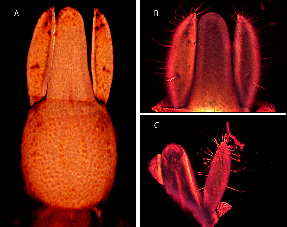
Color red. Oval in shape (Figure 1). Integument round papillate. Dorsum without rod-shaped dorsal plates, with a few sparse glandularia with setae (Figure 2A). Capitulum attached to a retractable long tubular extension of the integument. Idiosoma length up to the extreme of first coxae 1702 (1498); maximum width 1135 (1180).
Ocular plate (OC) total length 570 (477) (Figures 1, 2B). Fore-edge width 84 (61); ocular plate section anterior to eyes: lateral margins converging, length 44 (44); minimum width 46 (46). Eyes separated, length 90 (64); maximum width at eye level 119 (93). Keel length 206 (183). Four pairs of setae on the anterior part of the OC, the second one from the anterior edge somewhat bipectinate, the other three pairs simple. Lateral margins of the OC posterior to eyes slightly concave, terminal end rounded.
The anterior end of the first pair of coxae connected by engrossed integument (Figure 3A). A few setae along the anterior lateral edge of coxa I and on the suture line of coxa I and II. Posterior border of coxa II initially convex then straight, lateral border concave. First coxa complete lateral margin length 255 (222); medial 198 (159). Suture line between coxae I-II length, 173 (145). Second coxa lateral length, 138 (116); posterior 214 (168). Tip first coxa to end second coxa 367 (305).
A few simple setae near margins of coxae III and IV whose weak suture line is not clearly marked (Figures 3B, 4A, B). Total length coxa III-IV 470 (415).
Approximate acetabula diameter, 11 (8); acetabula stalk length, 11 (10); acetabula total length, 21 (19). Some 83 (71) acetabula on each side (Figure 4A).
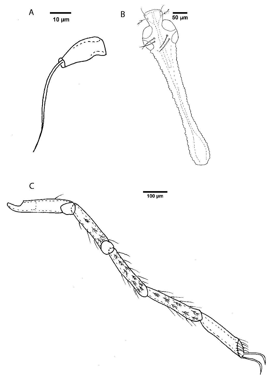
Rostrum slightly shorter than gnathosoma base (Figure 5A). Rostrum length 144 (116); width 74 (64). Gnathosoma base length 188 (174); width 145 (130). Mouth opening directed ventrally, diameter 38 (35). Approximate chelicera basal segment length 104 (-), claw 28 (28). P-II with numerous somewhat bipectinate setae distributed all along (Figures 5B, C). Dorsal length palp segments: P-I, 6 (-); P-II, 152 (135); width P-II, 32 (28).
Legs with numerous simple and bipectinate setae (Figure 2C). Length of leg segments: I-Leg-2, 231 (180); I-Leg-3, 185 (136); I-Leg-4, 146 (110); I-Leg-5, 162 (122); I-Leg-6, 131 (133); II-Leg-2, 246 (203); II-Leg-3, 185 (151); II-Leg-4, 162 (116); II-Leg-5, 169 (130); II-Leg-6, 123 (145); III-Leg-2, 223 (188); III-Leg-3, 185 (145); III-Leg-4, 169 (119); III-Leg-5, 192 (148); III-Leg-6, 146 (125); IV-Leg-2, 262 (206); IV-Leg-3, 223 (177); IV-Leg-4, 216 (162); IV-Leg-5, 231 (177); IV-Leg-6, 162 (139).
Approximate, number of bipectinate setae that could be counted on each leg segment, some may have been lost during manipulation of the specimen, (paratype): I-Leg-2, 5; I-Leg-3, 6; I-Leg-4, 5; I-Leg-5, 5; 2 I-Leg-6, 0; II-Leg-2, 4; II-Leg-3, 6; II-Leg-4, 5; II-Leg-5, 0; 2 II-Leg-6, 0; III-Leg-2, 0; III-Leg-3, 2; III-Leg-4, 4; III-Leg-5, 5; III-Leg-6, 0; IV-Leg-2, 4; IV-Leg-3, 5; IV-Leg-4, 6; IV-Leg-5, 5; IV-Leg-6, 0.
Etymology
The new species is named after Jesús Muñoz for his help during the stays of AGV in Ecuador.
Remarks
The bipectinate setae which, are very common on leg segments, besides the presence of simple setae, could be used as diagnostic, as they are abundant and frequently form a pattern of two rows, as already mentioned by Lundblad (1953). These setae are also common in other members of Limnocharidae (e.g. species of Limnochares) and may provide additional diagnostic value. However, they have been mentioned neither in the species described by Cook (1980) nor in the recent revision of the genus by Tuzovskij and Gerecke (2020), thus their possible diagnostic value could have been underestimated.
DNA barcoding
Not available for these specimens.
Rhyncholimnochares (Rhyncholimnochares) cristinae n. sp.
ZOOBANK: 1DF437DB-7ACF-45C5-BF20-F38DDFF9EF90 ![]()
Figures 6–8


Material examined
Holotype male. Tributary Napo river, kicking sample on gravel bottom. Jatun Sacha reserve (01°04.0' S, 77°37.0' W), Misahualli, Ecuador. 05/12/2008. Fixed in ethanol 100% in the field and mounted in glycerine jelly. Collection number: MNCN 20.02/19851.
Diagnosis
Rhyncholimnochares cristinae n. sp. can be distinguished from all previously described species of Rhyncholimnochares, among others, by the following combination of characters: a) A wide incision on the anterior part of the ocular plate; b) the straight lateral border of coxa II; c) an elongated and slightly concave coxa III; P-II terminating in a palm shaped seta.
Description
Color red. Oval in shape. Integument round papillate. Dorsum without rod-shaped dorsal plate but with sparse glandularia with setae (Figure 6F). Capitulum attached to a retractile long tubular extension of the integument. Idiosoma length up to the extreme of first coxae: 2225; maximum width 1544.
Ocular plate (OC) total length, 1022. OC fore-edge with a medial deep incision (Figures 6A, B and C), width 110. OC anterior part lateral margins converging; length 35; minimum width 72. Eyes separated; globular length, 130; maximum width at eye level, 194. Approximate keel length, 392.
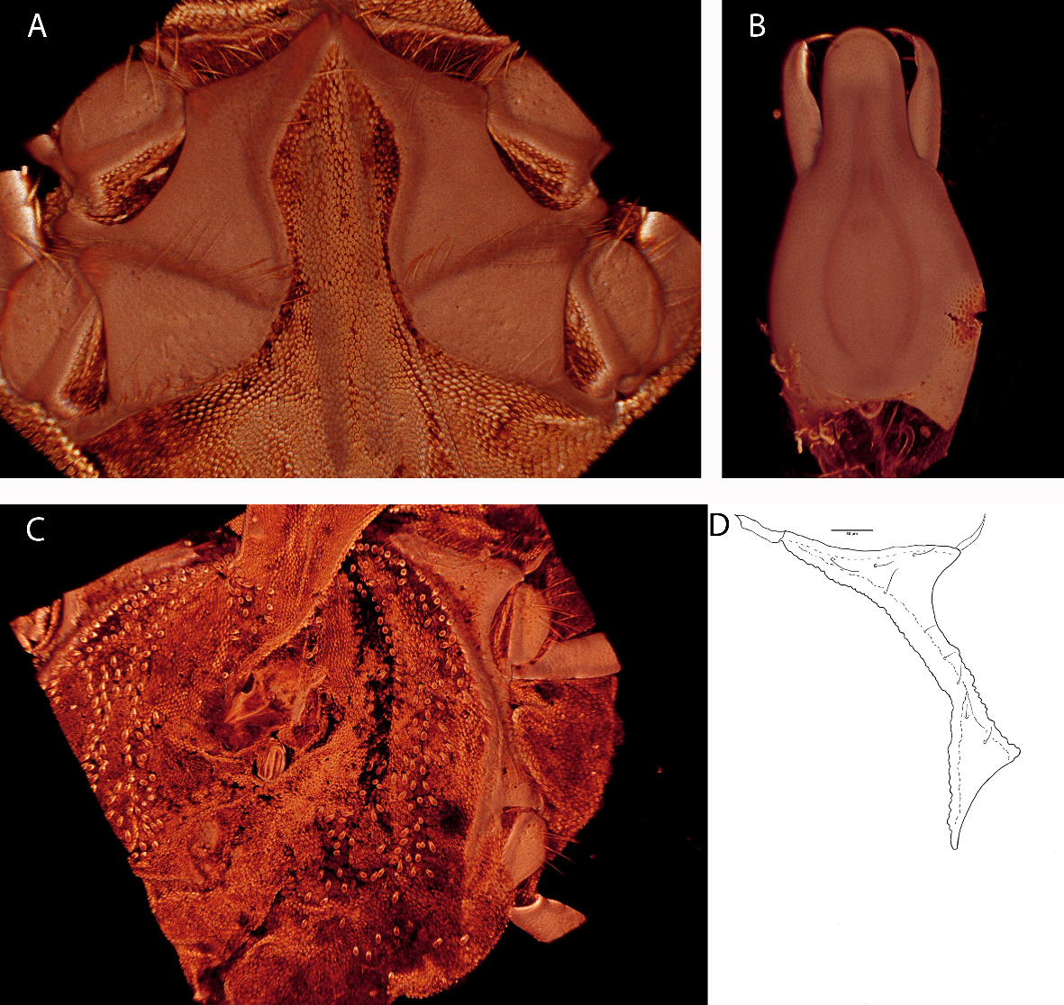
The anterior end of the first pair of coxae connected by engrossed tegument. A few (Figure 7A) setae along the anterior lateral and medial edge of coxa I and on the suture line of coxa I and II. The anterior part of coxa I elongated. Medial border of coxa II convex, lateral straight. First coxa lateral length, 378; medial, 333; suture line coxae I-II, 266. Second coxa lateral length, 258; medial, 297. Tip first coxa to end second coxa, 582.
A few simple setae at the margins of coxae III and IV (Figure 7C, D). Total length coxa III-IV, 770. Acetabula diameter, 11; acetabula stalk length, 10; acetabula total length, 23 (Figure 7C).
Rostrum much shorter than gnathosoma base (Figure 7B). Gnathosoma rostrum length 174; width, 110. Gnathosoma base length, 369; approximate width 293. Diameter mouth opening, 104. Approximate chelicera basal segment length 157, claw 75. Dorsal length palp segments: P-I, 9; P-II, 209; width P-II, 41 (Figure 8).
Legs segments with simple and bipectinate setae (Figures 6D, E). Length of leg segments: I-Leg-2, 362; I-Leg-3, 293; I-Leg-4, 293; I-Leg-5, 231; I-Leg-6, 185; II-Leg-2, 370; II-Leg-3, 285; II-Leg-4, 254; II-Leg-5, 254; II-Leg-6, 192; III-Leg-2, 331; III-Leg-3, 308; III-Leg-4, 277; III-Leg-5, 308; III-Leg-6, 200; IV-Leg-2, 385; IV-Leg-3, 354; IV-Leg-4, 331; IV-Leg-5, 362; IV-Leg-6, 231. Approximate number of bipectinate setae that could be count on each leg segment, some may have been lost during manipulation of the specimen, (`-' means that could not be seen properly): I-Leg-2, 5; I-Leg-3, 12; I-Leg-4, 0; I-Leg-5, 0; I-Leg-6, 0; II-Leg-2, 4; II-Leg-3, 7; II-Leg-4, -; II-Leg-5, 8; II-Leg-6, 0; III-Leg-2, 5; III-Leg-3, 11; III-Leg-4, 16; III-Leg-5, 14; III-Leg-6, -; IV-Leg-2, 5; IV-Leg-3, 7; IV-Leg-4, 13; IV-Leg-5, 5; IV-Leg-6, 0.
DNA barcoding
A 658 base pair fragment of the mitochondrial cytochrome C oxidase subunit I gene (COI) was sequenced and deposited in Genbank with accession number: MW940285 of the only specimen of R. cristinae.
Etymology
The species is named after the wife of the junior author (RGG), Cristina Quintana Colomo, for her support and encouragement to finish this work.
Remarks
As in the case of the previous species, there are plenty of bipectinate setae on legs whose pattern could be diagnostic. Lack of information in other taxa precludes their usefulness as comparative data.
Rhyncholimnochares sp.
Material examined
Incomplete specimen. Small puddle on the border of a road, Jatun Sacha reserve (01°04.0' S, 77°37.0' W), Misahualli, Ecuador. 04/12/2008. Fixed in ethanol 100% in the field and mounted in glycerine jelly. Collection number: MNCN 20.02/19852.
Only the morphology of coxae and legs and the DNA barcoding are provided for this specimen. The remaining parts of the body were lost in DNA isolation.
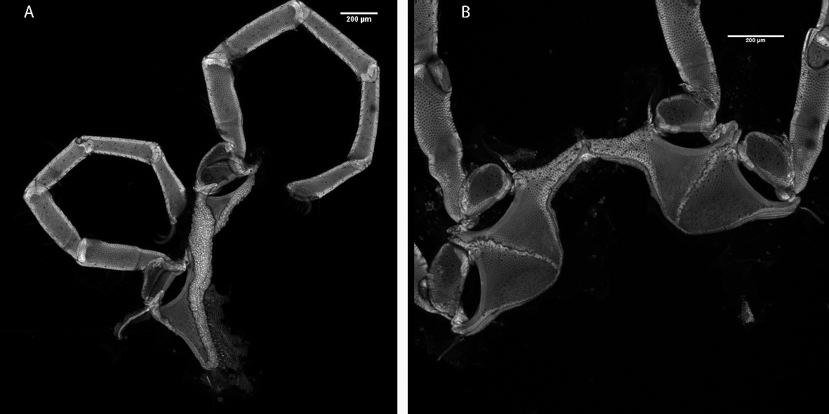
First coxa lateral length, 569; medial, 466; suture line coxae I-II, 422. Second coxa lateral length, 328; medial, 434. Tip first coxa to end second coxa, 822 (Figure 9A).
Length of leg segments: I-Leg-2, 608; I-Leg-3, 439; I-Leg-4, 393; I-Leg-5, 400; I-Leg-6, 339; II-Leg-2, 616; II-Leg-3, 454; II-Leg-4, 416; II-Leg-5, 400; II-Leg-6, 316; III-Leg-2, 554; III-Leg-3, 447; III-Leg-4, 408; III-Leg-5, 454; III-Leg-6, 339; IV-Leg-2, 647; IV-Leg-3, 570; IV-Leg-4, 531; IV-Leg-5, 531; IV-Leg-6, 339. Most of the setae were lost (Figure 9B)
DNA barcoding
We could sequence a 658 base pair fragment of the mitochondrial cytochrome C oxidase subunit I gene (COI) of this specimen and is deposited in Genbank with accession number: MW940284.
Final remarks
Morphology. The bipectinate setae which are very common on leg segments (besides the presence of simple setae) frequently form two rows, a fact already mentioned by Lundblad (1953) and Rosso de Ferradas (1975). These setae are easily lost during manipulation but, due to common occurrence in other members of the Limnocharidae (e.g., species of Limnochares), they may provide additional diagnostic value.
Barcoding
At the time of writing this manuscript (23/03/2021), the sequences of the genus Rhyncholimnochares were not available in the Genbank (https://www.ncbi.nlm.nih.gov/) and only 9 sequences were present in Bold Systems (https://www.boldsystems.org/), whereas only one was public. Using BOLD Specimen Identification option, the specimen of Rhyncholimnochares cristinae has a similarity between 82.95% and 83.26% with the Rhyncholimnochares sequences retrieved from BOLD. The specimen Rhyncholimnochares sp. has a similarity between 83.1% and 83.41% with BOLD Rhyncholimnochares sequences. The similarity of R. cristinae with Rhyncholimnochares sp. is 83.89%. All these levels of similarity point to the independent species identity of examined specimens (Blattner et al. 2019).
Acknowledgements
Jesus Muñoz (RJB Madrid) director of the Máster Oficial en Biodiversidad en Áreas Tropicales y su Conservación (CSIC-UIMP) made possible AGV several stays in Ecuador. Miguel Angel Alonso Zarazaga helped with nomenclatorial issues. Two anonymous referees and Joanna Makol provided a very useful revision of a previous version of this manuscript.
References
- Cook D.R. 1974. Water mite genera and subgenera. Mem. Amer. Entom. Inst., 21, 1-860.
- Cook D.R. 1980. Studies on neotropical water mites. Mem. Amer. Entom. Inst., 31: 1-645.
- Folmer O., Black M., Hoeh W., Lutz R., Vrijenhoek R. 1994. DNA primers for amplification of mitochondrial cytochrome c oxidase subunit I from diverse metazoan invertebrates. Mol. Mar. Biol. Biotechnol., 3: 294-299
- García-Jiménez R., Horreo J.L., Valdecasas A.G. 2017. Minimal barcode distance between two water mite species from Madeira Island: a cautionary tale. Exp. Appl. Acarol., 72: 133. doi:10.1007/s10493-017-0147-5
- Lundblad O. 1953. Die Hydracarinenfauna von Colombia. Ark. Zool. (s.2), 5 (8): 435-585.
- Rosso de Ferradas B.1975. Ácaros acuáticos (Acari, Hydrachnellae) de lagos y embalses y cuencas relacionadas de la provincia de Córdoba, República Argentina. Physis (B), 34 (88): 27-35.
- Tuzovskij P., Gerecke R. 2020. New water mite species of the genus Rhyncholimnochares Lundblad (Acariformes, Limnocharidae) from Central and South America, with a key to all known species of the genus. Ann. Limnol. - Int. J. Lim., 56: 15. doi:10.1051/limn/2020013
- Valdecasas A.G. 2019. A new species of Arrenurus (Acari, Parasitengona, Hydrachnidia) found in the rop of a Yellow-billed Teal Anas flavirostris in Bolivia. Acarologia 59(2): 253-260. doi:10.24349/acarologia/20194329
- Valdecasas A.G., Abad A. 2011. Morphological confocal microscopy in arthropods and the enhancement of auto fluorescence after proteinase K extraction. Microscopy and Microanalysis, 17: 109-113. doi:10.1017/S1431927610094213
This is the second version, uploaded on 2021-07-22. The Figure 9 caption has been changed from "Rhyncholimnochares sp. Confocal microscope image. A - Coxae I and II; B - Coxae III and IV." to "Rhyncholimnochares sp. Confocal microscope image. A - Coxae III and IV; B - Coxae I and II." The printed edition does not have the correction.


2021-01-21
Date accepted:
2021-04-25
Date published:
2021-04-29
Edited by:
Mąkol, Joanna

This work is licensed under a Creative Commons Attribution 4.0 International License
2021 Valdecasas, Antonio G. and García-Jímenez, Ricardo
 Download article
Download the citation
RIS with abstract
(Zotero, Endnote, Reference Manager, ProCite, RefWorks, Mendeley)
RIS without abstract
BIB
(Zotero, BibTeX)
TXT
(PubMed, Txt)