


New Leptogamasus mite species (Parasitiformes: Parasitidae) from Europe. I. Poland
Witaliński, Wojciech1
1✉ Department of Comparative Anatomy, Institute of Zoology and Biomedical Research, Jagiellonian University, Gronostajowa 9, 30-387 Kraków, Poland.
2020 - Volume: 60 Issue: 4 pages: 698-721
https://doi.org/10.24349/acarologia/20204397ZooBank LSID: 00C55073-9BB1-4A31-B394-86F3D0D95216
Original research
Keywords
Abstract
Introduction
Genus Leptogamasus (Parasitidae family) comprises four subgenera which differ among themselves depending on the length of peritreme in adults. In the Breviperigamasus subgenus Juvara-Balş, 1981 peritreme is vestigial, equal to the stigma diameter. In Medioperigamasus Witaliński, 2019, it ends anteriorly at the level of the opening for coxa III (Co III), whereas in Leptogamasus Trägårdh, 1936 s. str. and in Holoperigamasus Juvara-Balş, 1981, the anterior end of peritreme is located at the level of the opening for coxa II (Co II), or close to the coxa I (Co I), respectively. Species described in this study belong to Leptogamasus subgenus, and accordingly the peritremes terminate anteriorly in the midregion of the Co II opening. The podonotum features 21 pairs of podonotal setae – r1, s3 and z3 are lacking.
Methods
Mites were routinely extracted from forest litter using Berlese funnels into 70% ethanol, mounted in Hoyer's medium on glass slides, cured for several days in an oven (60 °C), and studied using an Olympus BX51 microscope fitted with a drawing equipment. All measurements are expressed in micrometres, taken as follows: idiosoma measurements were acquired along the sagittal line (length) and in the widest place (width) of holodorsal shield in the female, and at the margins of idiosoma in the male; setal lengths were measured from the alveolus to the apex of the seta. The drawings were made with the aid of Corel Draw X8 and a Wacom Intuos Graphic Tablet.
The legs and pedipalps are considered as protruding laterally from the idiosoma and the gnathosoma, respectively, thus antero- or posterolateral location of the setae or other structures is applied (Evans and Till 1979). The system of dorsal and ventral setal notations and poroidotaxy was based on Evans and Till (1979) and Lindquist and Moraza (1998), whereas adenotaxy – on Johnston and Moraza (1991) and Moraza and Peña (2005), with some necessary adjustments for Parasitidae.
Systematics
Family Parasitidae
Genus Leptogamasus Trägårdh, 1936 sensu Juvara-Balş, 1981
Type species Leptogamasus suecicus Trägårdh, 1936: 227. Subgenus Leptogamasus Trägårdh, 1936 s. str.
Leptogamasus (Leptogamasus) Trägårdh, 1936.
Type species Leptogamasus suecicus Trägårdh, 1936: 227.
Leptogamasus (Leptogamasus) bihamatus n. sp.
ZOOBANK: C423D349-F27B-411D-8611-09FB9802B47C ![]()
(Figures 1–5)
Diagnosis
Female and male — Gnathotectum trispinate with pointed prongs, shorter in the male; gland pore gv1 present; podonotum with 21 seta pairs, opisthonotum features 22 pairs of setae (R5 and R6 lacking); Tr IV without tubercle.
Female — Presternal plates distant; sternal shield anterior margin with shallow concavity; epigynial shield with anterior margins slightly convex, posterolateral margins short and arcuate, less pigmented band at the level of paragynial setae st4, the internal (dorsal) surface without teeth; endogynium with axially elongated spherules and lateral arcuate lines, adaxial spherule margins thickened and prolonged anteriad as a support for two backward directed hooks, richly dentate stipule broad and lamellar, growing from a distinctly thickened base.
Male — Genital lamina with rounded corners and slightly concaved anterior edge; presternal plates subrectangular; corniculi with elevation on the adaxial margin; cheliceral fixed digit straight and narrow, with a row of ca. 9 minute denticles behind and one tooth and 1-2 minute denticles in front of pilus dentilis; leg II with femur main spur finger-shaped and straight, axillary process slightly curved posterolaterally, rounded and symmetric (oval) in lateral perspective, spurs on genu and tibia conical, genual close to the distal article margin and tibial at some distance from the distal margin of the tibia.
Description
Female (Figures 1–3)


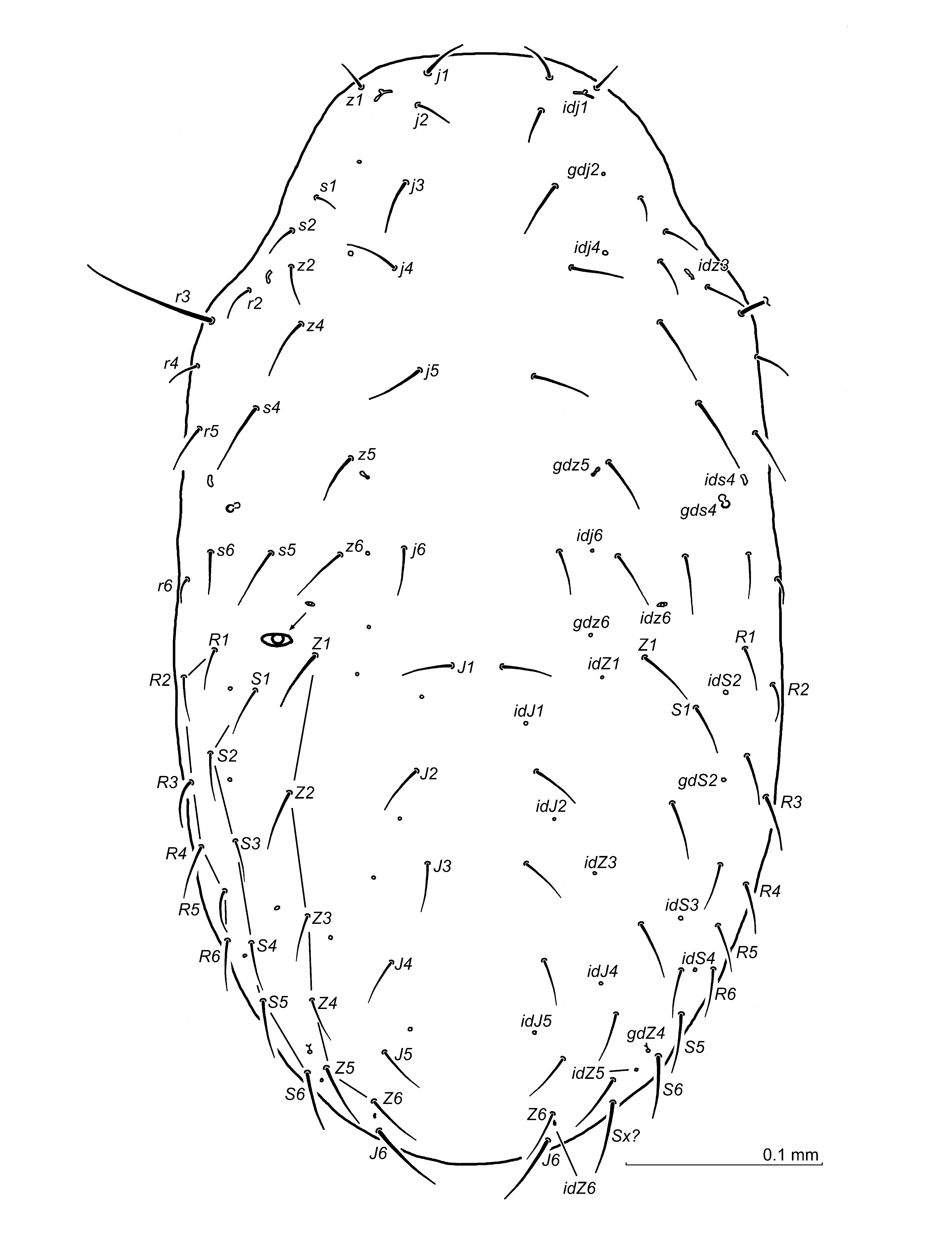
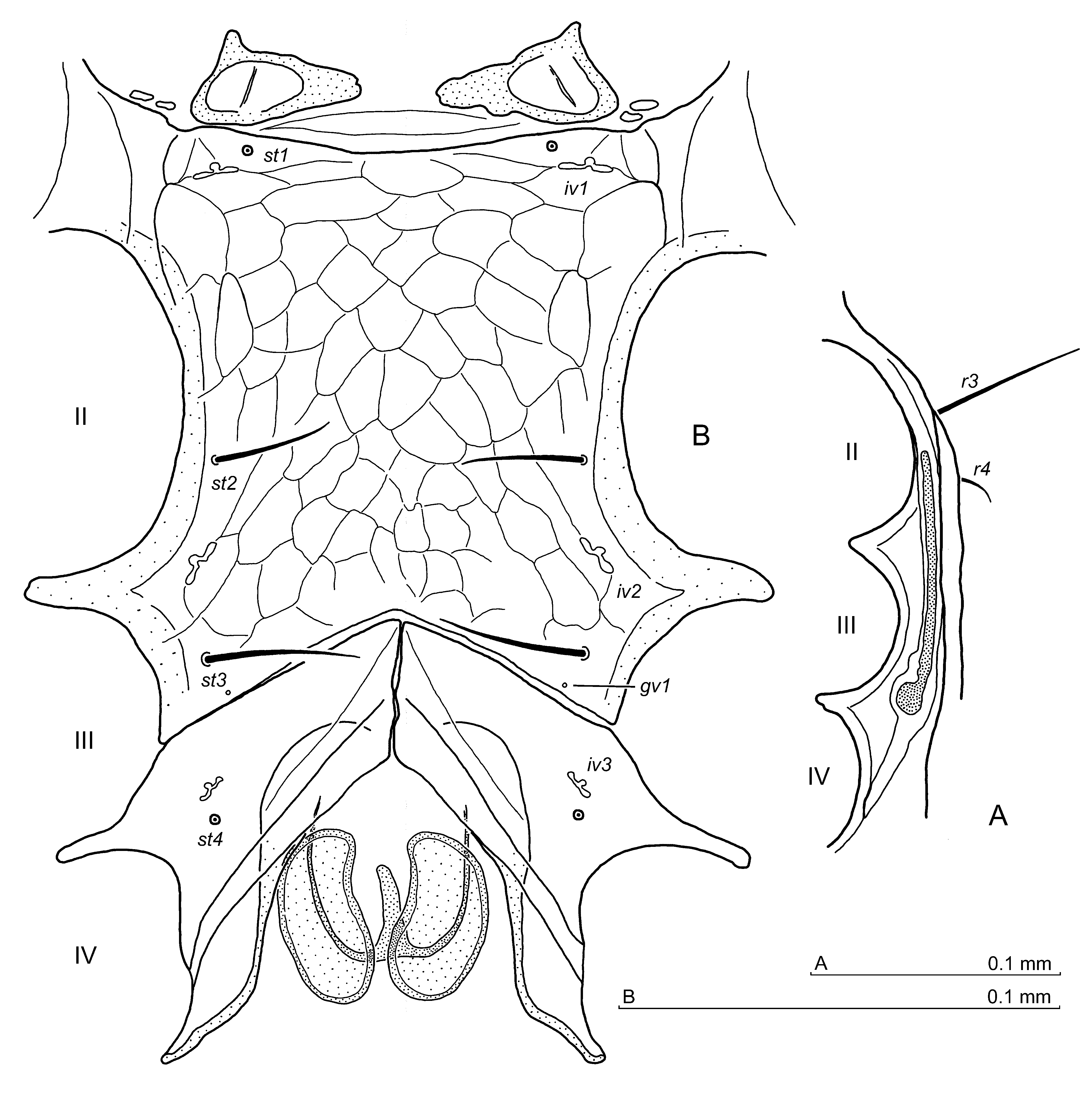
Idiosoma (Fig. 1) — Moderately sclerotised, 525–545 x 285–315 (length x width, n=5), holotype 545 x 320. Podonotum – setae length: 25–27 (j1), 27–31 (j2), 32–37 (j3), 26–31 (j4), 22–26 (j5), 20–24 (j6), 68–71 (r3), in holotype 25 (j1), 26 (j2), 33 (j3), 20 (j5), 21 (j6), 66 (r3), j4 broken. Opisthonotum – 22 pairs of setae (setae R5 and R6 lacking), setae length from ca. 20 up to 27, holotype 20–28, longer in the lateral parts. Dorsal setae simple, reticulation of podonotum poorly discernible, opisthonotum with a scale-like reticulation. Peritreme (Fig. 2) – length 97–107 including stigma (holotype 103), stigma diameter 10–12 (holotype 12), ending anteriorly in the midregion of the opening for Co II, i.e. behind the podonotal seta r3.
Ventral idiosoma — Setae length: 30–35 (st1), 38–43 (st2), 41–48 (st3), 30–33 (st4), 30–34 (st5), 30–34 (JV1), 17–22 (ZV1), in holotype 34 (st1), 38 (st2), 42 (st3), 31 (st4), 30 (st5), – (JV1 broken), 23 (ZV1). Ventral setae simple, reticulation of the sternum and opisthogaster scale-like. Anterior margin of the sternal shield (Fig. 2) with shallow concavity, presternal plates distant, gv1 pores close to the posterior margin of the sternum and far from each other. Paragynial shields (Figs 2, 3AB) metagynial sclerites indistinctive. Epigynial shield (Figs 2, 3B) the anterior margins slightly convex, posterolateral margins short and arcuate, the posterior one straight, in apical part of epigynium a less pigmented band present, denticles on internal (dorsal) surface absent. Endogynium (Figs 2, 3C) with axially elongated spherules and lateral arcuate lines. Adaxial margins of spherules thickened and prolonged anteriad to be a support for two backward directed hooks. Lamellar, broad and richly dentate stipule growing from a distinctly thickened base. Gland pores gv2 with two openings; iv5, ivo2, ivo3 and gv3 well discernible.
Gnathosoma — Gnathotectum (Fig. 3D) trispinate, all prongs narrow and acute, Corniculi conical, hypostome with 10 rows of denticles, hypostomatic and palpcoxa setae simple. Palptrochanter v1 seta simple, v2 barbed. Chelicera (Fig. 3E) movable digit with four teeth, the proximal one the largest. Fixed digit with 2 teeth in front of pilus dentilis and 2 teeth followed by the two tooth-like lamellar protrusions behind pilus dentilis. Between the teeth and the lamellar protrusions a minute tooth may be discernible.
Legs — Anterolateral setae (al1, al2) on Fe II short and conical, leg IV ventral setae on the tibia larger, posteroventral seta on the femur conical, some ventral and posterolateral setae on tarsus thickened, posterolateral seta on the basitarsus blunt terminally. Tr IV without a dorsal tubercle. Other aspects of legs I–IV unremarkable.
Male (Figures 4, 5)


Idiosoma — Sclerotized as in the female, 510–530 x 270–295 (length x width, n=5), body slightly incised at Co IV level. Podonotum – setae length: 26–30 (j1), 20–23 (j2), 22–25 (j3), 24–27 (j4), 22–25 (j5), 21–24 (j6), 64–69 (r3). Opisthonotum – setae length from ca. 18 up to 24. Peritreme – length including stigma 95–99 (stigma diameter 11–12), ending anteriorly, as in the females. Dorsal setae simple.
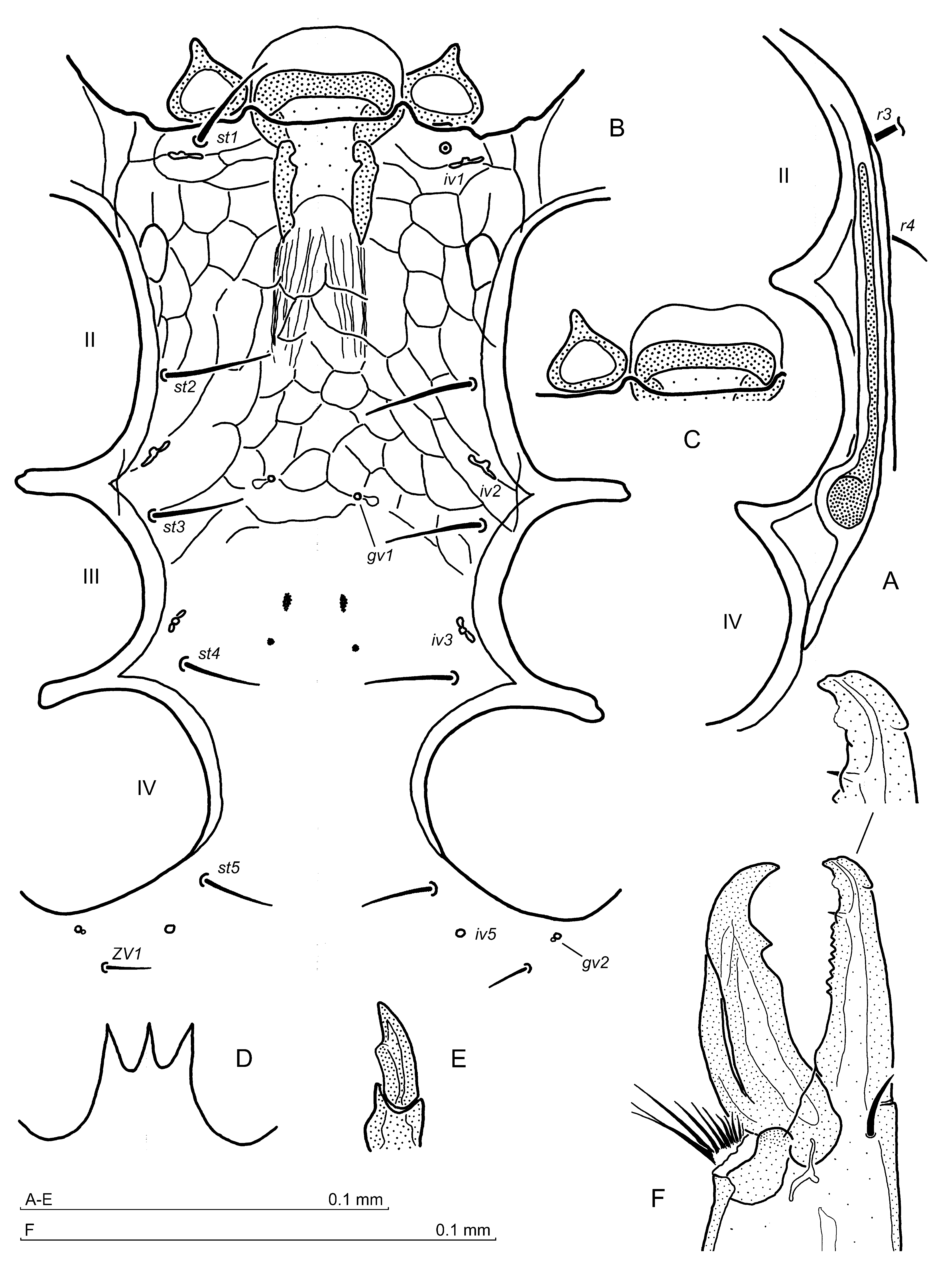
Ventral idiosoma — Setae length: 27–30 (st1), 26–30 (st2), 26–30 (st3), 18–21 (st4), 20–24 (st5), 24–26 (JV1), 12–14 (ZV1), other opisthogastral setae ca. 16–26. Ventral setae simple. Sternal region (Fig. 4A,B) – genital lamina with anterior margin slightly concaved and rounded anterior corners, flanked by subrectangular presternal plates. Sternum with gland pores gv1 at the st3 setae level, each one located at one-third of the distance between the setae bases, followed by two small elongated thickenings and two distinct circular thickenings of the sternal cuticle (Fig. 4B). Pores gv2 with a double channel, pore iv5 halfway between st5 and ZV1 setae. Sternum, opisthogaster and opisthonotum reticulation scale-like.
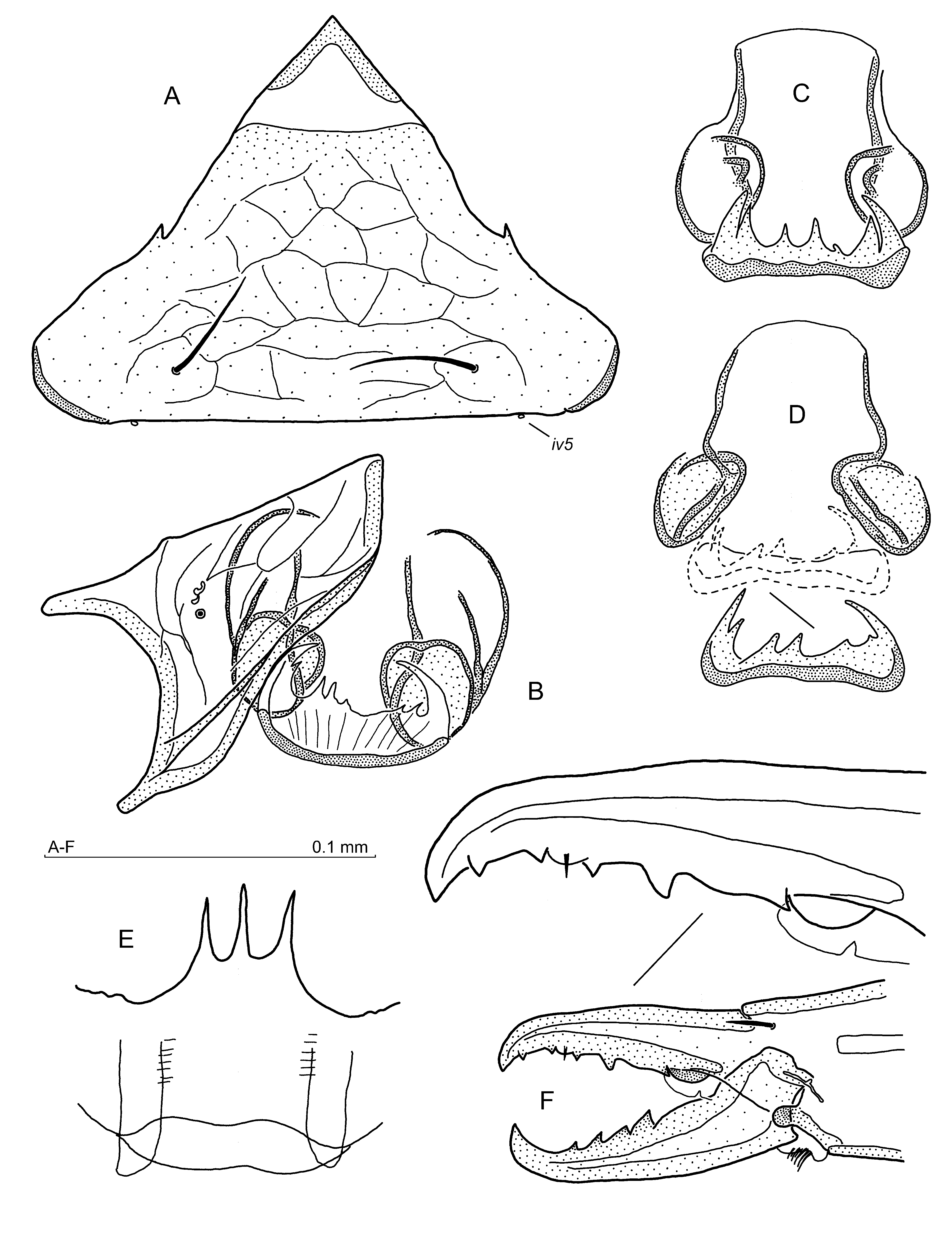
Gnathosoma — Gnathotectum (Fig. 4C) trispinate, prongs triangular and similar. Corniculi (Fig. 4D) with elevation on the adaxial margin, hypostome with 10 rows of denticles, hypostomatic setae simple, palpcoxal setae finely barbed. Palptrochanter v1 seta simple, v2 barbed. Chelicera (Fig. 4E,F): movable digit with one tooth followed by a sinuous edge proximally, fixed digit (Fig. 4F) narrow, with a tooth between the apex and pilus dentilis, followed by a row of 8–9 minute denticles; 1–2 similar denticles in front of pilus dentilis can be encountered.
Legs — Leg II (Fig. 5A–C) spurred as follows: when viewed from the ventral side, both the main spur and axillary process on femur slightly curved posterolaterally, the main spur with a low convexity on the posterolateral margin (Fig. 5A,B). Spurs on genu and tibia small (Fig. 5A), tip of genual spur ends closer to a distal article margin than the tibial spur. When the leg II is observed from a lateral perspective (Fig. 5C), femur main spur is finger-shaped, axillary process oval; spurs on genu and tibia conical, the one on the tibia slightly larger. Setae on leg II simple, setae al2 and ad2 on femur short and thick, ad1 normal, ad3 nidle-like. Seta al on Tr I short and thick, leg IV setation as in the female. Tubercle on Tr IV absent. Other aspects of legs I–IV unremarkable.
Type material
Holotype — female (slide no. 660 B), Rytro, southern Poland, 49.4767 °N, 20.6560 °E, ca. 490 m a.s.l., 11 Oct. 1970, beech forest litter. Paratypes — 6 females, 5 males (slides no. 660 C–H), ibid.; 13 females, 9 males (slides no. 1424, 1425), Jaworki, Czarna Woda, near Pieniny Mts., 49.4233 °N, 20.5771 °E, ca. 690 m a.s.l., 9 Sept. 2001, minor litter in a mixed forest; 9 females, 2 males (slides no. 1792, 1794), Hałuszowa, Pieniny Mts., 49.4264 °N, 20.3556 °E, ca. 675 m a.s.l., 10 Nov. 2003, spruce forest, a little hay stack at the forest edge.
Type deposition — Types are deposited in the Zoological Division of the Nature Education Centre, Jagiellonian University, Kraków, Poland.
Etymology
The specific name bihamatus (lat.: hamatus means hooked) refers to the endogynium which features two hooks located anteriorly.
Leptogamasus (Leptogamasus) montanus n. sp.
ZOOBANK: DF977E39-8961-4516-A35E-DA645199A5DD ![]()
(Figures 6–10)
Diagnosis
Female and male — Gnathotectum trispinate with pointed prongs, shorter in the male; gv1 present; podonotum with 21 seta pairs and opisthonotum with 23 seta pairs (1-3 extra setae can be present); Tr IV without tubercle.
Female — Presternal plates distant; the sternal shield anterior margin concaved; the epigynial shield with the anterior margins nearly straight, and with fine teeth facing coxae IV, the posterolateral margins convex and overlapping opisthogastral cuticle, the ventral epigynium surface without teeth, the margins of anterior epigynium tip more pigmented (an inverted-V); endogynium with roundish, distant spherules, stipule broad with lateral teeth the largest and curved adaxially, supported on a much sclerotized, usually posteriorly concaved basis.
Male — Genital lamina with rounded corners and a low convexity of anterior edge; presternal plates subrectangular; corniculi with the adaxial margin sinuous; chelicera fixed digit narrow and straight, apically blunt, with a row of 8–9 minute denticles behind pilus dentilis and 1–2 in front of it; leg II spurs in the ventral perspective: femur main spur is slightly directed posterolaterally and its apical part rounded and somewhat swollen, axillary process curved posterolaterally, spurs on the genu and tibia oval and at some distance from the distal article margin; Fe II laterally: the main spur straight, with a small elevation above seta pv1, axillary process tongue-shaped and directed towards the main spur.
Description
Female (Figures 6–8)


Idiosoma — Moderately sclerotised, 575–590 x 330–340 (length x width, n=2), holotype 576 x 351. Podonotum – setae length: 37–39 (j1), 42–44 (j2), 39–41 (j3), 41–42 (j4), 35–39 (j5), 33–34 (j6), 78–84 (r3), in holotype 40 (j2), 43 (j3), 34 (j5), 30 (j6), 75 (r3), j1 and j4 not available. Opisthonotum – setae length from ca. 27 up to 39, holotype 26–38, shorter along opisthonotum anterior margin. Opisthonotal setae and pores variable, both between specimens and specimen sides (Fig. 6). Dorsal setae simple, reticulation of podonotum not discernible except for some lines at the j4 setae level, opisthonotum with a scale-like reticulation. Peritreme (Fig. 7) – length 107–110 including stigma (holotype 110), stigma diameter 12–13 (holotype 12), ending anteriorly just behind the midregion of the opening for Co II, i.e. between the podonotal setae r3 and r4.
Ventral idiosoma — Setae length: 38–41 (st1), 38–43 (st2), 34–39 (st3), 29–34 (st4), 31–34 (st5), 34–38 (JV1), 21–24 (ZV1), in holotype 41 (st1), 42 (st2), 42 (st3), 35 (st4), 35 (st5), 39 (JV1), 25 (ZV1). Ventral setae simple, reticulation of the sternum and opisthogaster scale-like. Anterior margin of the sternal shield (Fig. 7) concaved, presternal plates distant, gv1 pores close to the posterior margin of the sternum and far from each other. Paragynial shields (Figs 7, 8B) metagynial sclerites arcuate and narrow. Epigynial shield (Figs 7, 8A) without teeth on the internal (dorsal) surface, the anterior margins nearly straight, with fine teeth facing coxae IV, posterolateral margins convex and overlapping the opisthogastral cuticle, the posterior margin slightly concaved. The margins of epigynium apex more pigmented in the form of inverted V. Endogynium (Figs 7, 8B–D) with roundish, distant spherules, stipule broad and dentate, with lateral teeth the largest and curved adaxially. The stipule grows from a much sclerotized, usually posteriorly concaved cuticle. Gland pores gv2 with two openings; iv5, ivo2, ivo3 and gv3 well discernible.
Gnathosoma — Gnathotectum (Fig. 8E) trispinate, prongs narrow and acute, Corniculi conical, hypostome with 9–10 rows of denticles, hypostomatic and palpcoxa setae simple. Palptrochanter v1 seta simple, v2 barbed on the external margin. Chelicera (Fig. 8F) movable digit with four teeth, the proximal one the largest. Fixed digit with 2 distant teeth in front, and 2 teeth behind pilus dentilis.
Legs — Setae al on Tr I and 2–3 setae on Fe I short and thick. Seta al2 on Fe II short and thick, whereas anteroventral seta thickened, but of normal length. Ti II with thickened ventral setae: anteroventral barbed, posteroventral simple. Leg IV: dorso- and posterolateral setae on femur thick and short, posteroventral seta on Ge IV and both ventral setae on the tibia thickened, out of those the posteroventral ones barbed. Some ventral and posterolateral setae on the tarsus thickened, posteroventral seta on the basitarsus terminally barbed. Tr IV without dorsal tubercle. Other aspects of legs I–IV unremarkable.
Male (Figures 9, 10)


Idiosoma — Sclerotized as in the female, 575 x 310 μm (length x width, n=1), Podonotum – the length of setae: 39 (j1), 44 (j2), 81 (r3), other podonotal setae j series not available. Opisthonotum – setae length from ca. 31 to 34, on the posterior margin up to 39. Peritreme – including stigma 107 long (stigma diameter 12), ending anteriorly as in the females. Dorsal setae simple.
Ventral idiosoma — Setae length: 39 (st1), 41 (st2), 36 (st3), 33 (st4), 32 (st5), 31 (JV1), 21 (ZV1), other opisthogastral setae ca. 25–40. Ventral setae simple. Sternal region (Fig. 9A) – genital lamina with a low convexity of the anterior edge and rounded anterior corners, flanked by subrectangular presternal plates. Sternum with gland pores gv1 at the st3 setae level, each one located from a seta at one-third of the distance between setae bases, followed by irregularly distributed four distinct circular thickenings of sternal cuticle. Pores gv2 with two openings, pore iv5 somewhat closer to st5 than ZV1 seta. Sternum, opisthogaster and opisthonotum reticulation scale-like.
Gnathosoma — Gnathotectum (Fig. 9B) trispinate, prongs triangular and similar. Corniculi (Fig. 9C) adaxial margin sinuous, hypostome with 8 rows of denticles, hypostomatic and palpcoxal setae simple. Palptrochanter v1 seta simple, v2 thicker and barbed. Chelicera (Figs 9C–E): movable digit with one tooth followed by a sinuous edge proximally, fixed digit with blunt apex, with 1–2 small teeth in front and a row of 8–9 minute denticles behind pilus dentilis.
Legs — Leg II (Fig. 10A–C) spurred as follows: when observed from the ventral side, femoral main spur is directed slightly posterolaterally and its apical part is rounded and somewhat swollen (Fig. 10B), axillary process curved posterolaterally. Spurs on the genu and tibia oval and located at some distance from the distal article margin, the one on the tibia is somewhat larger. When viewed from the lateral side, leg II (Fig. 10C) shows straight main spur with a small elevation above seta pv1, axillary process tongue-shaped and directed towards the main spur. Setae on leg II simple, setae al2 and ad2 on the femur short and thick, ad3 nidle-like, seta al1 also relatively short and thick. Seta al on Tr I short and thick, leg IV setation as in the female. Tubercle on Tr IV absent. Other aspects of legs I–IV unremarkable.
Type material
Holotype — female (slide no. 1979 A), Pańszczyca Valley, Tatra Mts., 49.2507 °N, 20.0383 °E, alt. ca. 1493 m a.s.l., 4 July 2005, sphagnum moss in a spruce forest. Paratypes — 3 females, 1 male (slides no. 1979 B–E), ibid.
Type deposition — Types are deposited in the Zoological Division of the Nature Education Centre, Jagiellonian University, Kraków, Poland.
Etymology
The specific name montanus refers to the type of specimens indigenous for a mountainous area.
Leptogamasus (Leptogamasus) renogynialis n. sp.
ZOOBANK: 19A88CAA-A7AD-4B3D-85F5-ADC4EF9774CB ![]()
(Figures 11–15)
Diagnosis
Female and male — Gnathotectum trispinate with similar, pointed prongs; gland pore gv1 present; podonotum with 21 seta pairs, opisthonotum features 24 pairs of setae (in some specimens one or two extra setae may be discernible); Tr IV with dorsal tubercle located distally.
Female — Presternal plates distant; sternal shield anterior margin slightly concaved; epigynial shield with anterior margins straight and a less pigmented lenticular area in apical part, the internal (dorsal) surface without teeth; endogynium with the kidney-shaped spherules and bacillary stipule.
Male — Genital lamina with convex margins; presternal plates subtriangular; corniculi with an elevation on the adaxial margin; chelicera fixed digit with a row of ca. 8 minute denticles behind pilus dentilis, and subapical incision on the dorsal edge; leg II with a femur main spur and axillary process curved posterolaterally in the ventral perspective, but straight when viewed from the side; spur on the genu conical, close to the distal article margin, tibial spur more elongated and more distant from the article margin; setae al1 and al2 on tibia and al1 on the genu barbed.
Description
Female (Figures 11–13)

Idiosoma (Fig 11) — Moderately sclerotised, 545–565 x 265–295 (length x width, n=5), holotype 530 x 245, usually with shallow lateral incisions at the Co IV level. Podonotum – the length of setae: 20–25 (j1), 15–18 (j2), 25–26 (j3), 25–27 (j4), 22–30 (j5), 22–26 (j6), 62–70 (r3), in holotype 26 (j1), 24 (j2), 26 (j3), 22 (j4), 20 (j5), 21 (j6), 65 (r3). Opisthonotum – setae length from ca. 20 up to 35, longest at the posterior margin. Dorsal setae simple, reticulation of podonotum poorly discernible, opisthonotum with a scale-like reticulation. Peritreme (Fig. 12A) – length 105–112 including stigma (holotype 108), stigma diameter 12–14 (holotype 13), ending anteriorly in the midregion of the opening for Co II, i.e. between the podonotal setae r3 and r4.
Ventral idiosoma — Setae length: 27–31 (st1), 34–38 (st2), 35–41 (st3), 25–29 (st4), 24–27 (st5), 30–35 (JV1), 18–22 (ZV1), in holotype 24 (st1), 31 (st2), 35 (st3), 20 (st4), 20 (st5), 27 (JV1), 13 (ZV1). Ventral setae simple, reticulation of opisthogaster scale-like. Anterior margin of the sternal shield (Fig. 12B) with shallow concavity, presternal plates distant, gv1 pores close to the posterior margin of the sternum and far from each other. Paragynial shields (Figs 12B, 13A) with narrow and arcuate metagynial sclerites. Epigynial shield (Fig. 13B), the anterior and posterior margins straight, the apical part with a less pigmented lenticular area, denticles on the internal (dorsal) surface absent. Endogynium (Figs 12B, 13C,D) with the kidney-shaped spherules and bacillary stipule, sometimes with a minute apical indentations. Gland pores gv2 with indistinctive double opening; iv5 poorly visible, whereas ivo2, ivo3, and gv3 well discernible.
Gnathosoma — Gnathotectum (Fig. 13E) trispinate, all prongs narrow and acute, similar in shape and size. Corniculi conical, hypostome with 10 rows of denticles, hypostomatic and palpcoxa setae simple. Palptrochanter v1 seta simple, v2 barbed. Chelicera (Fig. 13F) movable digit with four teeth, the proximal one the largest. Fixed digit with 3 teeth in front of pilus dentilis – the anteriormost located on adaxial digit side – and 2 teeth followed by the two tooth-like lamellar protrusions behind pilus dentilis.
Legs — Setae al on Tr I and 2–3 setae on Fe I short. Setae al1, al2 on Fe II short but thick, Ti II with the anteroventral seta barbed, and the posteroventral ones simple. Leg IV: posterodorsal setae on the femur short, both ventral setae on the tibia thickened, out of them the posteroventral ones barbed. Some ventral and posterolateral setae on the tarsus and basitarsus thickened, Tr IV (Fig. 13G) with moderately pronounced tubercle on the dorsal surface located distally. Other aspects of legs I–IV unremarkable.
Male (Figures 14, 15)

Idiosoma — Sclerotized and reticulated as in the female, 505–520 x 220–245 (length x width, n=5), body with lateral incisions at Co IV level. Podonotum – setae length: 25–28 (j1), 21–22 (j2), 26–31 (j3), 29–33 (j4), 20–24 (j5), 18–20 (j6), 60–65 (r3). Opisthonotum – setae length from ca. 20 up to 33, longer in the posterior part. Peritreme (Fig. 14A) – length including stigma 93–101 (stigma diameter 10–12), ending anteriorly as in the females. Dorsal setae simple.
Ventral idiosoma — Setae length: 24–28 (st1), 26–29 (st2), 25–29 (st3), 20–24 (st4), 18–22 (st5), 24–27 (JV1), 13–15 (ZV1), other opisthogastral setae ca. 20–25. Ventral setae simple. Sternal region (Fig. 14B) – genital lamina with the anterior margin arcuate, but sometimes wavy (Fig. 14C), lateral margins arcuate, flanked by subtriangular presternal plates. Sternum with gland pores gv1 slightly anterior to the st3 setae level, each one located at one-third of the inter-setae distance, followed by two pairs of distinct thickenings of the sternal cuticle, the anterior ones elongated, the posterior ones rounded. Pores gv2 with indistinctive double opening, pore iv5 halfway between st5 and ZV1 setae, ivo2, ivo3, and gv3 well discernible.
Gnathosoma — Gnathotectum (Fig. 14D) trispinate, prongs similar, triangular and acute. Corniculi (Fig. 14E) with an elevation on the adaxial surface, hypostome with 10–11 rows of denticles, hypostomatic and palpcoxal setae simple. Palptrochanter v1 seta simple, v2 finely barbed. Chelicera (Fig. 14F): movable digit with one tooth followed by a low edge proximally; fixed digit with a lamellar blunt denticle in front of pilus dentilis, a row of ca. 8 denticles behind pilus dentilis and an incision on an external (dorsal) margin in the apical part.
Legs — Leg II (Fig. 15A–C) spurred as follows: when viewed from the ventral side, both the main spur and axillary process slightly curved posterolaterally (Fig. 15A,B). Spurs on the genu and the tibia small (Fig. 15A), tip of genual spur ends closer to a distal article margin than the tibial spur. When the leg II is viewed from a lateral perspective (Fig. 15C), the femur main spur and axillary process are straight, finger-shaped; the spur on the genu conical, the one on the tibia finger-shaped. Femur main spur with a small ventral elevation in its basal part above the posteroventral seta (pv1). Setae on leg II simple except for the anterolateral setae (al1, al2) on the tibia and the distal anterolateral seta (al1) on the genu, which are finely barbed. Tr IV (Fig. 15D) with moderately pronounced dorsal tubercle. Ventral and some posterolateral setae on the tibia, basitarsus and tarsus of the fourth pair of legs thickened. Other aspects of legs I–IV unremarkable.
Material examined
Holotype — female (slide no. 2528), Ojców National Park, southern Poland, 50.2394 °N, 19.7867 °E, alt. ca. 400 m a.s.l., 20 Dec. 2014, beech forest litter. Paratypes — 21 females, 4 males (slide no. 605), Myślenice, southern Poland, 26 March 1970, litter in a spruce forest; 4 females, 6 males (slides no. 725A–725G), Pieniny National Park, Facimiech Mountain, 2 Oct. 1976, litter in a thermophilous fir and beech forest; 13 females, 6 males (slide no. 2537), Ojców National Park, southern Poland, 18 April 2015, beech forest litter. Other material — 27 females, 7 males (slides no. 606, 607), Myślenice, southern Poland, 26 March 1970, litter in a spruce forest; 1 female, 1 male (slide no. 732), Graz, Austria, 5 July 1975, litter in a beech forest, mixed with some pines and hazelnut bushes; 14 females, 13 males (slides no. 722, 723), Pieniny National Park, Mt. Facimiech, 2 Oct. 1976, litter in a thermophilous fir and beech forest.
Material deposition — Types are deposited in the Zoological Division of the Nature Education Centre, Jagiellonian University, Kraków, Poland, whereas all remaining material is held in the author's collection.
Etymology
The specific name renogynialis (lat.: ren + gr. gyne) refers to the kidney-shaped spherules in the female genitalia (endogynium).
Remarks on distribution
Leptogamasus (L.) renogynialis n. sp. is encountered as a relatively large populations in southern Poland in the two natural reserves, i.e. The Ojców National Park and Pieniny National Park, as well as in Gorce Mts, and across some hilly locations in south-eastern Poland, even though it was also encountered in Graz (Austria). The species seems to have its natural habitat in the beech or mixed forests, preferably in a mountainous terrain of moderate elevation.
New species differential taxonomy
In subgenus Leptogamasus females show more complex characteristics, so they are better for being selected as the holotypes. Most useful characteristics are the structure of the endogynium, epigynium, and paragynial features like metagynial sclerites, but also the dentation of the fixed digit which is not so readily available, though, as it requires sectioning. The males are in most cases very similar and their differential diagnosis is much weaker. It is postulated, therefore, to have the female specimens selected for identification purposes.
Leptogamasus (L.) bihamatus n. sp. female shows some features which should be compared with the two other species, i.e. Leptogamasus bidens (Sellnick, 1951) sensu Athias-Henriot, 1967 and Leptogamasus paracarpaticus Juvara-Balş, 1981. L. bidens described by Sellnick as Pergamasus bidens has been redescribed by Athias-Henriot (1967: 157) as Pergamasus (?) bidens Sellnick, 1950, although she put the identification into question. In fact, endogynium presented by Athias-Henriot (1967, fig. 706) differs from the one described by Sellnick (1951, fig. 3). Consequently, the specimen described by Athias-Henriot is purportedly a new species, even though a diligent examination of her specimen is still required. This species is similar to L. bihamatus n. sp. due to lateral tooth in the anterior part of the endogynial margins, but differs at least by the following characteristics: L. bidens has two teeth on the internal surface of the epigynium, which are absent in L. bihamatus n. sp., as well as a relative length of stipule, which is much shorter in a newly described species. On the other hand, L. bidens shows half-moon spherules with a large adaxial tooth each, whereas in L. bihamatus n. sp. spherules are edentated and elongated. Leptogamasus paracarpaticus presents epigynium edentate, short lamellar stipule and two teeth in the anterior part of the endogynium, although the spherules are of different shape than in L. bihamatus n. sp., anterior teeth are located on the endogynial sac wall rather than on the anteriorly elongated adaxial margins of the spherules.
Leptogamasus (L.) montanus n. sp. female shows a characteristic stipule richly dentated with marginal teeth, the longest and curved adaxially. Besides, stipule base is usually convex anteriorly. There are three species featuring a similar stipule and endogynium structure: Leptogamasus alstoni (Bhattacharyya, 1963), Leptogamasus doinae Juvara-Balş, 1981, and Leptogamasus nudiglobatus (Athias-Henriot, 1967). The first two species have teeth on the internal surface of the epigynium, whereas L. montanus n. sp. has the epigynium edentated. L. nudiglobatus differs mainly in view of its stipule, which is longer and devoid of any distinct base, as well as through the absence of evidently sclerotized walls of endogynial sac. Metagynial sclerite of paragynium is also different.
Leptogamasus (L.) renogynialis n. sp. is characterized mainly by the kidney-shaped spherules and bacillary stipule in the females. Such endogynium structure is absent in other Leptogamasus species. The most similar is Leptogamasus lossainti Athias-Henriot, 1972, but its spherules are shoe-shaped rather than kidney-shaped, and besides, the stipule is relatively wide instead of bacillary as in L. renogynialis n. sp.
The males of the newly described species have to be compared with the males belonging to most similar species of the females under study. Leptogamasus (L.) bihamatus n. sp. male differs from the males of L. bidens (Sellnick, 1951) sensu Athias-Henriot, 1967 and L. paracarpaticus by the structure of chelicera fixed digit, but also through the details of the second leg armature. The male of Leptogamasus (L.) montanus n. sp. can be compared with L. nudiglobatus male. Both males have similar chelicera structure, but differ with regard to the second leg armature and corniculi shape. Leptogamasus (L.) renogynialis n. sp. male differs from the male of L. lossainti by the shape of gnathotectum and the armature of leg II.
Acknowledgements
The present study was partly supported by a grant allocated by the Jagiellonian University (Grant Ref. No K/ZDS/008060).
References
Athias-Henriot, C. 1967. Observations sur les Pergamasus. I. Sous-genre Paragamasus Hull, 1918 (Zoologie Acariens anactinotriches, Parasitidae). Mémoires du Muséum National d$'$Histoire Naturelle, Paris, série A (Zoologie), 49: 1-197.
Athias-Henriot, C. 1972. Espèces françaises du sous-genre Leptogamasus s.s. (Arachnida, Gamasida, Parasitidae: genre Leptogamasus). Annales de la Société Entomologique de France, 8: 189-204.
Bhattacharyya, S.K. 1963. A revision of the British mites of the genus Pergamasus Berlese s.lat. (Acari: Mesostigmata). Bulletin of the British Museum (Natural History), Zoology, 11: 131-242. doi:10.5962/bhl.part.4717
Evans, G.O., Till, W.M. 1979. Mesostigmatic mites of Britain and Ireland (Chelicerata: Acari-Parasitiformes). An introduction to their external morphology and classification. Transactions of the Zoological Society of London, 35: 139-270. doi:10.1111/j.1096-3642.1979.tb00059.x
Johnston, D.E., Moraza, M.L. 1991. The idiosomal adenotaxy and poroidotaxy of Zerconidae (Mesostigmata: Zerconina). In: Dusbabek, F., Bukva, V. (Eds). Modern Acarology. The Hague: SPB Academie. p. 349-356.
Juvara-Balş, I. 1981. Nouvelle définition du genre Leptogamasus Trägårdh, 1936 (Acarina, Gamasida, Parasitidae) et description de six nouvelles espèces. Revue suisse de Zoologie, 88: 77-93. doi:10.5962/bhl.part.82355
Lindquist, E.E., Moraza, M.L. 1998. Observations of homologies of idiosomal setae in Zerconidae (Acari: Mesostigmata), with modified notation for some posterior body setae. Acarologia, 39: 203-226.
Moraza, M.L., Peña, M.A. 2005. The family Pachylaelapidae Vitzthum, 1931 on Tenerife Island (Canary Islands), with description of seven new species of the genus Pachylaelaps (Acari, Mesostigmata: Pachylaelapidae). Acarologia, 45: 103-129.
Sellnick, M. 1951. Zwei neue Milbenarten aus dem Marchfelde 1. Zeitschrift für Angewandte Entomologie 32: 275-278. doi:10.1111/j.1439-0418.1951.tb00626.x
Trägårdh, I. 1936. Leptogamasus, a new genus of Acari from Sweden. Entomologisk tidskrift, 57: 227-234.
Witaliński, W. 2019. Five new species of mites in the genus Leptogamasus Trägårdh, 1936, and a new subgenus Medioperigamasus (Parasitiformes: Parasitidae). Zootaxa, 4619: 487-517. doi:10.11646/zootaxa.4619.3.4


2020-08-12
Date accepted:
2020-09-29
Date published:
2020-10-05
Edited by:
Faraji, Farid

This work is licensed under a Creative Commons Attribution 4.0 International License
2020 Witaliński, Wojciech
 Download article
Download the citation
RIS with abstract
(Zotero, Endnote, Reference Manager, ProCite, RefWorks, Mendeley)
RIS without abstract
BIB
(Zotero, BibTeX)
TXT
(PubMed, Txt)