


Aceria species associated with Solanaceae worldwide with description of a new species
Tajaddod, Sadegh1 ; Lotfollahi, Parisa2 and de Lillo, Enrico3
1Department of Plant Protection, Faculty of Agriculture, Azarbaijan Shahid Madani University, Tabriz, Iran.
2✉ Department of Plant Protection, Faculty of Agriculture, Azarbaijan Shahid Madani University, Tabriz, Iran.
3Department of Soil, Plant and Food Sciences (Di.S.S.P.A.), University of Bari Aldo Moro, via Amendola, 165/a, 70126, Bari, Italy.
2020 - Volume: 60 Issue: 2 pages: 243-253
https://doi.org/10.24349/acarologia/20204365ZooBank LSID: 5F62B54F-AC4C-40AD-BF42-B3647A44D239
Original research
Keywords
Abstract
Introduction
The family Solanaceae is one of the largest and economically important families of flowering plants, including fruit, spice, and drug plants representing 8,400 scientific plant names of species (2,678 species names are accepted) within 115 plant genera (The Plant List on-line database 2013). This family includes evolutionarily successful and advanced taxa and shows high level of diversity reflected by the variety of life forms of its members, ranging from ephemeral herbs to large trees. They are cosmopolitan plants found throughout tropical and temperate regions, but with more focus in Australia and Latin America (Majaz Ganaie et al. 2018).
Due to the high host specificity of the eriophyoid mites, it seems that a large number of these mites must be found on Solanaceae. However, about 46 eriophyoid species have been collected on this plant family until now and 18 of them belong to the genus Aceria (Amrine and de Lillo unpublished database; Table 1). Six eriophyoid mite species are reported in Iran from Solanaceae including Aceria eucricotes (Nalepa), A. melongena (Zaher & Abou-Awad), A. paramacrodonis Kuang, Aculops lycopersici (Tryon), Tetra lycopersici Xue & Hong and Echinacrus ruthenicus Lotfollahi, de Lillo & Haddad (Sepasgozarian 1973; Baradaran-Anaraki and Daneshvar 1992; Ramazani et al. 2006; Jalilian et al. 2010; Kamali 2011; Xue and Hong, 2005, Xue et al. 2011; Lotfollahi et al. 2014, 2017; Delfan et al. 2015; Honarmand and Sadeghi 2016).


The seventh species from Solanaceae was collected from Lycium ruthenicum Murray in Iran. It is described and illustrated herein and a key to the Aceria species associated with Solanaceae plant species is given in order to assist species identification.
Material and Methods
Plant samples of Russian Box Thorn, L. ruthenicum, were collected in Ajabshir region of East Azerbaijan province (Iran), on July 2016. Eriophyoid mites were recovered from the plant samples by means of a modified washing method developed by Monfreda et al. (2007). The mites were slide mounted according to Baker et al. (1996) with some changes: specimens were directly placed in modified Hoyer's medium without previous clearing and fibers were interposed between slide and coverslip. Mounted specimens were cleared at 90°C for a few minutes. Then, the slides were dried for about four weeks in an oven at 47°C. The terminology and the setal notation in the morphological description of the mite follow mainly Lindquist (1996) and terminology of the internal female genital apparatus follows Chetverikov (2014) and Chetverikov et al. (2014). All morphological measurements were taken by means of a phase contrast microscope Olympus BX53, at 1,000 magnification (oil immersion), according to Amrine and Manson (1996) as modified by de Lillo et al. (2010), and are given in micrometers. Slight clarifications should be added as follows: dorsal semiannuli were counted from the first semiannulus behind the rear margin of the prodorsal shield; ventral semiannuli were counted from the first complete annulus after coxae II; coxigenital semiannuli were counted medially from the coxal region to the anterior margin of the external genitalia and were not included in the ventral semiannuli count. Measurements and means are rounded off to the nearest integer when required except of the minute characters. Measurements refer to the length of the morphological trait unless otherwise specified and are given in micrometers. In the female description, the holotype measurements are followed by range values, in parentheses, of the studied population (i.e. holotype and paratypes) and for males only the range values are given. The mean values of the paratypes are reported in the cases in which the measurements of the holotype could not be taken, due to the slide mounting position of the specimens and were marked by an asterisk (*) in the description. Line drawings were hand-drawn with a camera lucida according to de Lillo et al. (2010) and the abbreviations labelling schematic drawings follow mainly Amrine et al. (2003). The genus classification follows Amrine et al. (2003) but new genera described after 2003 were also considered. Host plant names and their synonymies are in accordance with ''The Plant List on-line database'' (2013).
Type materials are deposited at the Acarology Laboratory, Department of Plant Protection, Faculty of Agriculture, Azarbaijan Shahid Madani University, Tabriz (Iran).
Family Eriophyidae
Subfamily Eriophyinae
Tribe Acerini
Aceria ajabshiriensis n. sp.
ZOOBANK: 714B12BB-BC8E-43DA-BA85-BB6E61A48939 ![]()
Description — FEMALE (Figure 1; measured specimens n = 10).
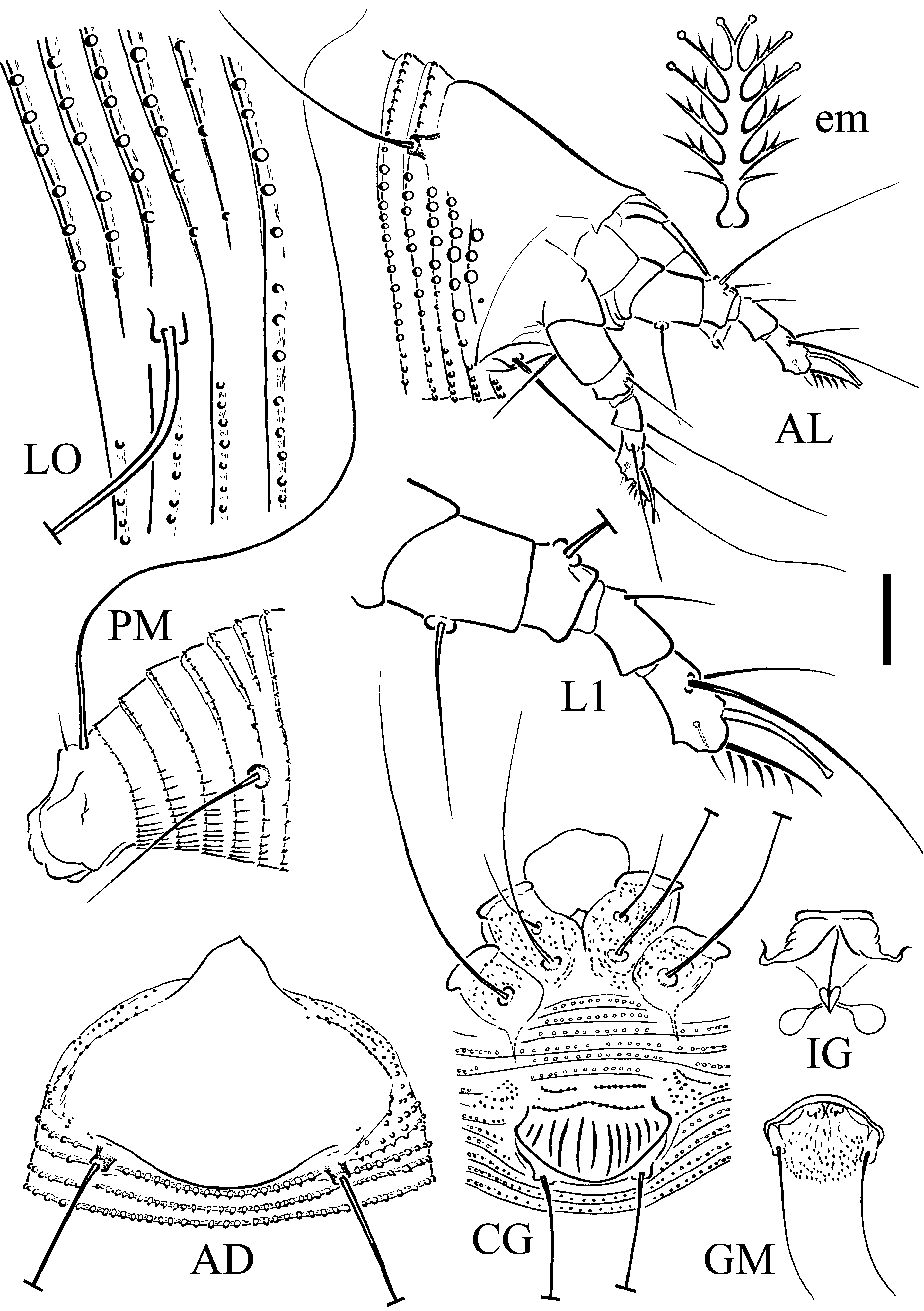
Body vermiform, 190 (173–205, excluding gnathosoma), 53* (52–54) thick, 55 (50–58) wide.
Gnathosoma projecting obliquely downwards, chelicerae 26 (26–30), palp 28 (26–35), palp coxal setae ep 3.5* (3–3.5), dorsal palp genual setae d 8 (7–10), unbranched.
Prodorsal shield 35 (24–35) including frontal lobe, 43* (40–45) wide, sub-circular; with a short flexible distally acuminate frontal lobe, 7 (5–7), over gnathosomal base, completely smooth. Tubercles of scapular setae sc on rear shield margin, 29 (27–29) apart, setae sc 30 (26–35), directed backward divergently.
Legs with all usual segments and setae. Leg I 31 (26–33), trochanter 6 (6–7), femur 8 (8–10), genu 6 (5–6), tibia 8 (6–9), tarsus 9 (7–9), tarsal solenidion ω6 (6–8) distally enlarged and tapered, empodium simple, 7.5 (6.5–9), 6-rayed; femoral setae bv 14* (12–16), genual setae l" 29 (25–33), paraxial tibial setae l' 8 (6–10), located in basal third of tibia, paraxial fastigial tarsal setae ft' 15 (10–19), antaxial fastigial tarsal setae ft" 26 (23–30), paraxial unguinal tarsal setae u' 4 (3–4.5). Leg II 31 (26–33), trochanter 5 (5–7), femur 10 (9–10), genu 5 (4–5), tibia 7 (5–8), tarsus 7 (7–8), tarsal solenidion ω8 (7–8.5) distally tapered, empodium simple, 6 (5.5–8), 6-rayed; femoral setae bv 12 (12–15), genual setae l" 11 (9–15), paraxial fastigial tarsal setae ft' 6 (6–9), antaxial fastigial tarsal setae ft" 24 (21–27), paraxial unguinal tarsal setae u' 4.5 (3.5–5.5).
Coxisternal region. Prosternal apodeme 5 (5–6.5), anterior setae on coxisternum I 1b 12* (10–19), 11 (9–11) apart; proximal setae on coxisternum I 1a 34 (27–41), 10 (7–10) apart; proximal setae on coxisternum II 2a 48 (44–54), 23 (18–23) apart; 7 (6–8) microtuberculate semiannuli between coxae and genital coverflap plus 3 (2–3) transversal rows of lined granules at the base of the coverflap. Coxae ornamented with numerous dots and dashes.
External genitalia 11 (10–15), 21 (21–22) wide, coverflap with 9 (9–10) longitudinal striae; setae 3a 22 (20–27), 14 (11–14) apart.
Internal genitalia: spermathecae ovoid, oriented posterolaterad; spermathecal tubes relatively short; transverse genital apodeme trapezoidal, distally folded.
Opisthosoma dorsally arched, with 48 (39–53) dorsal semiannuli, 64 (46–64) ventral semiannuli.
Microtubercles: subelliptical, on posterior margin of dorsal semiannuli, bigger on last 17–20th dorsal semiannuli and minute spiny on last 3 (no variation) dorsal semiannuli; circular, on posterior margin of ventral semiannuli, elongated and linear on last 5 (5–7) ventral semiannuli.
Setae c2 55 (37–57) on ventral semiannulus 11 (9–11), setae d 69 (69–86) on ventral semiannulus 23 (18–23); setae e 53 (47–72) on ventral semiannulus 40 (27–40); setae f 33 (28–36) on ventral semiannulus 59 (42–59); 5 (4–5) annuli posterior to setae f. Setae h2 110 (87–115) apically very fine, h1 3 (3–5).
MALE (measured specimens n = 3). Similar in shape and prodorsal shield arrangement to female. Body smaller than female, 125–150, 47–52 wide, 42 thick; palp genual setae d 7–8; prodorsal shield 24–32, 40–42 wide; setae sc 25–26, 20–25 apart. Opisthosoma with 35–41 dorsal semiannuli and 53–50 ventral semiannuli; 8 semiannuli between coxae and genitalia, with microtubercles similar to those of female. Setae: 1b 8.5–9, 1a 20–27, 2a 32–43, c2 37–47, d 42–44, e 33–41, f 22–29, h1 3, h2 55–67). Male genitalia 15–20 wide, setae 3a 15–21, 12 apart.
Type host plant — Lycium ruthenicum Murray (Solanaceae), Russian Box Thorn.
Type locality — Rahmanloo village, Ajabshir region, East Azerbaijan province, Iran (37°18'39.8'`N, 45°28'50.3''E), 1,290 m above sea level, coll. S. Tajaddod, late July 2016.
Type material — Holotype: single female on a microscope slide (LR-IEA-RO16T-1). Paratypes: 5 females and 3 males mounted singly on separate microscope slides (LR-IEA-RO16T-2–8).
Other material — Mites preserved in a vial (LR-IEA-RO16T) of Oudemans' fluid (Walter and Krantz, 2009) as extracted from the same sample as the type specimens.
Relation to the host plant — Vagrant; no apparent symptom was observed.
Etymology — This species is named after Ajabshir, the region where it was collected.
Differential diagnosis — The new species was compared with 18 Aceria species associated with the plants of family Solanaceae known to date. The new species closely resembles Aceria eucricotes (Nalepa) collected on Lycium europaeum L. from Algeria and, previously, also on L. ruthenicum from Iran (Lotfollahi et al. 2017). Both species have completely smooth prodorsal shields, similar number of empodial rays and body setal length. But these two species differ in number of dorsal semiannuli (39–53 in the new species versus 51–73 in A. eucricotes), number of semiannuli between coxae and genital coverflap (6–8 in the new species versus 3–5 in A. eucricotes). In addition, A. ajabshiriensis n. sp. has a short flexible distally acuminate frontal lobe, while A. eucricotes doesn't have a frontal lobe. Finally, the female genital coverflap of the new species is ornamented with 9–10 longitudinal striae, whereas A. eucricotes has a smooth coverflap.
Remarks — This is the third eriophyoid species collected on L. ruthenicum and all three species were collected from this host plant in Iran (Lotfollahi et al. 2014, 2017).
Key of the Aceria species associated to the Solanaceae plants
A key of the Aceria mite species collected on Solanaceae worldwide is proposed on the base of the most detailed published descriptions:
1. Female genitalia coverflap smooth
...... 2
— Female genitalia coverflap with ornamentations
...... 7
2. Empodium 6-rayed
...... 3
— Empodium 5-rayed
...... 4
3. Prodorsal shield completely smooth
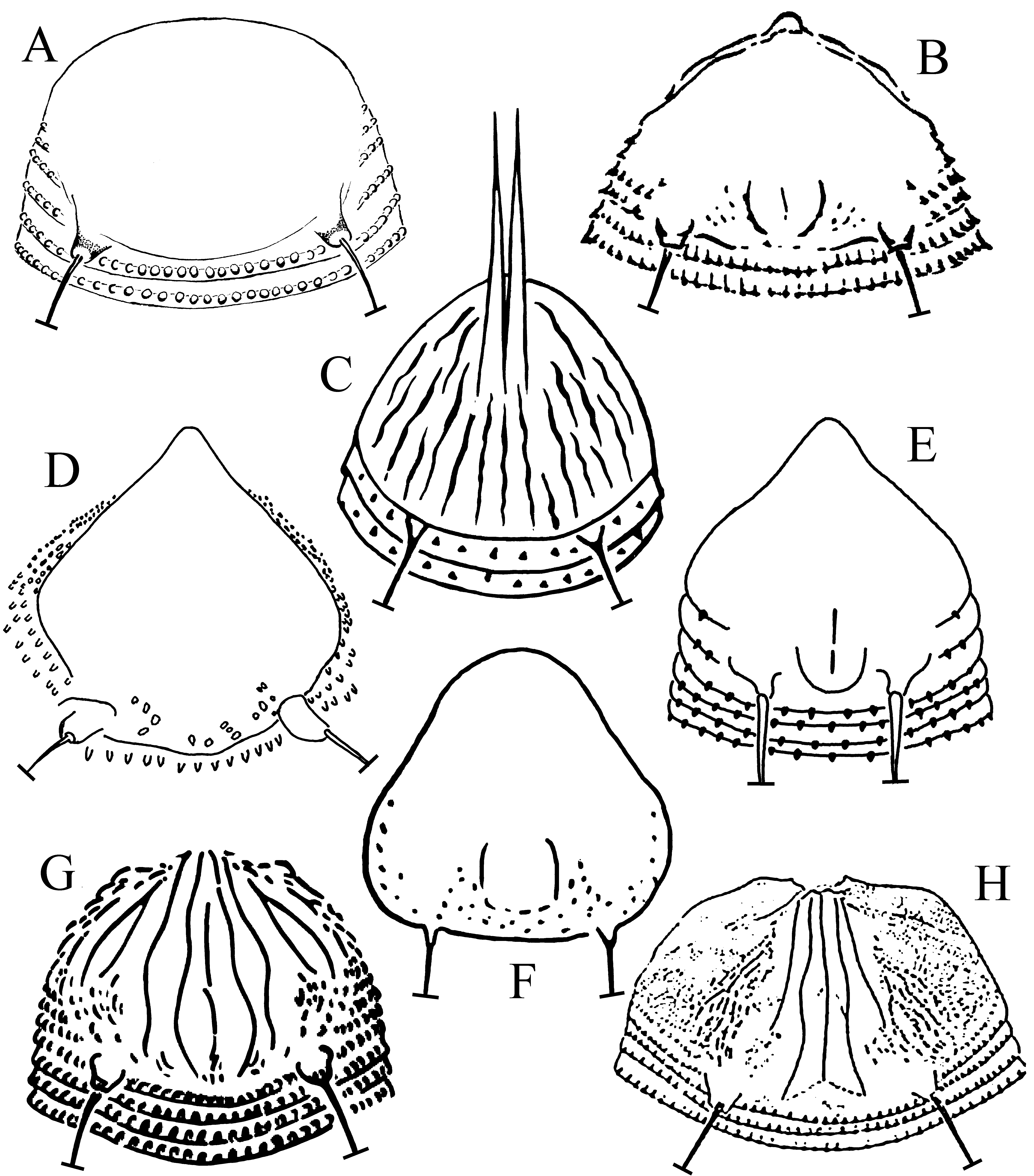
...... A. eucricotes (Figure 2A) (from Lotfollahi et al. 2017)
— Prodorsal shield with design almost absent, with very short median and admedian lines near rear prodorsal shield margin as short curved lines converging posteriorly, and surrounded outwardly by some granules
...... A. pallida (Figure 2B) (from Keifer 1964)
4. Prodorsal shield without distinct lines
...... 5
— Prodorsal shield with distinct design posteromedially
...... 6
5. Prodorsal shield very small, triangular, smooth or with obscure design
...... A. kendalli (no Figure available) (from Kendall 1929)
— Prodorsal shield with granules between setae sc tubercles near rear margin
...... A. kuko (Figure 2D) (from Ripka and Sanchez 2017)
6. Median line very short near rear prodorsal shield margin
...... A. parawagnoni (Figure 2E) (from Kuang 1983)
— Prodorsal shield with very short admedian lines and without median lines
...... A. paramacrodonis ( Figure 2F) (from Kuang 1988)
7. With two long horn-shaped projections anteriorly on prodorsal shield
...... A. bicornis (Figure 2C) (from Trotter 1900)
— Without the projections; frontal lobe normal if present
...... 8
8. Prodorsal shield completely smooth
...... A. ajabshiriensis n. sp. (Figure 1AD)
— Prodorsal shield with ornamentations
...... 9
9. Empodium 4-rayed
...... 10
— Empodium with more than 4 rays
...... 14
10. Setae e 4 and setae 3a 6
...... A. sodomaei (Figure 2G) (from Keifer 1976)
— Without this combination for setae e and length
...... 11
11. With more than 70 dorsal semiannuli
...... 12
— With less than 70 dorsal semiannuli
...... 13
12. Prodorsal shield with complete median line and first submedian lines in the middle of shield, quite close to admedian lines
...... A. melongena (Figure 2H) (from Zaher and Abou-Awad 1979)
— Prodorsal shield with incomplete median line (on posterior 2/3 of shield) and without first submedian lines
...... A. daturae (Figure 3A) (from Soliman and Abou-Awad 1978)
13. Opisthosoma with about 58 annuli and sc setae almost one and half the prodorsal shield length
...... A. baliotes (no Figure available) (from Nalepa 1921)
— Opisthosoma with about 70 annuli and sc setae almost twice the prodorsal shield length
...... A. lycopersici (Figure 3B) (from Farkas 1965)
14. Empodium 5-rayed
...... 15
— Empodium with more than 5 rays
...... 16
15. Prodorsal shield with numerous short dashes, obscuring the shield design
...... A. acnistii (Figure 3C) (from Keifer 1953)
— Prodorsal shield mostly smooth, with just a few dotted transverse lines near rear margin
...... Aceria dunaliae (Boczek & Oleczek, 1988) n. comb. (Figure 3D) (from Boczek and Oloczek 1988)
Note Boczek and Oleczek (1988) assigned this species to the genus Paraphytoptus. According to Amrine et al. (2003), members of this genus are characterized by wider annuli on the posterior opisthosoma. But in this species annuli of posterior opistosoma are continuous and subequal dorsoventrally and this species morphologically fits the diagnosis of the genus Aceria, and therefore we propose a new combination, Aceria dunaliae (Boczek and Oleczek, 1988) n. comb.
16. Prodorsal shield with submedian lines connected to admedian lines
...... A. annui (Figure 3E) (from Keifer 1977)
— Prodorsal shield without submedian lines
...... 17
17. Median and admedian lines very short near rear prodorsal shield margin; spiny microtubercles
...... A. wagnoni (Figure 3F) (from Keifer 1977)
— Without median lines, only short admedian lines present near rear prodorsal margin
...... 18
18. Prodorsal shield design close to the rear margin, consisting of short admedian lines subparallel, outwardly convex, surrounded laterally and posteriorly with granules; opisthosoma with about 70 annuli
...... A. macrodonis (Figure 3G) (from Keifer 1965)
— Prodorsal shield design weak and close to the rear margin, admedian lines represented by short centrally curved lines on rear 1/4; opisthosoma with 60 rings
...... A. caulicecis (Figure 3H) (from Keifer 1972)

Acknowledgements
This research was supported by Iran National Science Foundation (Iran) and partially by MIUR (Progetto ''Pietro Della Valle'') which are greatly appreciated.
References
Amrine J.W. Jr., Manson D.C.M. 1996. Preparation, mounting and descriptive study of Eriophyoid mites. In: Lindquist E.E., Sabelis M.W., Bruin J. (Eds), Eriophyoid Mites. Their Biology, Natural Enemies and Control. World Crop Pests, 6, Amsterdam, The Netherlands: Elsevier Science Publishers, p. 383-396. doi:10.1016/S1572-4379(96)80023-6 ![]()
Amrine J.W. Jr., Stasny TA.H., Flechtmann C.H.W. 2003. Revised keys to world genera of Eriophyoidea (Acari: Prostigmata). West Bloomfield, Michigan, USA: Indira Publish., pp. 244.
Baker E.W., Kono T., Amrine J.W. Jr., Delfinado-Baker M., Stasny T.A.H. 1996. Eriophyoid mites of the United States. West Bloomfield, Michigan, USA: Indira Publish., pp. 394 + i-viii.
Baradaran Anaraki P., Daneshvar H. 1992. Studies on the biology and chemical control of tomato russet mite, Aculops lycopersici (Acari: Eriophyidae), in Varamin. App. Entomol. Phytopath., 59(1-2): 25-27.
Boczek J.H., Oleczek M. 1988. Six new species of eriophyid mites (Acarida: Eriophyoidea). Rocz. Nauk Roln. Ser. E., Ochr. Rosl., 17(1): 107-118.
Chetverikov P.E., Craemer C., Vishnyakov A.E., Sukhareva S.I. 2014. CLSM anatomy of internal genitalia of Mackiella reclinata n.sp. and systematic remarks on eriophyoid mites from the tribe Mackiellini Keifer, 1946 (Eriophyoidea, Phytoptidae). Zootaxa, 3860(3): 261-279. doi:10.11646/zootaxa.3860.3.5 ![]()
Chetverikov P.E. 2014. Comparative confocal microscopy of internal genitalia of phytoptine mites (Eriophyoidea, Phytoptidae): new generic diagnoses reflecting host-plant associations. Exp. App. Acarol., 62: 129-160. doi:10.1007/s10493-013-9734-2 ![]()
de Lillo E., Craemer C., Amrine J.W. Jr., Nuzzaci G. 2010. Recommended procedures and techniques for morphological studies of Eriophyoidea (Acari: Prostigmata). Exp. App. Acarol., 51(1-3): 283-307. doi:10.1007/s10493-009-9311-x ![]()
Delfan A., Jafari S., Shakarami J. (2015) Identification of a part of plant mites of superfamily Eriophyoidea in Khoramabad region, Lorestan province. J. Entomol. Res., 7(2), 143-159.
Farkas H.K. 1965. Familie Eriophyidae, Gallmilben. Die Tierwelt Mitteleuropas, 3: 1-155.
Honarmand A, Sadeghi H. 2016. New records of eriophyoid mites (Eriophyoidea: Eriophyidae, Diptilomiopidae) from Iran. Entomofauna, 18: 297-308.
Jalilian F., Sheykholeslami M., Moini Naghadeh N., Mahjoob S.M., Tohidi M.T. 2010. Extensive contamination of tomato rust mite in greenhouses in Kermanshah province. 19th congress of Plant Protection of Iran, 31 July - 3 August, p. 387.
Kamali H. 2011. Current knowledge on Eriophyoidea (Acari: Prostigmata) as biological control agent of weeds in Iran. Proceeding of first Persian Congress of Acarology, 22-23 December, p. 72.
Keifer H.H. 1953. Eriophyid Studies XXI. Bull. Calif. Dept. Agr., 42: 65-79.
Keifer H.H. 1964. Eriophyid Studies B-12. Bur. Ent., Calif. Dept. Agric.: 1-20.
Keifer H.H. 1965. Eriophyid Studies B-16. Bur. Ent., Calif. Dept. Agric.: 1-20.
Keifer H.H. 1972. Eriophyid Studies C-7. ARS-USDA: 1-24.
Keifer H.H. 1976. Eriophyid Studies C-12. ARS-USDA: 1-24.
Keifer H.H. 1977. Eriophyid Studies C-13. ARS-USDA: 1-24.
Kendall J. 1929. Descriptions of four new forms of Eriophyes. Psyche, 36: 296-312. doi:10.1155/1929/85898 ![]()
Kishida K. 1927. [Illustrated Encyclopedia of the Fauna of Japan (Exclusive of Insects)]. The Hakuryukan Co. Ltd., Tokyo, 986 pp, 1900 figs (in Japanese).
Kuang H.-Y. 1983. [Notes on four species of eriophyid pests of wolfberry in China (Acarina: Eriophyoidea)]. J. Nanjing Agric. Univ., 12(4): 40-48.
Kuang H.-Y. 1988. [Two new specie of the genus Aceria from China (Acariformes: Eriophyidae)]. Acta Zool. Sinica, 13(1): 49-51.
Lindquist E.E. 1996. External anatomy and notation of structures. In: Lindquist E.E., Sabelis M.W., Bruin J. (Eds). Eriophyoid Mites. Their Biology, Natural Enemies and Control. World Crop Pests, 6, Amsterdam, The Netherlands: Elsevier Science Publishers, p. 3-31. doi:10.1016/S1572-4379(96)80003-0 ![]()
Lotfollahi P., de Lillo E., Haddad Irani-Nejad K. 2017. Contribution on Aceria spp. (Acari: Trombidiformes: Eriophyoidea) from southwest of East Azerbaijan province in Iran: new records and description of two new species. Syst. Appl. Acar., 22(8): 1167-1180. doi:10.11158/saa.22.8.4 ![]()
Lotfollahi, P., de Lillo, E., Haddad Irani-Nejad, K. 2014. Three new species from the subfamily Phyllocoptinae (Acari, Trombidiformes, Eriophyidae) in Iran. ZooKeys, 426: 17-27. doi:10.3897/zookeys.426.8087 ![]()
Majaz Ganaie M., Raja V., Ahmad Reshi Z., Verma V. 2018. Family Solanaceae: Taxonomy and modern trends. Ann. Plant Sci., 7(9): 2403-2414. doi:10.21746/aps.2018.7.9.1 ![]()
Monfreda R., Nuzzaci G., de Lillo E. 2007. Detection, extraction, and collection of Eriophyoid mites. Zootaxa, 1662: 35-43.
Nalepa A. 1892. Neue Gallmilben. 4. Fort. Anz. kais. Akad. Wiss., Math.-Natur Kl., Wien., 29(13): 128.
Nalepa A. 1921. Eriophyiden aus Java. III. Treubia, 2(1): 146-153.
Ramazani L., Mosaddegh M.S., Shishehbor P., Kamali K. 2006. Seven new records of eriophyoid mites on weeds from Iran. The Proceedings 17th Plant Protection Congress Iran, 185 p.
Ripka G., Sanchez I. 2017. A new Aceria species (Acari: Eriophyidae) from Spain on Pycnocomon rutifolium (Dipsacaceae) and supplementary descriptions of Aceria eucricotes and A. kuko from Lycium spp. (Solanaceae). Zootaxa, 4244 (2): 195-206. doi:10.11646/zootaxa.4244.2.2 ![]()
Sepasgozarian H. 1973. Mites and their economic important in Iran. Proceedings of the 3rd International Congress on Acarology, Dr. W. Junk Publishers Publisher, The Hague - Academia, 1971: 241-245. doi:10.1007/978-94-010-2709-0\_45 ![]()
Soliman Z.R., Abou-Awad A. 1978. Five new species of the genus Eriophyes in the A.R.E. (Acarina: Eriophyoidea: Eriophyidae). Acarologia, (1977) 19(4): 668-677.
The Plant List. 2013. Version 1.1. [Internet]. [Accessed 15 November 2019]. Available from: http://www.theplantlist.org/ ![]() .
.
Trotter A. 1900. Description d'une espece nouvelle d'Eriophyes (Acar.) de l'Amerique du Sud. Bull. Soc. Entomol. France, 11: 224-226.
Walter D.E., Krantz G.W. 2009. Collecting, rearing, and preparing specimens. In: Krantz G.W., Walter D.E. (Eds.), A Manual of Acarology, Third Edition. Texas Tech University Press, Lubbock Texas, USA: 83-96.
Wolffenstein O. 1879. Phytoptus lycopersici W. Monatsschrif des Vereins zur Befoerderung des Gartenbaues in den Konigl. Preuessischen Staaten, 22: 424-426.
Xue X.-F., Hong X.-Y. 2005. Five new species of the genus Tetra Keifer (Acari: Eriophyoidea) from China. Zootaxa, 1067: 37-48. doi:10.11646/zootaxa.1067.1.2 ![]()
Xue X.-F., Sadeghi H., Hong X.-Y., Sinaie S. 2011. Nine eriophyoid mite species from Iran (Acari, Eriophyidae). ZooKeys, 143: 23-45. doi:10.3897/zookeys.143.2162 ![]()
Zaher M.A., Abou-Awad B.A. 1979. Three new species of the genera Eriophyes and Phytoptus in Egypt. (Eriophyoidea: Eriophyidae). Acarologia, 20(4): 556-562.


2019-11-15
Date accepted:
2020-02-18
Date published:
2020-03-02
Edited by:
Roy, Lise

This work is licensed under a Creative Commons Attribution 4.0 International License
2020 Tajaddod, Sadegh; Lotfollahi, Parisa and de Lillo, Enrico
 Download article
Download the citation
RIS with abstract
(Zotero, Endnote, Reference Manager, ProCite, RefWorks, Mendeley)
RIS without abstract
BIB
(Zotero, BibTeX)
TXT
(PubMed, Txt)