


Contribution to systematics of the genus Eustigmaeus (Acari: Stigmaeidae) of Russia
Khaustov, Alexander A.1
1✉ Tyumen State University, Tyumen, Russia.
2019 - Volume: 59 Issue: 1 pages: 152-173
https://doi.org/10.24349/acarologia/20194320ZooBank LSID: 0554C6FA-E817-4CA9-858A-FF34D9109467
Original research
Keywords
Abstract
Introduction
The predatory mite family Stigmaeidae (Acari: Prostigmata) is the largest in the superfamily Raphignathoidea and includes about 598 species of 34 valid genera (Doğan et al. 2015; Fan & Ueckermann 2016; Fan et al. 2016; Khaustov 2016b; Paktinat-Saeij et al. 2016; Stathakis et al. 2016; Bingül & Doğan 2017; Bingül et al. 2017; Doğan et al. 2017; Khanjani et al. 2017; Khaustov et al. 2017; Nazari & Khanjani 2017; Akyol & Gül 2018; Da-Costa et al. 2018; Rehman et al. 2018; Khaustov & Tsurikov 2018). Among them, the genus Eustigmaeus Berlese, 1910 is the second largest genus with 128 species (Fan et al., 2016; Khaustov 2016b; Stathakis et al. 2016; Karasu et al. 2018; Khaustov & Tsurikov 2018).
In Russia 13 species of Eustigmaeus were recorded: E. anauniensis (Canestrini, 1889), E. clavatus (Canestrini and Fanzago, 1876), Eustigmaeus collarti (Cooreman, 1955), E. coronarius (Kuznetsov, 1977b), E. extremiorientalis Khaustov, 2016a, E. ioanninensis Kapaxidi and Papa-doulis, 1999, E. jiangxiensis Hu, Chen and Huang, 1996, E. parvisetus (Chaudhri, 1965), E. pinnatus (Kuznetsov, 1977a), E. plumifer (Halbert, 1923), E. rhodomela (Koch, 1841), E. segnis (Koch, 1836), and E. tjumeniensis Khaustov and Tolstikov, 2014 (Wainstein & Kuznetsov 1978; Khaustov & Tolstikov 2014; Khaustov 2016a).
During this study two new species, Eustigmaeus bochkovi n. sp. and E. grandis n. sp. were found from Khabarovsky and Primorsky Kray of Russia, respectively. The new species are described in this paper and new generic and species synonymies are also provided.
Materials and methods
The type materials of Eustigmaeus pinnatus and Paravillersia grata as well as specimens of Paravillersia grata and Eustigmaeus ioanninensis deposited in the collection of the Tyumen State University Museum of Zoology were examined. Specimens of Eustigmaeus bochkovi n. sp. and E. grandis n. sp. were collected from rotten wood and soil, respectively, using Berlese funnels and mounted on slides in Hoyer's medium.
Mite morphology was studied using a Carl Zeiss AxioImager A2 compound microscope with phase contrast and DIC objectives. Photomicrographs were taken with an AxioCam ICc5 digital camera. For SEM microscopy, alcohol-preserved mites were dried in freeze drying device JFD 320 (JEOL, Japan), dusted with gold and scanned with aid of a JEOL-JSM-6510LV SEM microscope.
In the description below, the palpal, idiosomal and the leg setation follows Grandjean (1939, 1944, 1946). The nomenclature of prodorsal setae follows Kethley (1990). All measurements are given in micrometers (μm) for the holotype and paratypes (in parentheses). In descriptions of leg setation the number of solenidia is given in parenthesis.
Taxonomy
Family Stigmaeidae Oudemans, 1931
Genus Eustigmaeus Berlese, 1910
Type species: Stigmaeus kermesinus Koch, 1841, by original designation.
Eustigmaeus bochkovi n. sp.
(Figs 1–13)
ZOOBANK: 904DD186-CC7C-4E6A-BC31-85B3EC6204C5 ![]()
Description
Female (Figs 1–5) (n=4)


Idiosoma oval. Length of idiosoma 345 (325–375), width 235 (225–270).
Idiosomal dorsum (Figs 1A, 4A, C, 5) — Eyes present. Idiosoma almost completely covered by 2 large shields. Shields with large round dimples (Figs 1A, 4C, 5A, B) and distinct subcuticular reticulation. Dorsal setae baculiform, with weakly developed hyaline sheaths distally; setae e1, f1, h1, and h2 with many small bards (Fig. 5D), other dorsal setae smooth or with 1-2 minute barbs. Setae h1 and h2 situated ventrally. Prodorsal shield subtriangular, with weak incisions laterally to bases of setae sci and 2 pairs of small apodemal marks near bases of setae ve and near posterior margin. Hysterosomal shield with 1 pair of small apodemal marks posteriorly to setae e1 and narrow incisions posterolaterally to setae e2. Lengths of dorsal setae: vi 40 (36–39), ve 48 (42–45), sci 35 (31–37), sce 40 (37–44), c1 34 (31–34), c2 46 (42–45), d1 34 (31–34), d2 39 (37–42), e1 35 (34–37), e2 39 (36–41), f1 44 (46–49), h1 34 (34–43), h2 39 (34–38).
Idiosomal venter (Figs 1B, 4B, E, F, 5C) — With 1 small oval callosity located on soft striated cuticle between endopodal plates of legs III and IV (Fig. 4E). Suranal plate situated ventrally, with distinct large dimples. Endopodal plates separated medially. Humeral plate subtriangular, with distinct large dimples. Most of ventral setae weakly barbed and pointed; with 3 pairs of simple subequal aggenital and 3 pairs of pseudanal setae, of which setae ps2 short, blunt-ended and smooth. Aggenital plate smooth, with very weak subcuticular reticulation posteriorly to setae ag1 (Fig. 4F). Coxal and endopodal plates of legs I-IV with weak subcuticular reticulation. Lengths of ventral setae: 1a 26 (22–24), 1b 27 (22–27), 1c 26 (20–24), 2b 26 (21–24), 2c 23 (21–23), 3a 28 (24–26), 3b 25 (22–25), 3c 22 (17–20), 4a 22 (20–23), 4b 21 (17–21), 4c 20 (18–19), ag1 17 (15–18), ag2 21 (18–20), ag3 24 (20–22), ps1 25 (23–25), ps2 7 (7–8), ps3 24 (18–20).
Gnathosoma (Figs 2, 4D) — Tibial claw well-developed. Seta l' on palpal tibia short, spine-like. Seta d of palpal femur blunt-ended, barbed; other palpal setae of femur, genu and tibia (except l'Ti) pointed and barbed; all setae of palptarsus smooth. Number of setae on palpal segments: Tr 0, Fe 3 (d, l', v''), Ge 2 (d, l''), Ti 3 (d, l', l''), Ta 8(1) (fused eupathidia ul', ul'', sul, eupathidion acm, ba, bp, lp, 1 solenidion ω). Palpal supracoxal setae (ep) needle-like, located dorsally (Fig. 5C). Rostrum of subcapitulum distinctly elongate. All subcapitular setae pointed; setae or1 and m smooth, other setae with 1-2 weak barbs. Basal part of subcapitulum weakly punctate and with subcuticular reticulation posterolaterally to setae n (Fig. 4D). Length of subcapitular setae: m 29 (27–30), n 20 (17–20), or1 13 (15–16), or2 20 (19–20). Chelicerae dorsally smooth, with long stylets.
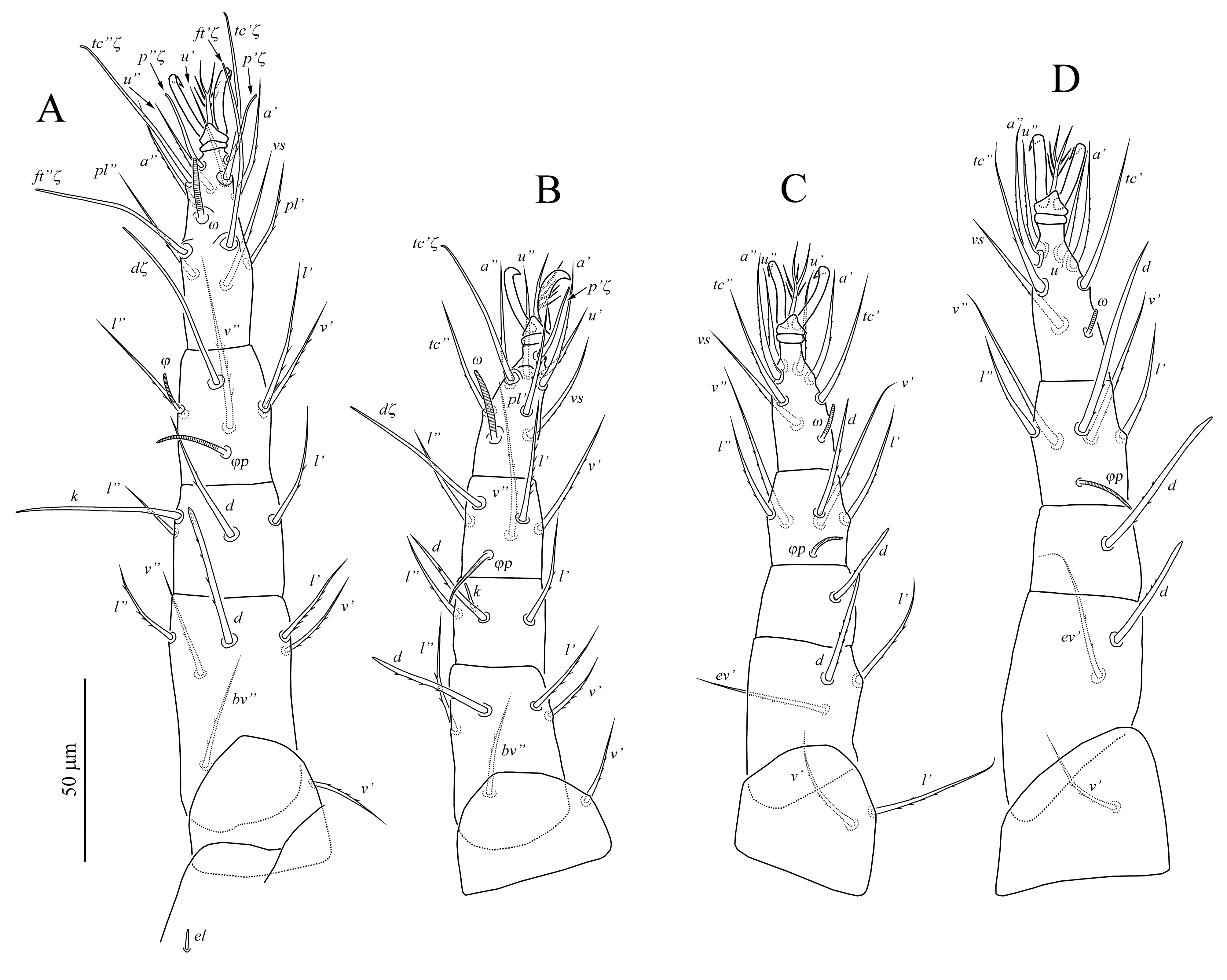
Legs (Fig. 3) — Empodial raylets not capitate. Leg I (Fig. 3A). Coxae I posterodorsally with needle-like leg supracoxal setae (el). Leg setation: Tr 1 (v'), Fe 6 (d, l', l'', v', v'', bv''), Ge 4 (d, l', l'', k), Ti 5(2) (dξ, l', l'', v', v'', φ, φp), Ta 13(1) (p'ξ, p'' ξ, tc' ξ, tc'' ξ, ft' ξ, ft'' ξ, u', u'', a', a'', pl', pl'', vs, ω). Setae d of tibia, (p), (tc) and (ft) of tarsus are eupathidia. Seta d of femur barbed, with weak hyaline sheath; seta k 45 (45–48) smooth, blunt-ended, subequal with seta d of genu; other dorsal setae (except eupathidia) pointed and barbed. Solenidion ω short 17 (17–18), finger-shaped; solenidion φ 11 (10–11) baculiform, solenidion φp 21 (18–22) attenuate. Leg II (Fig. 3B). Leg setation: Tr 1 (v'), Fe 5 (d, l', l'', v', bv''), Ge 4 (d, l', l'', k ), Ti 5(1) (dξ, l', l'', v', v'', φ), Ta 9(1) (p'ξ, tc'ξ, tc'', u', u'', a', a'', pl', vs, ω). Setae d of tibia, p' and tc' of tarsus represented by eupathidia. Seta d of femur blunt-ended and barbed; seta k 7 (7) of genu short, rod-like; other setae (except eupathidia) pointed and barbed. Solenidion ω 19 (19–21) finger-shaped; solenidion φp 17 (17–19) attenuate. Leg III (Fig. 3C). Leg setation: Tr 1 (v'), Fe 3 (d, l', ev'), Ge 1 (d), Ti 5(1) (d, l', l'', v', v'', φ), Ta 7(1) (tc', tc'', u', u'', a', a'', vs, ω). Solenidion ω 10 (9–10) finger-shaped; solenidion φp 12 (12–15) attenuate. All leg setae barbed. Setae d of femur and genu distinctly blunt-ended, seta d of tibia weakly blunt-ended; other setae pointed. Setae (u) of tarsus smooth, other tarsal setae weakly barbed. Leg IV (Fig. 3D). Leg setation: Tr 1 (v'), Fe 2 (d, ev'), Ge 1 (d), Ti 5(1) (d, l', l'', v', v'', φ), Ta 7(1) (tc', tc'', u', u'', a', a'', vs, ω). Solenidion ω 7 (5–6) baculiform; solenidion φp 18 (16–18) attenuate. All leg setae barbed (sometimes tc' smooth). Setae d of femur and genu distinctly blunt-ended, seta d of tibia weakly blunt-ended; other setae pointed.
Male(Figs 6–7) (n=3)

Idiosoma oval, but opisthosoma more narrower than in female. Length of idiosoma 260–280, width 165–185.
Idiosomal dorsum (Fig. 6A) — In general similar to female, but hysterosomal shield transversely divided into 2 shields; anterior shield more clearly reticulated than posterior, with setae c1, d1, d2; posterior shield with setae e1, e2, f1. Suranal shield and genital opening located dorsally. Genital opening with 2 well-sclerotized projections. Aedeagus long and narrow, weakly sclerotized. Setae ps1-3 short, smooth, spiniform, other dorsal setae weakly barbed, baculiform, without distinct hyaline sheaths. Lengths of dorsal setae: vi 26–29, ve 32–36, sci 23–32, sce 31–33, c1 25–27, c2 34–37, d1 26–27, d2 31–33, e1 21–22, e2 30–33, f1 32–35, h1 10–12, h2 32– 35, ps1 3–4, ps2 4–5, ps3 7–8.
Idiosomal venter (Fig. 6B) — Podosoma as in female. Opisthosoma with smooth, weakly sclerotized aggenital plate fused posteriorly with suranal plate. Aggenital plate with 3 pairs of smooth or weakly barbed aggenital setae. Lengths of ventral setae: 1a 23–24, 1b 23–24, 1c 20–21, 2b 21–24, 2c 22–23, 3a 24–25, 3b 23–24, 3c 19–20, 4a 19, 4b 19–20, 4c 18–19, ag1 17–18, ag2 18–20, ag3 19–20.
Gnathosoma — As in female.
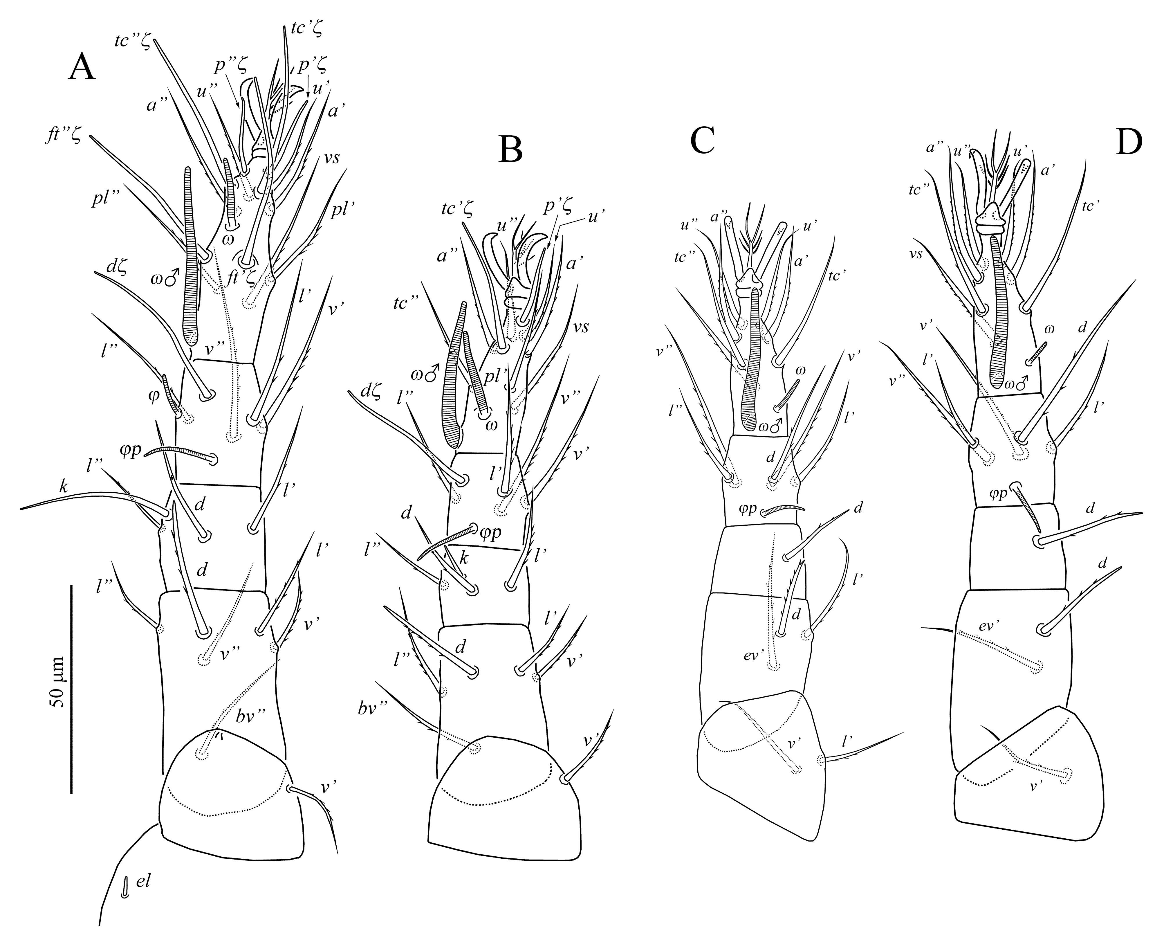
Legs (Fig. 7) — In general similar to those of female except presence of male solenidia on tarsi I–IV. Leg I (Fig. 7A). Seta d of femur barbed, blunt-ended, without hyaline sheath. Lengths of seta k 43 and solenidia: ω 16–17, ω♂ 44–46, ϕ 10, ϕp 18–20. Leg II (Fig. 7B). Lengths of seta k 8 and solenidia: ω 21–22, ω♂ 37–38, ϕp 15–16. Leg III (Fig. 7C). Lengths solenidia: ω 10, ω♂ 34–35, ϕp 13–14. Leg IV (Fig. 7D). Lengths solenidia: ω 6–7, ω♂ 35–36, ϕp 14–16.
Female deutonymph (Figs 8–10) (n=1)


Length of idiosoma 275, width 185.
Idiosomal dorsum (Figs 8A, 10A) — In general similar to female, but setae sce, d2, e2, f1 each located on separate plates; setae c1 and d1 on subrectangular central hysterosomal shield; setae e1 on unpaired shield. Suranal shield located dorsally. Dorsal setae weakly barbed or smooth, baculiform, without distinct hyaline sheaths, setae h2 pointed. Lengths of dorsal setae: vi 30, ve 37, sci 25, sce 34, c1 29, c2 36, d1 29, d2 31, e1 29, e2 33, f1 44, h1 34, h2 29.
Idiosomal venter (Figs 8B, 10B–D) — As in female. Lengths of ventral setae: 1a 23, 1b 23, 1c 23, 2b 20, 2c 20, 3a 20, 3b 19, 3c 19, 4a 18, 4b 19, 4c 17, ag1 14, ag2 13, ag3 16, ps1 23, ps2 4, ps3 15.
Gnathosoma — As in female.
Legs (Fig. 9) — In general similar to those of female except the absence of setae d on genu II, v' on femur II, d on genua III and IV, and v' on trochanter IV. Leg I (Fig. 9A). Lengths of seta k 37 and solenidia: ω 15, ϕ 8, ϕp 16. Leg II (Fig. 9B). Lengths of seta k 8 and solenidia: ω 19, ϕp 16. Leg III (Fig. 9C). Lengths solenidia: ω 8, ϕp 12. Leg IV (Fig. 9D). Lengths solenidia: ω 6, ϕp 13.
Male deutonymph (Fig. 11) (n=1)

Length of idiosoma 306, width 205.
In general, similar to female deutonymph except setae h1 short, smooth, spiniform and located dorsally (Fig. 11). Lengths of dorsal setae: vi 25, ve 33, sci 27, sce 28, c1 25, c2 30, d1 25, d2 27, e1 25, e2 26, f1 36, h1 28, h2 26. Lengths of ventral setae: 1a 24, 1b 19, 1c 16, 2b 17, 2c 17, 3a 21, 3b 17, 3c 17, 4a 15, 4b 14, 4c 14, ag1 12, ag2 13, ag3 15, ps1 3, ps2 3, ps3 9.
Leg I. Lengths of seta k 34 and solenidia: ω 12, ϕ 8, ϕp 14. Leg II. Lengths of seta k 8 and solenidia: ω 14, ϕp 12. Leg III. Lengths solenidia: ω 7, ϕp 11. Leg IV. Lengths solenidia: ω 4, ϕp 10.
Protonymph (Figs 12–13) (n=2)


Length of idiosoma 250–255, width 180.
Idiosomal dorsum (Fig. 12A) — In general similar to deutonymph, but suranal shield located ventrally, setae h1 pointed. Lengths of dorsal setae: vi 25–27, ve 30–33, sci 20–21, sce 27–30, c1 24–27, c2 30, d1 23–28, d2 28, e1 25–28, e2 27–29, f1 39–41, h1 28–31, h2 25.
Idiosomal venter (Fig. 12B) — Similar to deutonymph, except setae 4a, 4b, 4c absent, aggenital plate with 1 pair of setae. Lengths of ventral setae: 1a 20, 1b 20–21, 1c 18, 2b 16–18, 2c 16–17, 3a 21–24, 3b 17–19, 3c 17–19, ag1 16–21, ps1 12, ps2 3, ps3 11.
Gnathosoma — As in deutonymph, except absence of setae m.
Legs (Fig. 13) In general similar to those of deutonymph, except the absence of setae v', v'' on femur I, v' on trochanters I–III, and d on femur IV; also setae (ft) on tarsus I and d on tibia II not eupathidia. Leg I (Fig. 13A). Lengths of seta k 30–35 and solenidia: ω 14, ϕ 7–8, ϕp 14–15. Leg II (Fig. 13B). Lengths of seta k 7 and solenidia: ω 16–18, ϕp 12. Leg III (Fig. 13C). Lengths solenidia: ω 6–7, ϕp 11. Leg IV (Fig. 13D). Lengths solenidia: ω 5, ϕp 9.
Type material
Female holotype, slide N° ZISP T-St-002, Russia: Khabarovsky Kray, Nanayskiy region, 48°54'N, 136°17'E, in rotten log, 16 August 2018, coll. A.A. Khaustov. Paratypes: 3 females, 1 female deutonymph, 1 male deutonymph, 2 protonymphs, same data as holotype.
Etymology
The specific name is given after the prominent Russian acarologist, Andrey Bochkov who passed away in 2018.
Differential diagnosis
The new species differs from all known species of Eustigmaeus by the presence of callosity located between endopodal plates of legs III and IV (absent in other species).
Type deposition
The holotype is deposited in the collection of the Zoological Institute of Russian Academy of Sciences, St. Petersburg, Russia. All paratypes are deposited in the collection of the Tyumen State University Museum of Zoology, Tyumen, Russia.
Eustigmaeus grandis n. sp.
(Figs 14–17)
ZOOBANK: 37D41070-3037-45B2-9EC7-E703BDE85E54 ![]()
Description
Female (Figs 14–17) (n=8)


Idiosoma broadly oval. Length of idiosoma 405 (385–435), width 330 (325–345).
Idiosomal dorsum (Figs 14A, 17A, D, E) — Eyes present. Idiosoma almost completely covered by 2 large and well sclerotized plates. Plates punctate, with large round dimples (Figs 17D, E) and distinct subcuticular reticulation. Dorsal setae subequal, brush-like distally. Setae h1 and h2 situated ventrally. Lengths of dorsal setae: vi 47 (44–48), ve 50 (47–53), sci 42 (38–43), sce 37 (34–3)9, c1 41 (34–43), c2 30 (29–31), d1 43 (41–44), d2 37 (36–37), e1 47 (46–47), e2 44 (41–45), f1 52 (51–53), h1 45 (42–46), h2 32 (27–33).
Idiosomal venter (Figs 14B, 17F) — With 2 oval callosities located anterolaterally and posterolaterally to humeral plate; anterior callosity distinctly larger than posterior. Suranal plate situated ventrally, with distinct large dimples, subcuticular reticulation and punctate in central part. Endopodal plates separated medially. Humeral plate weakly sclerotized, subtriangular, with distinct large dimples. Ventral setae smooth and pointed; with 2 pairs of simple subequal aggenital, and 3 pairs of pseudanal setae. Aggenital plate smooth, with weak subcuticular reticulation posteriorly to setae ag1. All coxal plates distinctly punctate. Coxal and endopodal plates of legs I-IV with weak subcuticular reticulation. Lengths of ventral setae: 24 (20–26), 1b 23 (21–25), 1c 17 (16–18), 2b 18 (16–18), 2c 18 (16–19), 3a 25 (24–26), 3b 19 (17–19), 3c 16 (15–17), 4a 20 (18–20), 4b 19 (18–20), 4c 20 (19–21), ag1 17 (16–18), ag2 17 (15–18), ps1 16 (15–17), ps2 16 (14–17), ps3 15 (14–17).
Gnathosoma (Figs 15, 17B, C) — Tibial claw well-developed. Setae l' on palpal tibia short, spine-like, with slightly angulate margin. All palpal setae of femur, genu and tibia (except l'Ti) pointed and barbed; setae of palptarsus smooth, except weakly barbed va. Number of setae on palpal segments as in E. bochkovi n. sp. Palpal supracoxal setae (ep) needle-like, slightly curved, located ventrolaterally. Rostrum of subcapitulum short. All subcapitular setae smooth. Setae or2 distinctly blunt-ended and curved, other subcapitular setae pointed. Basal part of subcapitulum distinctly punctate and with indistinct subcuticular reticulation posterolaterally to setae n (Fig. 17C). Length of subcapitular setae: m 18 (17–19), n 17 (16–18), or1 15 (13–15), or2 17 (16–18). Chelicerae dorsally distinctly punctate (Fig. 17B) with short stylets.
Legs (Fig. 16) — Empodial raylets not capitate. Leg setation as in E. bochkovi n. sp. Leg I (Fig. 16A). Coxae I posterodorsally with needle-like leg supracoxal setae (el). Setae (p), (tc) and (ft) of tarsus are eupathidia. Setae d, l'' of femur, d, (l) of genu, and d, l' of tibia brush-like distally, located on small protuberances; other setae (except eupathidia) pointed; setae (u) of tarsus smooth, other setae (except eupathidia) sparsely barbed. Seta k 10–11 smooth, blunt-ended, more than twice shorter than seta d of genu. Solenidion ω long 25 (23–27), narrow, finger-shaped; solenidion φ 8 (8) baculiform, solenidion φp 16 (15–17) attenuate. Leg II (Fig. 16B). Setae p' and tc' of tarsus represented by eupathidia. Setae d, l'' of femur, d, (l) of genu, and d, l' of tibia brush-like distally, usually located on small protuberances; other setae (except eupathidia) pointed; setae (u) of tarsus smooth, other setae (except eupathidia) sparsely barbed. Seta k 7 (6–7) of genu short, rod-like. Solenidion ω 17 (14–18) finger-shaped; solenidion φp 14 (12–15) attenuate. Leg III (Fig. 16C). Solenidion ω 6 (5–6) short, baculiform; solenidion φp 10 (9–10) attenuate. Setae d, l' of femur, d of genu and tibia brush-like distally, seta l' of tibia weakly blunt-ended and strongly barbed; other setae pointed. Setae (u) of tarsus smooth, other tarsal setae barbed. Leg IV (Fig. 16D). Solenidion ω 5 (4–5) short, baculiform; solenidion φp 9 (8–9) rod-like. Setae d of femur, d of genu and tibia, and (l) of tibia brush-like distally, other setae pointed. Setae (u) of tarsus smooth, other tarsal setae barbed.
Type material
Female holotype slide N° ZISP T-St-003, and 7 female paratypes, Russia: Primorsky kray, Vladivostok, Botanical Garden-Institute, Far Eastern Branch of the Russian Academy of Sciences, 43°13'N, 131°59'E, from soil, 10 September 2015, coll. A.V. Tolstikov.
Etymology
The name of the new species is derived from Latin "grandis" meaning "large" and refers to very large body size.
Differential diagnosis
By the distinctly reticulate dorsal shields, similar shape of dorsal idiosomal setae and presence of two pairs of aggenital setae, the new species is most similar to E. changbaiensis (Bei and Yin), described from China by Bei & Yin (1995). The new species can be distinguished from E, changbaiensis by the presence of two pairs of callosities (only one pair in E. changbaiensis) and by much larger idiosomal length (385-435 vs. 285 in E. changbaiensis).
Type deposition
The holotype and 2 paratypes are deposited in the collection of the Zoological Institute of Russian Academy of Sciences, St. Petersburg, Russia. Other paratypes are deposited in the collection of the Tyumen State University Museum of Zoology, Tyumen, Russia.
Synonymy of the genera Paravillersia and Eustigmaeus
Kuznetsov (1978) created monotypic genus Paravillersia with type species P. grata Kuznetsov, 1978. He noted that the genus Paravillersia has intermediate position between the genera Eustigmaeus Berlese, 1910 and Villersia Oudemans, 1927. According to Kuznetsov (1978) the genus Paravillersia differs from Eustigmaeus by the location of setae sce on separate plate (on prodorsal shield in Eustigmaeus), and from Villersia by the location of setae d2 on hysterosomal shield (on separate plate in Villersia). Khaustov (2014) examined the holotype of P. grata and provided supplementary description of legs, gnathosoma and some idiosomal setae.
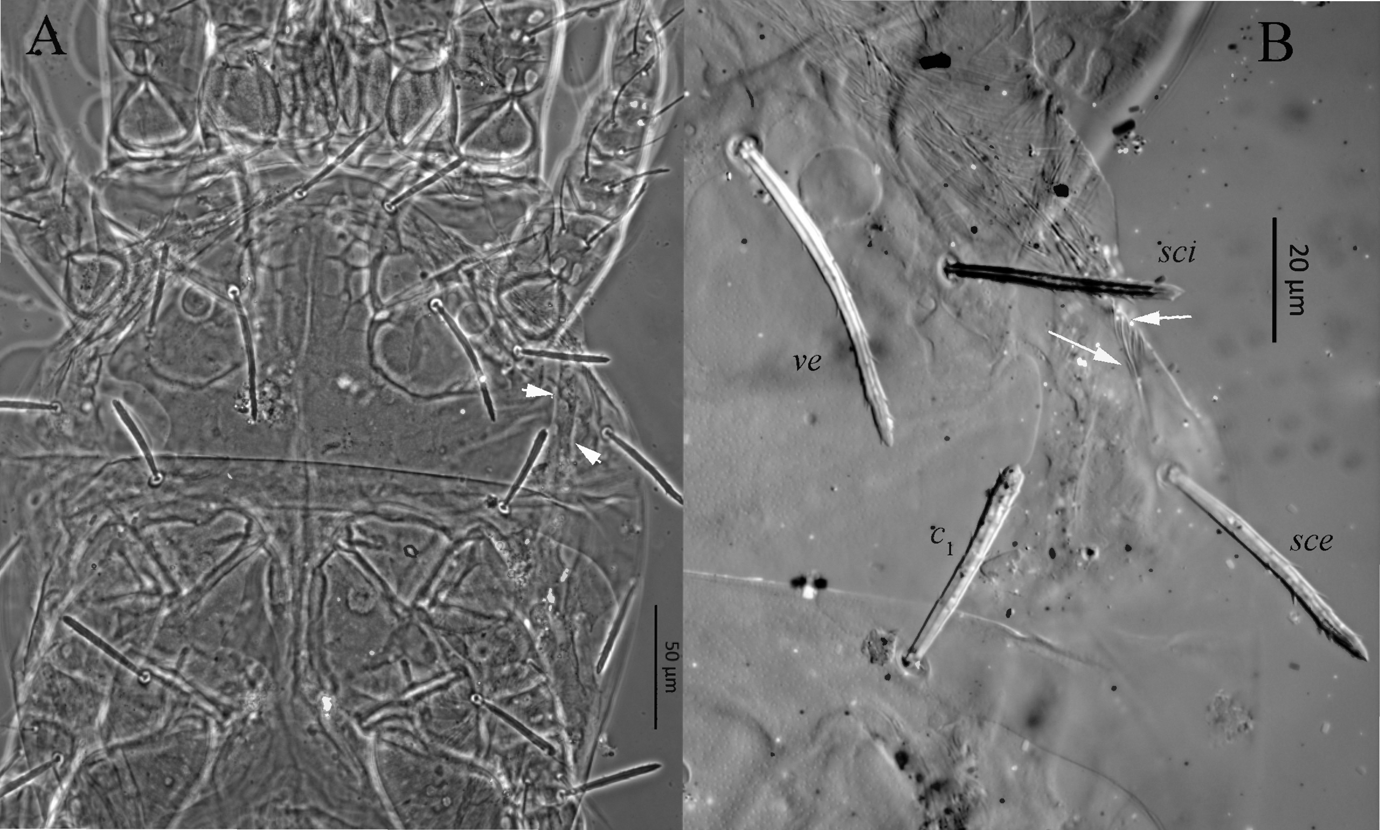
During this study, I examined 6 female paratypes of P. grata deposited in the collection of the Tyumen State University Museum of Zoology, Tyumen, Russia. All type specimens of P. grata are squeezed and strongly flattened. Thus, some ventral and dorsal structures are visible almost in the same plane, especially in phase-contrast objective. In some type specimens of P. grata it seems that seta sce is located on separate plate (Fig. 18A). However, in DIC objective it is clearly visible that seta sce is located on prodorsal shield and only thin striated incision of the prodorsal plate visible anteriorly to seta sce (Fig. 18B). Numerous specimens of this species, collected from Western Siberia are also confirmed that seta sce is located on prodorsal shield. Based on the absence of morphological differences between the genera Paravillersia and Eustigmaeus, I consider the genus Paravillersia as a junior synonym of Eustigmaeus. The specific epithet of Eustigmaeus gratus (Kuznetsov, 1978) comb. nov. is modified according to masculine gender of the generic epithet. The second described species in the genus Paravillersia, P. jamaliensis Khaustov, 2014 moved to the genus Villersia Oudemans, 1927, because seta sce of this species is located on separate plate as in the genus Villersia. However, in Villersia jamaliensis (Khaustov, 2014) comb. nov. seta d2 located on hysterosomal shield (on separate plate in Villersia), but other characters are typical for Villersia.
Synonymy of Eustigmaeus gratus (Kuznetsov, 1978) comb. nov. and E. ottavii (Berlese, 1910)
Eustigmaeus gratus (Kuznetsov, 1978) comb. nov. is characterized by the unique shape and location of callosities (see Fig. 7A in Khaustov 2014), baculiform and sparsely barbed dorsal idiosomal setae, presence of 3 pairs of aggenital setae and almost smooth dorsal idiosomal shields. Such combination (especially shape and location of callosities) of characters is known only in Eustigmaeus ottavii (Berlese, 1910) redescribed by Stathakis et al. (2016) and in E. isfahanensis Khanjani et al., 2014. Comparison of specimens of E. gratus from Russia with description of E. ottavii from Greece do not revealed any sufficient differences between these species. Therefore, I consider Eustigmaeus gratus (Kuznetsov, 1978) comb. nov. as a junior synonym of E. ottavii (Berlese, 1910). Potentially E. isfahanensis also could be a junior synonym of E. ottavii, but examination of the type material of this species is necessary.
Synonymy of Eustigmaeus ioanninensis Kapaxidi and Papadoulis, 1999 and E. pinnatus (Kuznetsov, 1977a)

Eustigmaeus pinnatus (Kuznetsov, 1977a) was described from European Russia based on two females (Kuznetsov 1977a). This species is unique in having 4 pairs of pseudanal setae. I examined the female holotype of this species. It has abnormal number of aggenital and pseudanal setae. Left side of ano-genital area with 2 aggenital and 3 pseudanal setae, while right side with 3 aggenital and 4 pseudanal setae (Fig. 19A). Undoubtedly, the presence of unpaired additional pseudanal seta (ps on Fig. 19A) is abnormal. The normal number of pseudanal setae in Eustigmaeus is 3 pairs (Fan & Zhang 2005). The most similar species to E. pinnatus with normal 3 pairs of pseudanal setae is E. ioanninemsis Kapaxidi and Papadoulis, 1999. I compared female holotype of E. pinnatus with description of E. ioanninensis and specimens reported from Western Siberia (Khaustov & Tolstikov 2014) and did not find any sufficient differences. One female from Western Siberia identified as E. ioanninensis also has abnormal number of aggenital and pseudanal seta. Left side of aggenital area with 1 aggenital and 2 pseudanals, while right side with normal 3 aggenital and 3 pseudanal setae (Fig. 19B). Variability in number of setae in ano-genital area was also observed in Turkish specimens of E. ioanninensis (Bingül et al. 2017b). Based on the variability in number of setae in ano-genital area, I consider E. ioanninensis as a junior synonym of E. pinnatus.
Discussion
Eustigmaeus bochkovi n. sp. is remarkable because the presence of callosity located between endopodal plates of legs III and IV. The external surface of the callosity has numerous tiny pores (Fig. 4E) and looks like a "sponge". Among approximately 125 described species of Eustigmaeus (Khaustov & Tsurikov 2018), at least 13 species (E. acidophilus (Wood, 1972), E. baguioensis Rimando and Corpuz-Raros, 1997, E. bali Doğan and Ayyildiz, 2003, E. changbaiensis (Bei and Yin, 1995), E. erciyesiensis Doğan et al., 2003, E. erzincanensis Doğan, 2005, E. etruscus (Berlese, 1910), E. granulosus (Wood, 1966), E. kauaiensis Swift, Gerson, Goff, 1985, E. parakauaiensis Kapaxidi et al., 2013, E. parvisetus (Chaudhri, 1965), E. schusteri (Summers and Price, 1961), E. zhengyii Hu and Zhu, 1994) have one pair of callosities usually located laterally to prodorsal shield, and at least 12 species (E. absens Dogan, 2005, E. extremiorientalis Khaustov, 2016a, E. frigidus (Habeeb, 1958), E. gersoni (Wood, 1972), E. isfahanensis Khanjani et al., 2014, E. grandis n. sp., E. lacuna (Summers, 1961), E. najeba (Habeeb, 1973), E. ottavii (Berlese, 1910), E. rhodomela (Koch, 1841), E. rotundus (Wood, 1072), E. tjumeniensis Khaustov and Tolstikov, 2014) have 2 pairs of callosities usually located laterally to humeral plate. Also all species of the genus Villersia have 2 pairs of callosities. The function of callosities is unknown. In light microscope numerous dimple-like round structures visible on callosities (see Fig.5C in Khaustov 2016a). The shape and location of callosities is good highly specific taxonomic character. It is not clear is the callosity found in E. bochkovi n. sp. homologous to callosities in other Eustigmaeus species because its location is very unusual. However, callosities found in the genus Villersia undoubtedly homologous to those found in Eustigmaeus species with 2 pairs of callosities. Potentially the genus Villersia could be synonymized to Eustigmaeus because it is differs only by the location of seta sce on separate plate.
Acknowledgements
Author thanks to Dr. Q.-H. Fan (Plant Health and Environment Laboratory, Ministry for Primary Industries, Auckland, New Zealand) for valuable comments. Author also thanks to Dr. A.V. Tolstikov (Tyumen State University, Russia) for the collecting of soil samples from Vladivostok and A.N. Bobylev (Tyumen State University, Russia) for the SEM photos.
References
Akyol M., Gül M.P. 2018. A new species of Zetzellia Oudemans (Acari, Stigmaeidae) from Turkey. Syst. Appl. Acarol., 23: 463-467. doi:10.11158/saa.23.3.5 ![]()
Berlese A. 1910. Acari Nuovi, Manipulus V. Redia, 6: 199-214.
Bei N.-X., Yin S.-G. 1995. A new species and a new record of the genus Ledermuelleria from China (Acari: Stigmaeidae). Acta Zootaxon. Sin., 20: 185-188.
Bingül M., Doğan S. 2017. Zetzellia erzincanica sp. nov., an intermediate species between the genera Zetzellia and Agistemus (Acari, Stigmaeidae). Syst. Appl. Acarol., 22: 14-20. doi:10.11158/saa.22.1.3 ![]()
Bingül M., Doğan S., Dilkaraoğlu, S. 2017a. Contributions to the knowledge of the mite genus Stigmaeus Koch, 1836 (Acari: Stigmaeidae) of Turkey. Europ. J. Taxon., 307: 1-16. doi:10.5852/ejt.2017.307 ![]()
Bingül M., Doğan S., Doğan S. 2017b. Morphological abnormalities in some stigmaeid species of Eustigmaeus, Stigmaeus and Storchia (Acari: Raphignathoidea: Stigmaeidae). Syst. Appl. Acarol., 22: 2119-2126. doi:10.11158/saa.22.12.7 ![]()
Canestrini G. 1889. Prospetto dell' Acarifauna Italiana, Famiglia dei Tetranychini. Atti R. 1st. Veneto Sci. Lett. Arti, 7: 493-531.
Canestrini G., Fanzago, F. 1876. Nuovi acari Italiana. Famiglia dei Tetranychini. Atti Soc. Veneto-Trent. Sci.Nat., 5: 135-140.
Chaudhri W.M. 1965. New mites of the genus Ledermuelleria. Acarologia, 7: 467-486.
Cooreman J. 1955. Notes sui quelques Acariens des Alpes françaises, Mém. Soc. Roy. Entomol. Belg., 27: 162-170.
Da-Costa T, Rocha M.S., Ferla N.J., Johann L. 2018. A new species of Stigmaeus Koch (Acari: Stigmaeidae) from southern Brazil. Syst. Appl. Acarol., 23: 715-723. doi:10.11158/saa.23.4.10 ![]()
Doğan S. 2005. Eustigmaeus mites from Turkey (Acari: Stigmaeidae). J. Nat. Hist., 39: 835-861. doi:10.1080/00222930400001558 ![]()
Doğan S., Ayyıldız N. 2003. New species of Eustigmaeus Berlese, 1910 (Acari: Stigmaeidae) from Turkey. J. Nat. Hist., 37: 2113-2117. doi:10.1080/00222930210133282 ![]()
Doğan S., Ayyıldız N., Fan Q.-H. 2003. Description of two new species and a newly recorded species of Eustigmaeus from Turkey (Acari: Stigmaeidae). Syst. Appl. Acarol., 8: 131-144. doi:10.11158/saa.8.1.15 ![]()
Doğan S., Bingül M., Dilkaraoğlu S., Fan Q.-H. 2015. Description of a new species of the genus Stigmaeus Koch (Acari: Stigmaeidae) from Turkey, with a list of described species in the world. Internat. J. Acarol., 41: 4, 290-299. doi:10.1080/01647954.2015.1028441 ![]()
DOI: 10.1080/01647954.2015.1028441 doi:10.1080/01647954.2015.1028441 ![]()
Doğan S., Doğan S., Erman O. 2017. Description of five new species of the genus Stigmaeus Koch (Acari: Raphignathoidea: Stigmaeidae) from Turkey. Zootaxa, 4276: 451-478. doi:10.11646/zootaxa.4276.4.1 ![]()
Fan Q.-H., Flechtmann C.H., De Moraes G.J. 2016. Annotated catalogue of Stigmaeidae (Acari: Prostigmata), with a pictorial key to genera. Zootaxa, 4176: 1-199. doi:10.11646/zootaxa.4176.1.1 ![]()
Fan Q.-H., Ueckermann E.A. 2016. Resurrection of the genus Nonocaligus Habeeb with redefinition of Nonocaligus and Mullederia Wood (Acari: Stigmaeidae). Syst. Appl. Acarol., 21: 1447-1449. doi:10.11158/saa.21.11.1 ![]()
Fan Q.-H., Zhang Z.-Q. 2005. Raphignathoidea (Acari: Prostigmata). Fauna of New Zealand, 52: 1-400.
Grandjean F. 1939. Les segments postlarvaires de l'hysterosoma chez les oribates (Acariens). Bull. Soc. Zool. Fr., 64: 273-284.
Grandjean F. 1944. Observations sure les Acariens de la famille des Stigmaeidae. Arch. Sci. Phys. Natur., 26: 103-131.
Grandjean F. 1946. Au sujet de l'organe de Claparède, des eupathides multiples et des taenidies mandibulaires chez les Acariens actinochitineux. Arch. Sci. Phys. Natur., 28: 63-87.
Habeeb H. 1958. New mites from New Brunswick. Leafl. Acadian Biol., 18: 1-4.
Habeeb H. 1973. Notes on water-mites. V. Leafl. Acadian Biol., 54: 1-2.
Halbert J.N. 1923. Notes on Acari, with description of new species. J. Linn. Soc. London, Zool., 35: 363-392.
Hu S., Chen X., Huang L. 1996. Mites of the genus Eustigmaeus from Jiangxi Province (Acari: Stigmaeidae). Entomol. Sini., 3: 314-322.
Hu C.-Y., Zha G.-C., Zhu J.-T. 1994. Two new species and two new records of the genus Eustigmaeus Berlese from China (Acari: Stigmaeidae). Acta Arachnol. Sin., 3: 81-85.
Kapaxidi E.V., Papadoulis G.Th. 1999. New records of stigmaeid mites from Greece with description of a new species (Acari: Stigmaeidae). Internat. J. Acarol., 25: 141-144. doi:10.1080/01647959908683625 ![]()
Kapaxidi E.V., Stathakis T.I., Papadoulis G.Th. 2013. New species and new records of the genus Eustigmaeus Berlese (Acari: Stigmaeidae) from Greece. Internati. J. Acarol., 39: 400-407. doi:10.1080/01647954.2013.794860 ![]()
Karasu N., Doğan S., Kuzucu M., Çankaya M. 2018. Genetic variations based on RAPD-PCR in Eustigmaeus erciyesiensis (Acari: Stigmaeidae) populations inhabiting Erzincan (Turkey). North-Western J. Zool., 14: 122-126.
Kethley J.B. 1990. Acarina: Prostigmata (Actinedida). In: D.L. Dindal (Ed.). Soil Biology Guide. Wiley, New York, 667-756.
Khanjani M., Najaf-Abadi P.R., Khanjani M. 2014. A new species of the genus Eustigmaeus (Acari: Stigmaeidae) from Isfahan province, Iran. Pers. J. Acarol., 3: 17-26.
Khanjani M., Khanjani M., Nardi A., Mohammadi L., Nazari A. 2017. A new species of the genus Stigmaeus Koch (Acari: Stigmaeidae) and re-description of Cheylostigmaeus howellsi Evans from Iran. Syst. Appl. Acarol., 22: 815-823. doi:10.11158/saa.22.6.7 ![]()
Khaustov A.A. 2014. A new species of the genus Paravillersia (Acari: Prostigmata: Stigmaeidae) from Western Siberia, with supplementary description of Paravillersia grata Kuznetsov, 1978. Zootaxa, 3873: 62-72. doi:10.11646/zootaxa.3873.1.5 ![]()
Khaustov A.A. 2016a. Two new species and a new record of mites of the family Stigmaeidae (Acari: Prostigmata) collected from mosses in Russia. Acarologia, 56: 321-339. doi:10.1051/acarologia/20162249 ![]()
Khaustov A.A. 2016b. New species and records of mites of the family Stigmaeidae (Acari: Prostigmata) collected from mosses in Southern Chile. Acarologia, 56: 639-679. doi:10.1051/acarologia/20164150 ![]()
Khaustov A.A., Tolstikov A.V. 2014. A new species and new records of the genus Eustigmaeus (Acari: Prostigmata: Stigmaeidae) from Western Siberia. Zootaxa, 3861: 531-553. doi:10.11646/zootaxa.3861.6.2 ![]()
Khaustov A.A., Tsurikov S.M. 2018. A new species of Eustigmaeus (Acari: Prostigmata: Stigmaeidae) from Vietnam. Pers. J. Acarol.: 7: 235-244.
Khaustov A.A., Ueckermann E.A., Theron P.D. 2017. A new species of Stigmaeus (Acari: Prostigmata: Stigmaeidae) from South Africa. Syst. Appl. Acarol., 22: 1413-1421. doi:10.11158/saa.22.9.8 ![]()
Koch C.L. 1833-1841. Deutschlves Crustaceen, Myriapoden und Arachniden. Heft Regensburg, 1-40.
Kuznetsov N.N. 1977a. A contribution to the fauna of mites of the family Stigmaeidae (Acariformes) in the central-chernozem zone. Zool. Zh., 56: 953-956. [in Russian]
Kuznetsov N.N. 1977b. New species of the family Stigmaeidae from Crimea. Zool. Zh., 56, 635-638. [in Russian]
Kuznetsov N.N. 1978. New records of raphignathoid mites (Raphignathoidea, Acariformes). Biol. Nauki, 12: 49-54. [in Russian]
Nazari A., Khanjani M. 2017. A new species of the genus Ledermuelleriopsis (Acari: Stigmaeidae) from Markazi province, Iran. Pers. J. Acarol., 6: 193-201.
Oudemans A. C. 1927. Acarologische Aanteekeningen LXXXVIII. Entomol. Ber., 7: 257-263.
Oudemans A.C. 1931. Acarologische aanteekeningen CVIII. Entomol. Ber., 8: 251-263.
Paktinat-Saeij S, Bagheri M., Marticorena J.L.M., de Moraes G.J. 2016. A new species of Stigmaeus (Acari: Trombidiformes: Stigmaeidae) from Brazil. Pers. J. Acarol., 5: 281-289.
Rehman M.U., Kamran M., Alatawi F. 2018. Genus Agistemus Summers (Acari: Trombidiformes: Stigmaeidae) from Saudi Arabia and a key to the world species. Syst. Appl. Acarol, 23: 1051-1072. doi:10.11158/saa.23.6.5 ![]()
Rimando L.C., Corpuz-Raros L.A. 1997. Some Philippine Raphignathoidea (Acari). III. Revision of the genus Eustigmaeus Berlese sensu latu (Stigmaeidae). Philipp. Entomol., 11: 1-24.
Stathakis Th.I., Kapaxidi E.V., Papadoulis G.Th. 2016. The genus Eustigmaeus Berlese (Acari: Stigmaeidae) from Greece. Zootaxa, 4191: 1-102. doi:10.11646/zootaxa.4191.1.1 ![]()
Summers F.M., Price D.W. 1961. New and redescription species of Ledermuelleria from North America (Acarina: Stigmaeidae). Hilgardia, 31: 369-387. doi:10.3733/hilg.v31n10p369 ![]()
Swift S.F., Gerson U., Goff M.L. 1985. A new species of Eustigmaeus (Acari: Prostigmata: Stigmaeidae) from Kaua'I Island, Hawaiian Islands. Interna. J. Entomol., 27: 375-381.
Wainstein B.A., Kuznetsov N.N. 1978. Family Stigmaeidae. In: M.S. Gilarov (Ed.). Opredelitel pochvoobitayushchikh kleshchey. Trombidiformes, 153-168. [in Russian]
Wood T.G. 1966. Mites of the genus Ledermuelleria Oudms (Prostigmata, Stigmaeidae) from New Zealand, with records of one species from some Southern Pacific Islands. N.Z. J Sci., 9: 84-102.
Wood T.G. 1972. New and redescribed species of Ledermuelleria Oudms, and Villersia Oudms (Acari: Stigmaeidae) from Canada. Acarologia, 13: 301-318.


2018-12-10
Date accepted:
2019-03-03
Date published:
2019-03-08
Edited by:
Faraji, Farid

This work is licensed under a Creative Commons Attribution 4.0 International License
2019 Khaustov, Alexander A.
 Download article Download low definition
Download the citation
RIS with abstract
(Zotero, Endnote, Reference Manager, ProCite, RefWorks, Mendeley)
RIS without abstract
BIB
(Zotero, BibTeX)
TXT
(PubMed, Txt)