


Eriophyoid mites from ferns: description of a new Leipothrix Keifer species (Eriophyidae: Phyllocoptinae) from the Arasbaran forests (Iran) and a key to the world species
Lotfollahi, Parisa1 and de Lillo, Enrico2
1✉ Department of Plant Protection, Faculty of Agriculture, Azarbaijan Shahid Madani University, Tabriz, Iran.
2Department of Soil, Plant and Food Sciences (Di.S.S.P.A.), Entomology and Zoology Section, University of Bari Aldo Moro, via Amendola, 165/a, 70126 Bari, Italy.
2017 - Volume: 57 Issue: 4 pages: 731-745
https://doi.org/10.24349/acarologia/20174179ZooBank LSID: 1C144CF2-E413-425D-8A76-06F5754F840B
Keywords
Abstract
The biosphere reserve of the Arasbaran forests constitutes the limited territory of Kaleybar, Ahar and Jolfa with an area of 160,000 ha on the Caucasus Iranian Highlands (Talebi et al., 2014). Different climates and various physiographic conditions of these forests induced very variable plant diversity and fauna associated with those plants. Considering the relevance of evaluating mite faunas in highly biodiverse areas like Iran (de Lillo and Skoracka, 2010), eriophyoid mites of ferns were surveyed in the Arasbaran forests.
About 36 eriophyoids have been recorded on ferns worldwide (Amrine & de Lillo unpublished databases; table 1), including three Leipothrix species: L. triquetra [Meyer (Smith), 1990], L. minidonta [Meyer (Smith), 1990] and L. serbicus (Petanović, 2001). The latter species was recently moved from Epitrimerus to Leipothrix (Petanović et al., 2015) after a careful examination of the morphology with scanning electron microscopy which clarified the bifurcated shape of the palp seta d (R. Petanović, unpublished micrographs and pers. comm., on 18 Sept. 2015).


Two Leipothrix species are known from Iran: L. retidorsi Lotfollahi, Haddad & de Lillo, 2014 from Rubia tinctorum L. (Rubiaceae) in Azarshahr, East Azerbaijan province (Lotfollahi et al., 2014), and L. liroi (Roivainen, 1947) from Primula sp. (Primulaceae), Aliabad-eKatul, Golestan province (Gol et al., 2015). Leipothrix liroi was moved from Epitrimerus to Neoleipothrix by Soika and Labanovki (2009) and, later, to Leipothrix by Jočić and Petanović (2012). The synonymy of L. liroi with L. jaceae (Liro) proposed by Chetverikov (2005) was a typographic mistake (P.E. Chetverikov, pers. comm., on 17 Aug. 2016).
In the current study a new Leipothrix species was collected and described on Polypodium vulgare L. (common polypody) and Gymnocarpium dryopteris (L.) Newman (common oak fern). This is the third Leipothrix species found in Iran, and the fourth from ferns worldwide.
In order to start a survey on the mite fauna of ferns, plants of the families Polypodiaceae and Cystopteridaceae were sampled during September 2010 in the Arasbaran forests (East Azerbaijan, Iran). Eriophyoid mites were recovered from the plant material by means of a modified washing method developed by Monfreda et al. (2007). The mites were slide mounted according to Baker et al. (1996).
The terminology and the setal notation in the morphological description of the mite follow mainly Lindquist (1996). All morphological measurements were taken by means of a phase contrast microscope Olympus BX50 according to Amrine and Manson (1996) as modified by de Lillo et al. (2010), and are given in micrometers. Slight clarifications should be added as follows: ventral semiannuli were counted from the first entire annulus behind the prodorsal shield; coxigenital semiannuli were counted medially from the coxal region to the anterior margin of the external genitalia and were not included in the ventral semiannuli count.
Measurements and means are rounded off to the nearest integer when required. Measurements refer to the length of the morphological trait unless otherwise specified. The holotype measurements are followed by range values, in parentheses, of the studied population (i.e. holotype and paratypes). The mean values of the paratypes are reported in the few cases in which the measurements of the holotype could not be taken, due to the slide mounting position of the specimens.
Line drawings were hand-drawn through a camera lucida according to de Lillo et al. (2010) and the abbreviations labelling schematic drawings in figure 1 follow mainly Amrine et al. (2003). The generic classification follows Amrine et al. (2003) and comparisons were also made with new genera described since that publication. Host plant names and their synonymies are in accordance with "The Plant List on-line database" (2013).
Type materials are deposited at the Acarology Laboratory, Department of Plant Protection, Faculty of Agriculture, Azarbaijan Shahid Madani University, Tabriz (Iran), and at the Department of Soil, Plant and Food Sciences (Di.S.S.P.A.), section of Entomology and Zoology, University of Bari Aldo Moro (Italy).
ZOOBANK: 86B001AA-AD6E-41FE-822C-564498CEA366 ![]()

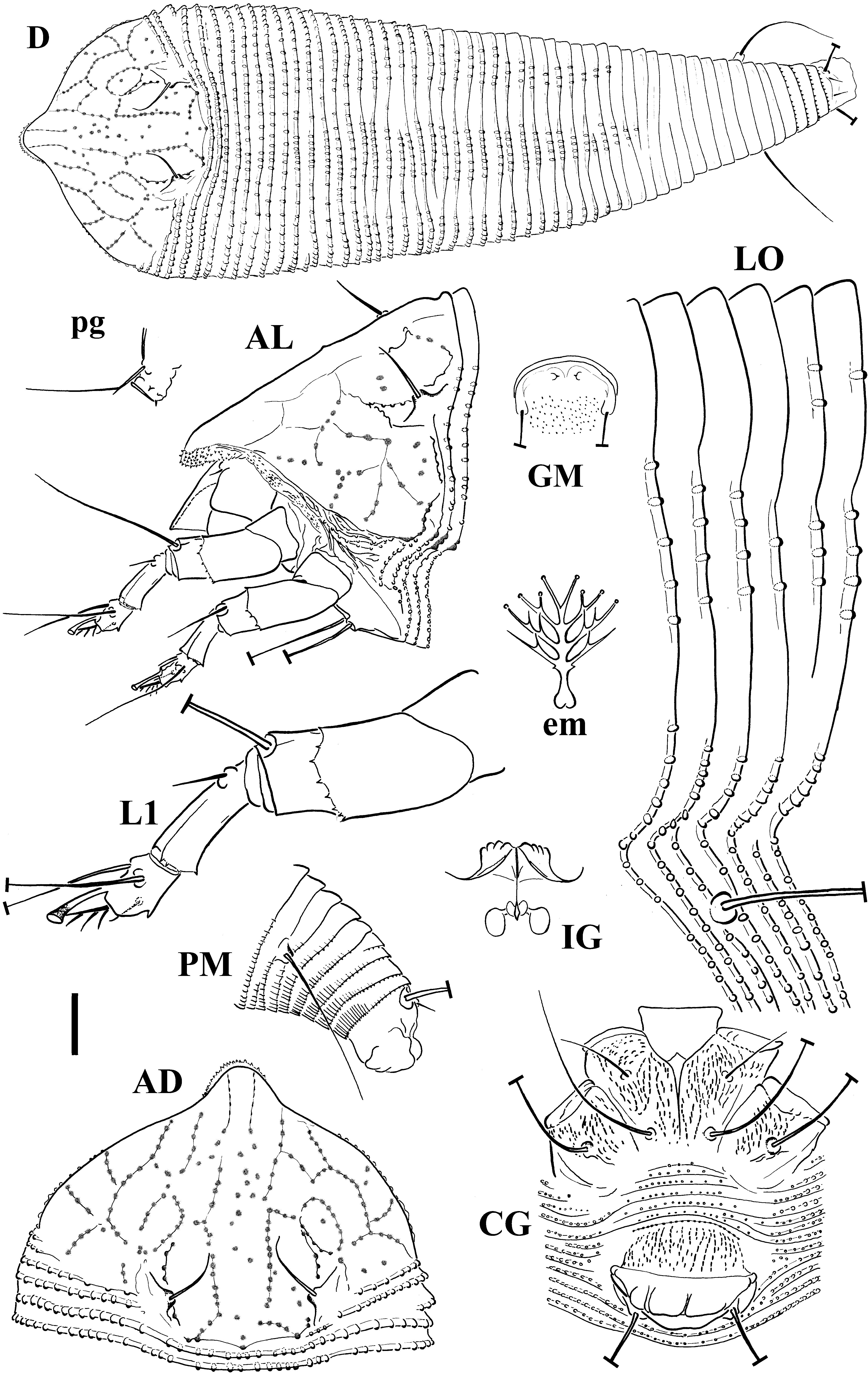
Diagnosis — Prodorsal shield with granulated lines forming a cell-like network; coverflap ornamented only on the basal part with 1-2 transverse rows of dashes in vertical lines.
Female (measured specimens n: 10)
Body — fusiform, 223 (216 – 235, excluding gnathosoma), 67 (62 – 73) thick, 72 (70 – 76) wide.
Gnathosoma — 25 (25 – 26) projecting obliquely downwards, chelicerae 20 (18 – 21), palp coxal setae ep 3 (2 – 4), palp genual setae d 21 (it is the average value of the measured population; 18 – 23), branched; branch 1 (no variation), at 5 (4 – 6) from the base of setae d.
Prodorsal shield — 54 (48 – 54) including frontal lobe, 61 (60 – 70) wide, sub-semicircular; with a broad-based frontal lobe, 13 (10 – 14), over gnathosomal base; frontal lobe with numerous very short and spiny protuberances. Shield pattern distinct, consisting of granulate lines forming a cell-like network; sparse granules on median shield area; complete admedian lines, short first submedian lines, extended ¼ ahead of tubercles of sc setae, complete second submedian lines; cell pattern of lateral and transverse lines on lateral shield areas. Tubercles of sc setae 5 (5 – 6) ahead of rear shield margin, 27 (23 – 27) apart, setae sc 13 (10 – 14), directed antero-medially.
Leg I — 40 (36 – 41), femur 11 (10 – 12), genu 5 (5 – 6), tibia 10 (9-11), tarsus 8 (8 – 9), ω 5 (no variation) distally funnel shaped, empodium simple, 6 (5 – 6.5), 4-rayed; femoral setae bv absent, genual setae l" 30 (28 – 31), tibial setae l' 5 (3 – 5), tarsal setae ft' 18 (15 – 19), ft" 22 (19 – 24).
Leg II — 38 (36 – 40), femur 12 (10 – 13), genu 5 (5 – 7), tibia 9 (9 – 10), tarsus 8 (8 – 10), ω 5 (5 – 5.5) distally funnel shaped, empodium simple, 4 (no variation), 4-rayed; femoral setae bv absent, genual setae l" 7 (7 – 13), tarsal setae ft' 3 (3 – 5), ft" 20 (18 – 22). Coxae with distinct dashes arranged in lines; setae 1b 12 (10 – 15), tubercles 1b 15 (14 – 16) apart, setae 1a 26 (24 – 36), tubercles 1a 8 (no variation) apart, setae 2a 51 (48 – 68), tubercles 2a 27 (26 – 30) apart. Prosternal apodeme 11 (9 – 12).
Opisthosoma — with 53 (52 – 59) dorsal semiannuli, 78 (65 – 81) ventral semiannuli (counted from first complete annulus after coxae II); dorsal median ridge, extended on the anterior 37 (35 – 37) dorsal semiannuli, flanked by two lateral ridges; 7 (5 – 7) semiannuli between coxae and genital coverflap.
Microtubercles — elliptical, on posterior margin of dorsal semiannuli, more distinct on ridges and faint on the space between median and lateral ridges of 15 – 17 anterior dorsal semiannuli, no microtubercles visible on the space between median and lateral ridges of remaining posterior dorsal semiannuli; circular, on posterior margin of ventral semiannuli; spiny on the rear margin of the last 4 dorsal semiannuli and elongated and linear on last five ventral semiannuli. Setae c2 25 (20 – 26) on ventral semiannulus 13 (11 – 15), setae d 47 (43 – 49) on ventral semiannulus 27 (27 – 32); setae e 17 (12 – 18) on ventral semiannulus 46 (46 – 55); setae f 30 (25 – 33) on ventral semiannulus 69 (66 – 74); 5 annuli posterior to setae f. Setae h2 75 (65 – 75) apically very fine, h1 5 (4 – 5). Genital coverflap 16 (15 – 20), 26 (20 – 28) wide, basal part ornamented with 1-2 transverse rows of dashes arranged in vertical lines, distal part smooth and apparently soft with a few folds on the edge; setae 3a 15 (13 – 17), 17 (16 – 18) apart.
Male (n = 1)
Similar in shape and prodorsal shield arrangement to female. Body 125, 51 wide; palp genual setae d 11; prodorsal shield 43, 40 wide; setae sc 13, 16 apart. Opisthosoma with 47 dorsal semiannuli and 51 ventral semiannuli (counted from first complete annulus after coxae II). Setae: 1b 7, 1a 16, 2a 35, c2 12.5, d 22, e 10, f 19, h2 60, h1 3. Male genitalia 12 wide, setae 3a 10, 14 apart.
Type host plant — Polypodium vulgare L. (Polypodiaceae), common polypody.
Type locality — Arasbaran forests, Kaleybar, East Azerbaijan, Iran (38°50'44.9''N, 47°00'27.8''E), 1,616 m above sea level, coll. P. Lotfollahi, late September 2010.
Type material — Holotype: single female on a microscope slide (PV-IEA-AN10L-1). Paratypes: 33 females, 1 male and 1 nymph mounted singly on separate microscope slides.
Other host plant — 38 females and 2 nymphs mounted singly on separate microscope slides, collected from Gymnocarpium dryopteris (L.) Newman (Cystopteridaceae), oak fern, Western oak fern, common oak fern or Northern oak fern in the same localities and on the same dates, as mentioned above.
Other material — Mites preserved in Oudemans' fluid (Walter & Krantz, 2009) as extracted from the same sample as the type specimens.
Relation to the host plant — Vagrant on leaves, in high density. The protocol applied for the mite extraction did not allow establishing the side of leaves colonized by the mite population. No apparent damage was observed.
Etymology — The specific epithet, pterisfolii, is the genitive case of a combined name composed from the Greek pteris, meaning fern, and the Latin folii, meaning leaf.
Differential diagnosis — The new species herein described was compared with all Leipothrix species currently known and similarities were observed only with those species known on other ferns: L. minidonta, L. triquetra and L. serbicus.
The new species differs from L. triquetra in the prodorsal shield pattern (complete median line in L. triquetra vs no median line in L. pterisfolii), frontal lobe (shorter and smooth in L. triquetra vs larger and with numerous spiny protuberances in L. pterisfolii), ornamentation of the female coverflap (seven slightly diagonal lines on distal part in L. triquetra vs smooth distal part in L. pterisfolii), number of the empodial rays, length of palp genual setae d, and setae 1a, e, f, h1 and 3a, number of dorsal and ventral semiannuli, and of semiannuli between coxae and genital coverflap (Fig. 2A, Table 2).


The new species differs from L. minidonta in the prodorsal shield pattern (incomplete and broken median line of L. minidonta vs no median line for L. pterisfolii; different arrangement for admedian lines and lines on the lateral sides), ornamentation of the female coverflap (8 inconspicuous slightly diagonal lines on the distal part in L. minidonta vs smooth distal part in L. pterisfolii), length of setae sc, c2 and d, number of dorsal semiannuli and of semiannuli between coxae and genital coverflap (Fig. 2B, Table 2).


The new species differs from L. serbicus in the prodorsal shield pattern (the published line drawing of the prodorsal shield does not fit perfectly with SEM pictures taken by R. Petanović, pers. comm., on 18 Sept. 2015); according to the new data (unpublished) also the prodorsal shield of L. serbicus is provided of a net-like ornamentation made by granulated lines and it differs from the prodorsal shield of the Iranian species on a complete median line which cannot be seen on L. pterisfolii. Other differences concern the length of palp genual setae d, scapular setae sc, setae 1a, c2, d, e and 3a (all of them are longer in L. pterisfolii) (Table 2).
Remarks — This is the first record of a species belonging to the tribe Phyllocoptini on a Polypodiaceae plant species, the first record of an eriophyoid mite from ferns of the family Cystopteridaceae and the first record of an eriophyoid mite from ferns in Iran. The mites (38 females) found on G. dryopteris showed highly similar morphometric details and variability in respect to the mites (34 females) found on P. vulgare. This latter fern was selected as type host plant because of the collection of male and nymphs within the collected mite population. The close cohabitation of the two host plant species does not allow to understand if G. dryopteris is an alternative or accidental host species due to mite dispersal or if they refer to two populations of a complex of species. DNA and biological studies are needed to determine the host plant range of this new mite species. In addition, a multifactorial morphometric analysis should be done on the two populations in order to give evidences of minor differences which could be caused by the plant genotype.
A key of the eriophyoid mite species collected on ferns worldwide is proposed on the base of the most detailed published descriptions and including a previous species key of the genus Litaculus (Manson & Gerson, 1986).
1. Gnathosoma large in comparison to the body; chelicerae abruptly curved and bent down near their base; prodorsal shield with a net-like pattern: median and admedian lines well distinct on the posterior half, admedian and submedian lines delimit 11 cells on the anterior half along with a pair of transverse lines; femoral setae bv present on both legs; empodium entire; opisthosoma with subequal dorsoventrally annuli
...... Diptilomiopidae Keifer, 1944 – Rhyncaphytoptinae Roivainen, 1953 – Rhinophytoptus plagiogyrus Huang, 2001c
— Gnathosoma small in comparison to the body; chelicerae straight or slightly curved down
...... Eriophyidae Nalepa 1898 – 2
2. Coxae I often fused with sternal line faint or absent; tibiae reduced or completely fused with tarsi; tibial setae l′ absent
...... Nothopodinae Keifer, 1956 – 3
— Combination of characters not as above
...... 7
3. Prodorsal shield with blunt frontal lobe and lobes at each anterolateral angles; tubercles sc ahead of the rear margin of the prodorsal shield; coxal setae 1b present; coxae I usually weakly divided; tibiae I completely fused with tarsi; empodium divided; opisthosoma with broad dorsal semiannuli and a dorsal median ridge; female coverflap with two rows of longitudinal striae
...... Colopodacini Mohanasundaram, 1984 – Dicolopodacus pterpterus (Huang, 1991)
— Coxal setae 1b absent; coxae and tibiae I variable
...... Nothopodini Keifer, 1956 – 4
4. Setae sc directed up and converging
...... Nothopoda footei (Keifer, 1969)
— Setae sc directed posteriorly
...... 5
5. Tubercles sc cylindrical, near the rear margin of the prodorsal shield
...... 6
— Tubercles sc plicate, ahead of the rear margin of the prodorsal shield; smooth dorsal semiannuli
...... Cosella pteridiae Huang, 2001b
6. Median line complete and connected to the admedian lines by two transverse lines on basal 1/3 and 1/2 of the prodorsal shield; dorsal semiannuli fully microtuberculated
...... Floracarus biseratae Huang, 2001a
— Median line on 2/3 of the prodorsal shield and not connected to the admedian lines; dorsal semiannuli with sparse spinulated microtubercles
...... Floracarus perrepae Knihinicki & Boczek, 2002
7. Female genital apodeme bent up and shortened, usually appearing as a heavy transverse line in ventral view; female coverflap appressed to coxae II and often with ridges typically in 2 uneven ranks; coxae I usually narrowly connate with a short sternal line; coxae often with curved lines outlining tubercles of setae 1a; prodorsal shield with two large tuberosity, medially contiguous, extending from the rear margin anteriorly to cover an half; tubercles sc well ahead of the rear margin of the prodorsal shield; empodium divided; opisthosoma with broad dorsal semiannuli, narrow ventral semiannuli, a middorsal furrow and two subdorsal rounded ridges
...... Cecidophyinae Keifer, 1966b – Esalquia centennaria Fletchmann, 2002
— Female genital apodeme extending moderate distance forward and not appearing as a heavy transverse bar in ventral view; female coverflap well far from coxae II, variably sculptured and striae rarely occurring in 2 ranks; sternal line usually evident; coxae usually without curved lines outlining setal tubercles
...... 8
8. Body vermiform, opisthosoma with annuli subequal dorsoventrally, at least on anterior 1/2 to 2/3; prodorsal shield lacking a frontal lobe or if frontal lobe present, it is narrow, basally flexible, and combined with narrow annuli
...... Eriophyinae Nalepa, 1898 – 9
— Body usually fusiform; prodorsal shield usually with a broad-based and rigid frontal lobe; opisthosoma typically with broad dorsal semiannuli and narrow microtuberculate ventral ones; if frontal lobe absent or only a slight one present, then semiannuli differ dorsoventrally, at least in broader dorsal ones
...... Phyllocoptinae Nalepa, 1892 – 21
9. Empodium divided, 4-rayed
...... Diphytoptini Amrine & Stasny, 1994 – Diphytoptus nephroideus Huang, 1991
— Empodium entire
...... 10
10. Tubercles and coxal setae 1b absent
...... 11
— Tubercles and setae coxal 1b present
...... 12
11. Prodorsal shield unornamented except for a few basal granules and faint, short lines on posterior half; coxae smooth; female coverflap sometimes with few faint markings on the distal part and granules on the proximal part
...... Acerimina pyrrosiae Manson, 1984
— Prodorsal shield design obscure, with many short dash-like lines, particularly near to the posterior margin; coxae with few granules; female coverflap with short distal striae and proximal granules
...... Acerimina shuishensis Huang, 1991
12. Setae sc directed posteriorly; tibial setae l′ present
...... 13
— Setae sc directed upwards and centrad or laterad
...... 16
13. Prodorsal shield unornamented except for a weak longitudinal, dash-like line directed anteriorly from each tubercle sc, and marks close to the rear margin of the prodorsal shield at the base between tubercles sc
...... Aceria gersoni Manson, 1984
— Prodorsal shield ornamented by distinct lines and granules
...... 14
14. Median and admedian lines complete and broken
...... Aceria pteridii Kuang & Gong, 1996
— Median line shorter than the anterior half of the prodorsal shield, admedian lines complete or broken
...... 15
15. Median line short on the anterior half of the prodorsal shield, admedian lines running about 2/3 length of shield, 3-4 submedian lines; sc setae longer than prodorsal shield; with about 94-104 microtuberculated annuli
...... Aceria gleicheniae Manson, 1984
— Median line short and broken, admedian lines as long as prodorsal shield length; sc setae shorter than prodorsal shield; with about 68 microtuberculated rings
...... Aceria pauropa (Nalepa, 1909)
16. Setae sc directed upwards and laterad; frontal lobe distinctly large and weak
...... Stenacis biserratae Huang, 2001a
— Setae sc directed upwards and centrad; frontal lobe absent or very short
...... 17
17. Prodorsal shield design made up of continuous lines
...... 18
— Prodorsal shield design made up of lined short dashes and granules
...... 19
18. Coxae I fused; setae bv absent; empodium 5-rayed
...... Eriophyes eckloniae Meyer (Smith) & Ueckermann, 1989
— Coxae I not fused; setae bv present; empodium 4-rayed
...... Eriophyes parapteridis Livshits, Mitrofanov & Sharonov, 1983
19. Both coxae completely granulated
...... 20
— Coxae I granulated and coxae II almost completely smooth; empodium 3-rayed
...... Eriophyes helicantyx Keifer, 1966a
20. Setae sc longer than half of the prodorsal shield length; empodium 4-rayed
...... Eriophyes quadrifidus Meyer (Smith) & Ueckermann, 1989
— Setae sc less long than half of the prodorsal shield; empodium 5-rayed
...... Eriophyes equiseti (Farkas, 1960)
21. Empodium divided
...... Acaricalini Amrine & Stasny, 1994 – 22
— Empodium entire
...... 28
22. Femur two-segmented and setae bv on the basal one; tibial setae l′ absent; opisthosoma with alternate dorsal annuli bearing longitudinal scorings laterally and small punctures centrally; intervening annuli smooth; wax plates laterally and around the shield
...... Cymeda zealandica Manson & Gerson, 1986
— Tibial setae l′ present; femur and opisthosoma annuli not as above
...... 23
23. Frontal lobe of the prodorsal shield acutely pointed, triangular; opisthosoma with 34 – 42 dorsal semiannuli
...... Litaculus acutus Manson & Gerson, 1986
— Frontal lobe of the prodorsal shield rounded or truncate
...... 24
24. Genital coverflap with two rows of striae; prominent protuberance at the base of genua I setae l″; opisthosoma with at most 30 dorsal semiannuli
...... Litaculus squarrosus Manson & Gerson, 1986
— Not as above
...... 25
25. Palp genual setae d with an abrupt right angle bend ('flag-shaped'); opisthosoma with at most 25 dorsal semiannuli
...... Litaculus antapicus Manson & Gerson, 1986
— Palp genual setae d normal
...... 26
26. All dorsal semiannuli (except the first three) with a rectangular or diamond shaped pattern at the mid lateral position; opisthosoma with at most 30 dorsal semiannuli
...... Litaculus khandus Manson & Gerson, 1986
— Dorsal semiannuli not as above
...... 27
27. Frontal lobe bluntly rounded; opisthosoma with more than 40 dorsal semiannuli and with a weak middorsal ridge
...... Litaculus gillianae Manson & Gerson, 1986
— Frontal lobe truncate, with a jagged anterior margin; opisthosoma with at most 30 dorsal semiannuli and without a ridge
...... Litaculus pennigerus Manson & Gerson, 1986
28. Setae and tubercles sc absent
...... Calacarini Amrine & Stasny, 1994 – 29
— Setae and tubercles sc present
...... 30
29. Prodorsal shield without median line; dorsal semiannuli evenly rounded; empodium 3-rayed
...... Jutarus plagiogyrus Huang, 2001c
— Prodorsal shield with median line on the rear 1/4 of shield; dorsal semiannuli with three equal ridges; empodium 6-rayed
...... Calacarus dicranopteris Wei & Feng, 2002
30. Tubercles sc on or very near the rear margin of the prodorsal shield, directing setae to rear, usually divergently; tubercles sc either subcylindrical or the alignment of their bases is transverse to the body
...... Anthocoptini Amrine & Stasny, 1994 – 31
— Tubercles sc usually well formed, often plicate and placed ahead of the rear margin of the prodorsal shield, directing setae sc forward, up or centrad; if tubercles sc are near the rear margin of the prodorsal shield, then tubercles are subcylindrical and bent forward or the alignment of their bases is longitudinal or diagonal to the body
...... Phyllocoptini Nalepa, 1892 – 32
31. Tubercles and coxal setae 1b absent
...... Metaculus tanythrix Flechtmann & De Queiroz, 2010
— Tubercles and coxal setae 1b present; prodorsal shield smooth; empodium 5-rayed
...... Aculus beevori (Manson & Gerson, 1986)
32. Palp genual setae d unbranched
...... 33
— Palp genual setae d bifurcate
...... 34
33. Prodorsal shield pattern distinct, with strongly granulated and complete median, admedian and two pairs of submedian lines; female coverflap with granules on the proximal part and striae on the distal part
...... Phyllocoptes pteridii Petanović, 2001
—Prodorsal shield pattern with finely granulated lines, median line incomplete, joint by a transverse line to the admedian line close to the rear margin of the prodorsal shield, admedian lines complete forming cells along with the first pair of submedian lines; female coverflap with a double row of striae
...... Phyllocoptes dimorphus Keifer, 1940
34. Median line complete; empodium 5-rayed
...... Leipothrix triquetra [Meyer (Smith), 1990]
— Median line incomplete or absent; empodium 4-rayed
...... 35
35. Distal part of the female coverflap smooth and with a few folds
...... Leipothrix pterisfolii n. sp.
— Distal part of the female coverflap ornamented by striae
...... 36
36. Median line short, on the posterior half, made by lined granules; distal part of the female coverflap with 8 inconspicuous slightly diagonal striae
...... Leipothrix minidonta [Meyer (Smith), 1990]
— Median line replaced by lined granules; distal part of the female coverflap with about 6-7 striae
...... Leipothrix serbicus (Petanović, 2001)*
The authors are grateful to Prof. Petanović R.U. and Dr. Vidović B. (Department of Entomology and Agricultural Zoology, Faculty of Agriculture, University of Belgrade, Serbia) for their critical review of this manuscript; to Dr. Chetverikov P.E. (Department of Invertebrate Zoology, Biological and Soil Sciences Faculty, Saint-Petersburg State University, Russia) for providing us explanation on Leipothrix liroi Roivainen and L. jaceae (Liro). This research was partially supported by Azarbaijan Shahid Madani University (Iran) and University of Bari Aldo Moro (Italy).
Amrine J.W.Jr., Stasny T.A. 1994 — Catalog of the Eriophyoidea (Acarina: Prostigmata) of the world — West Bloomfield, Michigan, USA: Indira Publish. House. pp. 804.
Amrine J.W. Jr., Manson D.C.M. 1996 — Preparation, mounting and descriptive study of Eriophyoid mites — In: Lindquist E.E., Sabelis M.W., Bruin J. (Eds), Eriophyoid Mites. Their Biology, Natural Enemies and Control. World Crop Pests, 6, Amsterdam, The Netherlands: Elsevier Science Publishers. p. 383-396. doi:10.1016/S1572-4379(96)80023-6 ![]()
Amrine J.W. Jr., Stasny TA.H., Flechtmann C.H.W. 2003 — Revised keys to world genera of Eriophyoidea (Acari: Prostigmata) — West Bloomfield, Michigan, USA: Indira Publishing House. pp. 244
Baker E.W., Kono T., Amrine J.W. Jr., Delfinado-Baker M., Stasny T.A.H. 1996 — Eriophyoid mites of the United States — West Bloomfield, Michigan, USA: Indira Publishing House. pp. 394 + i-viii.
Chetverikov P.E. 2005 — Eriophyid mites (Acari, Eriophyoidea: Eriophyidae) of the genus Leipothrix Keifer, 1966 from sedges (Cyperaceae) — Acarina, 13(2): 145-154.
de Lillo E., Skoracka A. 2010 — Whats "cool" on Eriophyoid Mites? — Exp. Appl. Acarol., 51(1-3): 3-30. doi:10.1007/s10493-009-9297-4 ![]()
de Lillo E., Craemer C., Amrine J.W. Jr., Nuzzaci G. 2010 — Recommended procedures and techniques for morphological studies of Eriophyoidea (Acari: Prostigmata) — Exp. Appl. Acarol., 51(1-3): 283-307. doi:10.1007/ S10493-009-9311-x ![]()
Farkas H.K. 1960 — Uber die Eriophyiden (Acarina) Ungarns. I. Beschreibung neuer und wenig bekannter Arten — Acta Zool. Acad. Sci. Hung., 6: 315-339.
Flechtmann C.H.W. 2002 — Esalquia centennaria, a new genus and a new species of Eriophyidae (Acari) from ferns in Brazil — Int. J. Acarol., 28(1): 41-44. doi:10.1080/01647950208684278 ![]()
Flechtmann C.H.W., De Queiroz D.L. 2010 — New taxa in the Eriophyidae (Acari, Prostigmata) from forest trees in southern Brazil — Zootaxa, 2337: 18-30.
Giesenhagen K. 1919 — Entwicklungsgeschichte einer Milbengalle an Nephrolepis biserrata Schott — Jahrb. wiss. Botanik. 58: 66-104.
Gol A., Sadeghi Namaghi H., Soika G. 2015 — Three new records of eriophyoid mites (Acari: Eriophyoidea) from Iran — Turk. J. Zool., 39: 535-540. doi:10.3906/zoo-1403-14 ![]()
Huang K.-W. 1991 — Three new eriophyoid mites recovered from ferns in Taiwan (Acarina: Eriophyoidea) — Chin. J. Entomol., 11: 324-329.
Huang K.-W. 2001a — Eriophyoid mites of Taiwan: description of twenty-three species from Lanyu — Bull. Nat. Mus. Nat. Sci., 13: 37-63.
Huang K.-W. 2001b — Eriophyoid mites of Taiwan: description of twenty-five species from Walapi — Bull. Nat. Mus. Nat. Sci., 13: 65-94.
Huang K.-W. 2001c - Eriophyid mites of Taiwan: description of eighty-six species from the Tengchih Area. Bull. Nat. Mus. Nat. Sci., 14: 1-84.
Keifer H.H. 1940 — Eriophyid Studies VIII — Bull. Calif. Dept. Agr., 29: 21-46.
Keifer H.H. 1944 — Eriophyid Studies XIV — Bull. Calif. Dept. Agr., 33: 18-38.
Keifer H.H. 1956 — Eriophyid Studies XXIV — Bull. Calif. Dept. Agr., 44: 159-164.
Keifer H.H. 1966a — Eriophyid Studies B-18 — Bur. Ent., Calif. Dept. Agric.: 1-20.
Keifer H.H. 1966b — Eriophyid Studies B-20 — Bur. Ent., Calif. Dept. Agric.: 1-20.
Keifer H.H. 1969 — Eriophyid Studies C-1 — ARS-USDA: 1-20.
Keifer H.H. 1972 — Eriophyid Studies C-6 — ARS-USDA: 1-24.
Knihinicki D., Boczek J. 2002 — New eriophyoid mites (Acari: Eriophyoidea) from Australia — Int. J. Acarol., 28(3): 241-249. doi:10.1080/01647950208684298 ![]()
Kuang H.-Y., Gong G. 1996 — Four new species of the family Eriophyidae from China (Acari: Eriophyoidea) — Acta Ent. Sin., 39(2): 208-213.
Jočić I., Petanović R. 2012 — Checklist of the Eriophyoid mite fauna of Montenegro (Acari: Prostigmata: Eriophyoidea) — Acta Entomol. Serb., 17(1/2): 141-166.
Lindquist E.E. 1996 —External anatomy and notation of structures — In: Lindquist E.E., Sabelis M.W., Bruin J. (Eds). Eriophyoid Mites. Their Biology, Natural Enemies and Control. World Crop Pests, 6, Amsterdam, The Netherlands: Elsevier Science Publishers. p. 3-31. doi:10.1016/S1572-4379(96)80003-0 ![]()
Livshits I.Z., Mitrofanov V.I., Sharonov A.A. 1983 — [A new tetrapod mite species (Acari: Eriophyoidea) from Ambrosia, Arbutus and Pteridium] — Vestnik Zool., (6): 74-77.
Lotfollahi P., Haddad Irani-Nejad K., Heydari Rikan M., de Lillo E. 2014 —Two new eriophyid mites (Acari: Trombidiformes: Eriophyoidea) on Rubia tinctorum L. in Iran —Int. J. Acarol., 40(3): 225-229. doi:10.1080/01647954.2014.904436 ![]()
Manson D.C.M. 1984 — Eriophyinae (Arachnida: Acari: Eriophyoidea) — Fauna of New Zealand, 5: 1-123 pp
Manson D.C.M., Gerson U. 1986 — Eriophyoid mites associated with New Zealand ferns — N. Zeal. J. Zool., 13: 117-129. doi:10.1080/03014223.1986.10422653 ![]()
Meyer M.K.P. (Smith) 1990 — African Eriophyoidea: the genus Flechtmannia Keifer, 1979 (Acari: Eriophyidae) — Phytophylactica, 22: 393-396.
Meyer M.K.P. (Smith), Ueckermann E.A. 1989 — African Eriophyoidea: the genus Eriophyes von Siebold, 1851 (Acari: Eriophyidae) — Phytophylactica, 21(4): 331-342.
Mohanasundaram M. 1984 — New Eriphyid mites from India (Acarina: Eriophyoidea) — Orient. Insects, 18: 251-283. doi:10.1080/00305316.1984.10432206 ![]()
Molliard M. 1898 — Notes de pathologie vegetale — Rev. Gen. Bot., 10: 87-101.
Monfreda R., Nuzzaci G., de Lillo E. 2007 — Detection, extraction, and collection of Eriophyoid mites — Zootaxa, 1662: 35-43.
Nalepa A. 1892 — Neue Arten der Gattung Phytoptus Duj. und Cecidophyes Nal. — Denkschr. kaiser. Akad. Wiss. Math.-Naturwiss., Wien., 59: 525-540 + 4 pls.
Nalepa A. 1898 — Zur Kenntnis der Gattung Trimerus Nal. — Zool. Jahrb., Abt. Syst., Geogr., Biol. Tiere, Jena, 11(5): 405-411 + 1 pl.
Nalepa A. 1909 — Ch. VI. Eriophyiden — Verh. kaiser.-koniglichen, Zool.-Bot. Gesell., Wien., 84: 523-536 + 2 pls.
Petanović R. 2001 — Three new species of Eriophyoid mites (Acari: Eriophyoidea) from Serbia with the notes on new taxa for the fauna of Yugoslavia — Acta Ent. Serb., (1999), 4 (1/2): 127-137.
Petanović R., Amrine J.W.Jr., Chetverikov P.E., Cvrković T.K. 2015 — Eriocaenus (Acari: Trombidiformes: Eriophyoidea), a new genus from Equisetum spp. (Equisetaceae): morphological and molecular delimitation of two morphologically similar species — Zootaxa, 4013(1): 51-66. doi:10.11646/zootaxa. 4013.1.3 ![]()
Roivainen H. 1947 — Eriophyid news from Finland — Acta Ent. Fenn., 3: 1-49.
Roivainen H. 1953 — Subfamilies of European Eriophyid mites — Ann. Ent. Fenn., 19(2): 83-87.
Soika G., Łabanovki G. 2009 — New eriophyoid mites occurring on perennial plants in Poland, with description of a new species Neoleipothrix valerianae (Acari: Eriophyoidea) — Genus, 20(3): 545-555.
Talebi K.S., Pourhashemi M., Sajedi T. 2014 — Forest of Iran: a treasure from the past, a hope for the future — Plant and Vegetation, Vol. 10, Dordrecht Heidelberg: Springer. pp. 152. doi:10.1007/978-94-007-7371-4 ![]()
The Plant List. 2013 — Version 1.1. [Internet] — [Accessed 23 September 2016]. Available from: http://www.theplantlist.org/ ![]()
Walter D.E., Krantz G.W. 2009 — Collecting, rearing, and preparing specimens. — In: Krantz G.W., Walter D.E. (Eds.), A Manual of Acarology, Third Edition. Texas Tech University Press, Lubbock Texas, USA: 83-96.
Wei S.-G., Feng Y.-B. 2002 — Three new species of Phyllocoptinae from China (Acari: Eriophyoidea) — Acta Zootax. Sin., 27(4): 729-734.
Dear readers, please substitute the downloaded file of this paper with this new and corrected version. Unfortunately, a course lapse was found few days after the on-line publishing of the paper. Sorry for this inconvenient.


2016-12-12
Date accepted:
2017-02-06
Date published:
2017-07-06
Edited by:
Navia, Denise

This work is licensed under a Creative Commons Attribution 4.0 International License
2017 Lotfollahi, Parisa and de Lillo, Enrico
 Download article
Download the citation
RIS with abstract
(Zotero, Endnote, Reference Manager, ProCite, RefWorks, Mendeley)
RIS without abstract
BIB
(Zotero, BibTeX)
TXT
(PubMed, Txt)