


Two new and a newly recorded species of the genus Pergalumna (Acari, Oribatida, Galumnidae) from China
Zheng, Qian-Fen1 ; Liang, Wen-Qin2 ; Ren, Guo-Ru3 and Yang, Mao-Fa4
1Institute of Entomology, Guizhou University, Guizhou Provincial Key Laboratory for Agricultural Pest Management of the Mountainous Region, Guiyang, 550025, China.
2Institute of Entomology, Guizhou University, Guizhou Provincial Key Laboratory for Agricultural Pest Management of the Mountainous Region, Guiyang, 550025, China & Guizhou Provincial Center for Disease Control and Prevention, Guiyang, 550004, China.
3Institute of Entomology, Guizhou University, Guizhou Provincial Key Laboratory for Agricultural Pest Management of the Mountainous Region, Guiyang, 550025, China.
4✉ Institute of Entomology, Guizhou University, Guizhou Provincial Key Laboratory for Agricultural Pest Management of the Mountainous Region, Guiyang, 550025, China.
2021 - Volume: 61 Issue: 1 pages: 154-172
https://doi.org/10.24349/acarologia/20214424ZooBank LSID: E7C19999-19AB-4EB0-8E8B-D92670CB2684
Original research
Keywords
Abstract
Introduction
Pergalumna was proposed by Grandjean, 1936 with Oribata nervosa Berlese, 1914 as type species. The genus is one of the largest genera of the family Galumnidae, which comprises two subgenera, 169 species and 14 subspecies, and collectively have a cosmopolitan distribution (Ermilov and Klimov, 2017; Subías, 2004, updated 2020). At present, 15 species and 4 subspecies of Pergalumna were recorded in China (Chen et al., 2010; Zheng et al., 2019; Subías, 2020).
During taxonomic identification of oribatid mites from China, we found two new species, Pergalumna clava n. sp. and Pergalumna pilosus n. sp. and a newly recorded species, Pergalumna amamiensis Aoki, 1984. The main goal of this study is to describe the new species, give a supplementary description to Pergalumna amamiensis and illustrate these species.
Material and methods
Samples, such as, deciduous leaves, moss, humus and grassland soil were collected by a soil-corer. Mites were extracted by using Tullgren-funnels, and then they were immersed in lactic acid and placed in DHG Series Heating and Drying Oven ''DHG-9013A'' at 50 °C for 8 to 72 hours, the time required varies depending on the size. Afterwards, the transparent mites were placed on a concave slide with glycerol for measurement and observation, which were made with a camera lucida using a Nikon digital microscope imaging system ''NIKON NI-E+DS-RI2+NIS-AR''. All body measurements are presented in micrometers. The body length was measured in lateral view, from the tip of the rostrum to the posterior edge of the ventral plate, to avoid discrepancies caused by different degrees of notogastral distension. Notogastral width refers to the maximum width in dorsal aspect. Lengths of body setae were measured in lateral aspect. Formulas for leg setation are given in parentheses according to the sequence trochanter-femur-genu-tibia-tarsus (famulus included). Formulas for leg solenidia are given in square brackets according to the sequence genu-tibia-tarsus.
The specimen (for scanning electron microscopy) is treated to a fully dehydrated state and dried, specimens were coated film by used rotation gold-plated-palladium alloy membrane, observed under the JEOL JCM-6000 scanning electron microscopy.
General terminology used in this paper follows that of F. Grandjean (see Travé and Vachon, 1975 for a complete list of Grandjean's references, Norton & Behan–Pelletier, 2009 for overview, and Ermilov and Klimov, 2017 for a concise overview of the general morphology of Galumnoidea).
In the figures and text, the following abbreviations are used: rostral tooth (rt); lamellar line (L), sublamellar line (S), prodorsal leg niche (N); lateral ridges of prodorsum (E, T), rostral setae (ro), lamellar setae (le), interlamellar setae (in), bothridial setae (bs), , dorsophragmata (D), prodorsal porose area (Ad), notogastral porose areas (Aa, A1, A2, A3), notogastral setae (c, la, lm, lp, h-, p-series), notogastral lyrifissure (ia, im, ip, ih, ips), opisthonotal gland openings (gla), median pore (mp), subcapitular setae (a, h, m), adoral setae (or), palp setae (v, l, d, cm, acm, ul, sul, vt, lt), palp and leg solenidion (ω), cheliceral setae (cha, chb), pedotecta I, II (Pd I, II), epimeral setae (a, b, c-series), discidium (dis), adanal and anal setae (ad, an-series), genital setae (g1‒g6) , aggenital setae (ag), adanal lyrifissure (iad), postanal porose (Ap), leg trochanter, femur, genu, tibia, tarsus, respectively (Tr, Fe, Ge, Ti, Ta), leg solenidia (σ, φ), leg famulus (ɛ), leg setae (v, ev, bv, l, d, ft, tc, it, p, u, a, s, pv, pl).
Results
Galumnidae Jacot, 1925
Pergalumna Grandjean, 1936
Pergalumna clava n. sp.
ZOOBANK: 6A6F527D-8E3E-4B0F-A13C-6AB896B8C83F ![]()
(Figures 1‒4)
Diagnosis
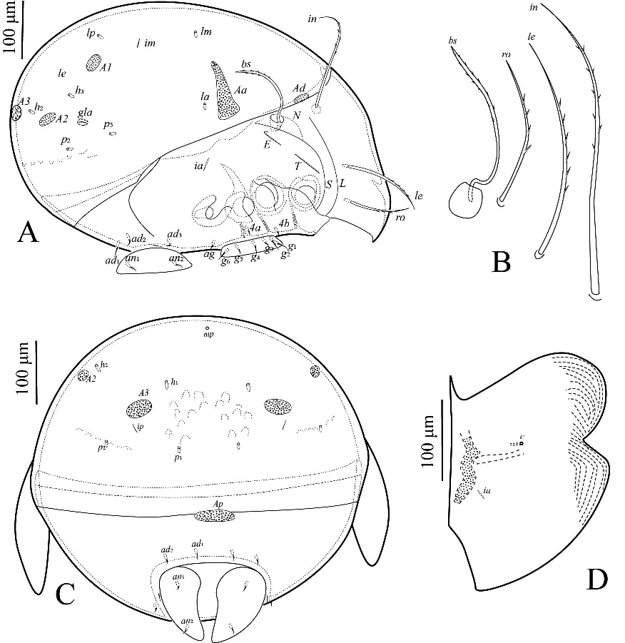
Surface of notogaster smooth, prodorsum, pteromorphs outer edge and epimeral region with granules. Rostrum pointed. Lamellar and sublamellar lines present. Rostral and lamellar setae setiform, smooth. Interlamellar seta short, slightly barbed. Bothridial seta clavate, barbed head. Dorsosejugal porose areas and dorsosejugal suture present. Four pairs of notogastral porose areas developed, Aa transverse irregular wedge, A1 rounded, A2 and A3 oval. Median pore area present. Postanal porose area transversely elongate oval.

Description
Measurements — Body length: 710 (holotype), 600–750 (53 paratypes); notogaster width: 540 (holotype), 450–570 (53 paratypes). No distinct differences between females and males in body size.

Integument — (Figures 1A–B, 2A–D, 4A–F). Body color brown to black. Prodorsum, pteromorphs outer edge and epimeral region with granules and striations, rest smooth.
Prodorsum — (Figures 1A, 2A, 2C, 4A-D). Rostrum pointed. Lamellar and sublamellar lines present, curving backwards at ventral end. Rostral ro (43–50) and lamellar le (78–86) setae setiform, smooth. Interlamellar seta in (23–30) short, setiform, slightly barbed. Bothridial seta bs (72–82) clavate, stalk smooth, head surface with slightly barbed and inside with particulate matter. Dorsosejugal porose area Ad (8–10 × 20–26) located transversely, elongate oval.
Notogaster — (Figures 1A, 2B, 2D, 4A). Dorsosejugal suture developed, complete. Notogaster rounded posteriorly. Notogastral setae represented by 10 pairs of alveoli or microsetae. With four pairs of notogastral porose areas, Aa (16–35 × 60–68) transverse irregular wedge, pointing mediad; A1 (diameter 22–27) rounded; A2 (10–14 × 26–30) and A3 (8–12 × 43–48) oval. Median pore present in females and males, located posterior to imaginary line connecting porose areas A1. Lyrifissure im located medial to setal alveoli lm and lp. Opisthonotal gland gla openings located lateral to A1.
Gnathosoma — (Figures 1D‒F, 4B). Subcapitulum size: 122–127 × 113–118, three pairs of setiform, slightly barbed, setae: h (20‒24), m (18‒22) and a (24‒28). Two pairs of setiform, smooth adoral setae or1 (8‒12) and or2 (8‒12). Length of palps 105–110. Palp with setation 0-2-1-3-9 (+ solenidion ω). Length of chelicerae 160–165. Cheliceral setae cha (60–65) and chb (43–48) setiform, barbed bilaterally. Trägårdh's organ (Tg) long, elongate triangular.
Epimeral and lateral podosomal regions — (Figures 1B, 4B). Epimeral region with granules and striations. Pedotecta I and II rounded in ventral view. Discidium triangular, circumpedal carina distinct. Epimeral setal formula: 1-0-1-2. Four pairs (1a, 3b, 4a and 4b, 5‒15) of epimeral setae setiform, thin, smooth.
Anogenital region — (Figures 1B, 2B, 4B, 4D‒E). Six pairs of genital setae (g1–g2, 12–22; g3–g6, 2–12), setiform, short, smooth; g1 and g2 parallel to each other at anterior edges of genital plate, other four pairs represented by alveoli or microsetae, arranged vertically in middle of genital plates. One pair of aggenital seta (ag, 0–4) located close to genital aperture. Two pairs of anal and three pairs of adanal setae represented alveoli or microsetae. Adanal lyrifissures located close and lateral to anal plates. Adanal setae ad1 and ad2 located in postanal position, ad3 anterolateral to iad. Distance between ad1–ad2 distinctly shorter than that of ad2–ad3. Postanal porose area Ap (22–26 × 58–62) elongate oval.


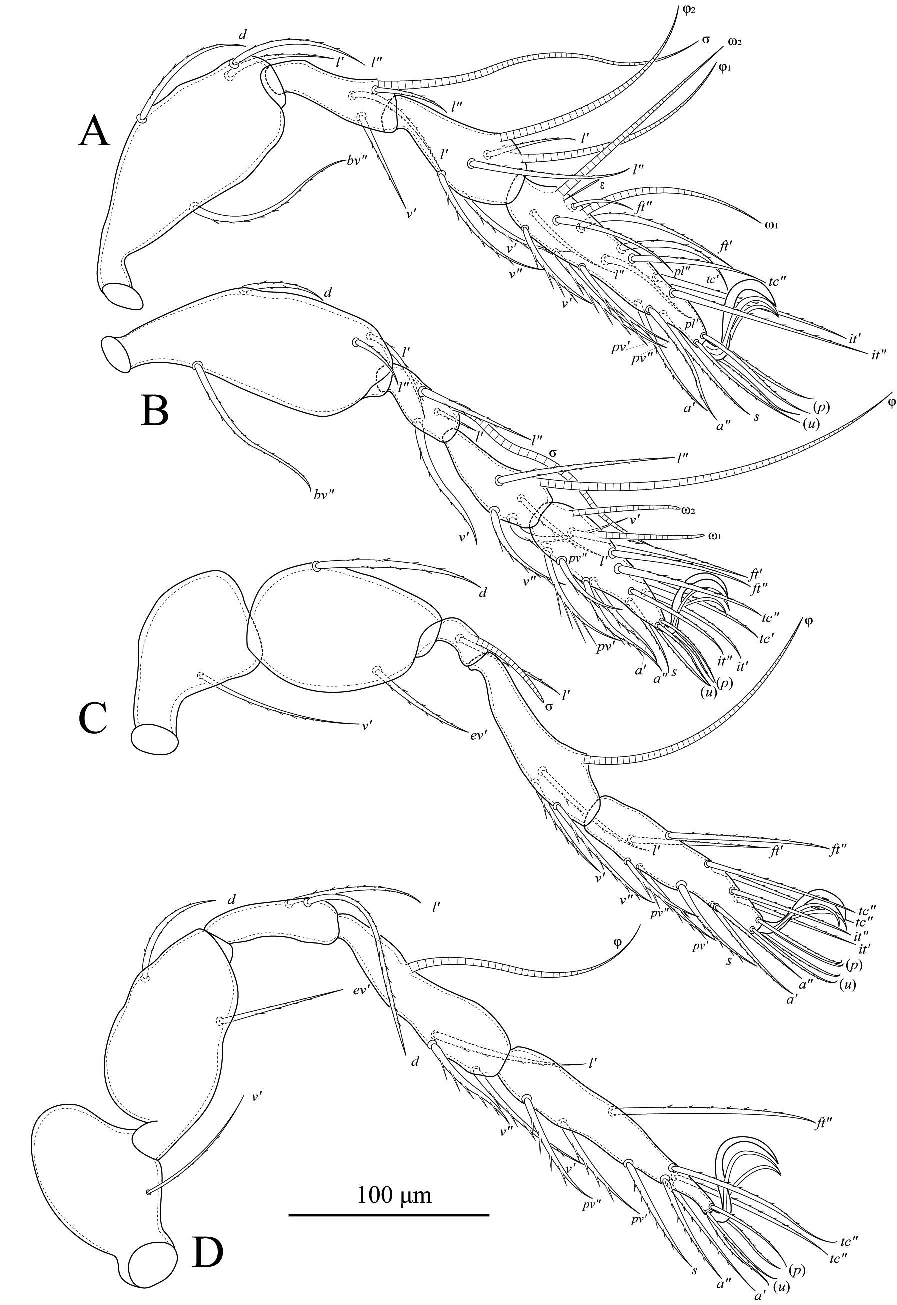
Legs — (Figure 3) All legs tridactylous, median claw distinctly thicker than lateral claws. Formulas of leg setation and solenidia: I (1-4-3-4-20) [1-2-2], II (1-4-3-4-15) [1-1-2], III (1-2-1-3-15) [1-1-0], IV (1-2-2-3-12) [0-1-0]. Famulus on tarsi I inserted between solenidia ω1 and ω2. Homology of setae and solenidia indicated in Table 1.
Material examined
Holotype (male), Mudanfeng National Nature Reserve (44°22' N, 129°53' E), Mudanjiang City, Heilongjiang Province, 20 Jul. 2010, Lixia Xie and Rong Huang, in soil. 53 paratypes: 8 (5 females 3 males) same data as holotype; 7 (5 females 2 males), Liangshui National Nature Reserve (47°9' N, 128°52' E), Yichun City, Heilongjiang Province, 25 Jul. 2010, Lixia Xie and Rong Huang, in soil; 38 (15 females 23 males), Baishilazi National Nature Reserve (40°56' N, 124°53' E), Dandong City, Liaoning Province, 3 Aug. 2010, Lixia Xie and Rong Huang, in soil.
Type deposition
The holotype and 7 paratypes are deposited in the Institute of Entomology, Guizhou University, Guiyang, China (GUGC) (Zhang 2018). 46 paratypes are deposited in the Guizhou Provincial Center for Disease Control and Prevention, Guiyang, China.
Etymology
The name of the new species comes from the Latin word ''clava'' meaning ''clavate'' which means that the new species bothridial seta clavate.
Remarks
In having dorsosejugal suture complete; developed four pairs notogastral porose areas, Aa transverse irregular wedge; short interlamellar seta and bothridial seta clavate, Pergalumna clava n. sp. is morphologically similar to Pergalumna akitaensis Aoki, 1961 and Pergalumna formicaria Berlese, 1914, redescribed by Mahunka, 1992. Differs from Pergalumna akitaensis Aoki, 1961 by the following characteristics: (1) larger porose area Aa and A3; Aa and A3 significantly larger than A1 (versus smaller porose area Aa, A1 and A3; and sizes not significantly different in Pergalumna akitaensis). (2) Median pore and postanal porose area present (versus median pore and postanal porose area absent). (3) Lyrifissure im located medial to lm and lp (versus lyrifissure im located closer to lp and distanced from lm). Differs from Pergalumna formicaria Berlese, 1914, by the following characteristics: (1) Rostral and lamellar setae smooth (versus rostral and lamellar setae slightly barbed in Pergalumna formicaria), (2) Prodorsum and pteromorphs with granules (prodorsum and pteromorphs smooth). (3) Lyrifissure im located medial to lm and lp (versus lyrifissure im closer to A1 and distanced from Aa).
Pergalumna pilosus n. sp.
ZOOBANK: 6E649612-AD54-4A21-8C4A-769F9A25BD46 ![]()
(Figures 5‒8)
Diagnosis
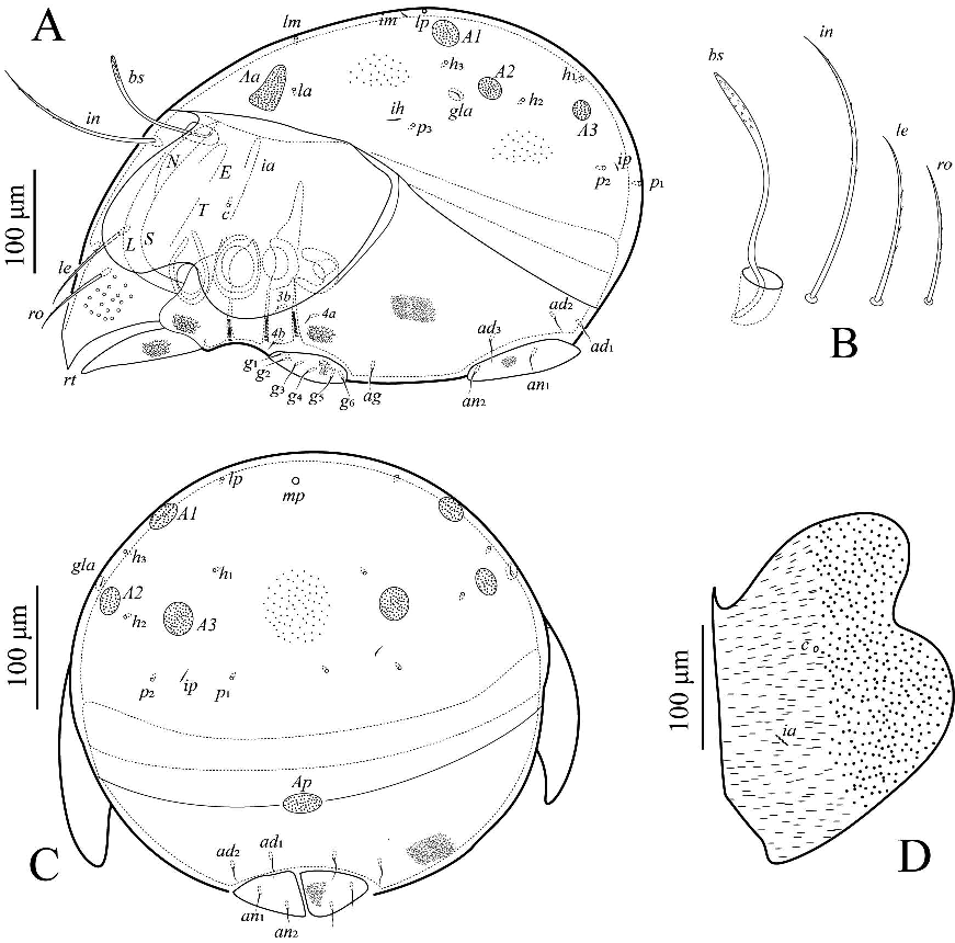
Body surface smooth, pteromorphs outer edge short striae, middle part of genital plates with striate. Rostrum pointed. Lamellar and sublamellar lines present. Rostral, lamellar, interlamellar and bothridial setae developed, setiform, slightly barbed. Dorsosejugal porose areas and dorsosejugal suture present. Four pairs of notogastral porose areas, Aa transverse long wedge, A1 rounded, A2 and A3 oval. Median pore and postanal porose area present.

Description
Measurements — Body length: 630 (holotype), 560–670 (26 paratypes); notogaster width: 450 (holotype), 400–480 (26 paratypes). No distinct differences between females and males in body size.
Integument — (Figures 5A–B, 6A, 6C–D, 8A–E). Body color brown to black. Body surface smooth, pteromorphs outer edge short striae, the middle part of genital plates with striate, the middle of pteromorphs bilobed.
Prodorsum — (Figures 5A, 6A–B, 8A, 8F). Rostrum pointed. Lamellar and sublamellar lines parallel, curving backwards at ventral end. Rostral ro (70–80) and lamellar setae le (95–105) setiform, barbed unilaterally. Interlamellar seta in (130–140) long, setiform, first half almost smooth and the second half has a few bilaterally slightly barbed. Bothridial seta bs (120–130) setiform, slightly barbed. Dorsosejugal porose areas Ad (9–13 × 30–33) located under anterior notogastral margin posterior to in, elongate oval.
Notogaster — (Figures 5A, 6A, 6C–B, 8A). Dorsosejugal suture developed, complete. Notogastral setae represented by10 pairs of alveoli. Four pairs porose areas, Aa (10–46 × 100–103) located between la and lm, transverse long wedge; A1 (diameter 25–30) rounded; A2 (10–13 × 25–28) and A3 (18–21 × 40–43) oval. Median pore present in females and males, located posterior to imaginary line connecting porose areas A1. Lyrifissure im located between alveoli lm and lp. Opisthonotal gland gla openings located posterolateral to A1, lateral to h3 .
Gnathosoma — (Figures 5C–E, 8D). Subcapitulum size: 161–166 × 153–158, three pairs of setiform, smooth, curved subcapitular setae: h (24‒28), m (36‒40) and a (32‒36). Two pairs of setiform, smooth adoral setae or1 (5‒9) and or2 (5‒9). Length of palps 140–145. Palp with setation 0-2-1-3-9 (+ solenidion ω). Length of chelicerae 192–197. Cheliceral setae cha (73–78) and chb (50–55) setiform, barbed bilaterally. Trägårdh's organ long, elongate triangular.
Epimeral and lateral podosomal regions — (Figures 5B, 6A, 8B). Epimeres smooth. Pedotecta I and II rounded in ventral view. Discidium triangular, circumpedal carina distinct. Epimeral setal formula: 1-0-1-2. Four pairs (1b, 3b, 4a and 4b (3‒7) of epimeral setae setiform, thin, smooth.
Anogenital region — (Figures 5B, 6A, 6C, 8B, 8C, 8E). Middle part of genital plates with striate. Six pairs of genital setae (g1–g6, 10–15), anterior edges of genital plates with two pairs of setae. One pair of aggenital seta (ag, 4–8) located between genital and anal plates, closer to genital than to anal plates. Two pairs of anal and three pairs of adanal setae short, thin, smooth. Adanal lyrifissures located close and lateral to anal plates. Adanal setae ad1 and ad2 postanal, ad3 located anterolateral to iad. Distance between ad1–ad2 distinctly shorter than that of ad2–ad3. Postanal porose area oval (15–20 × 60–65).
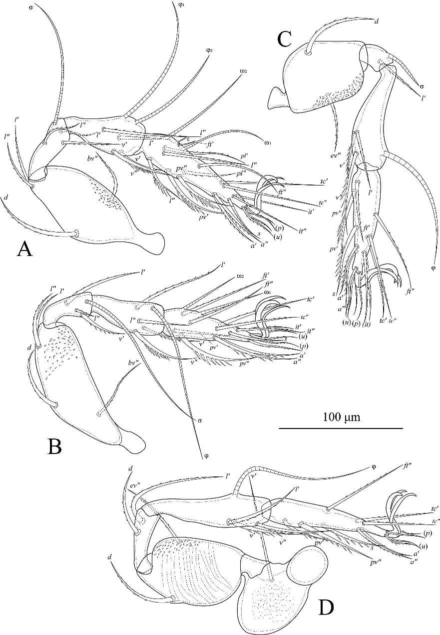
Legs — (Figure 7). All legs tridactylous, median claw distinctly thicker than lateral claws. Formulas of leg setation and solenidia: I (1-4-3-4-20) [1-2-2], II (1-4-3-4-15) [1-1-2], III (1-2-1-3-15) [1-1-0], IV (1-2-2-3-12) [0-1-0]. Famulus on tarsi I inserted between solenidia ω1 and ω2. Solenidion on tibiae IV inserted in anterior part of the segment. Homology of setae and solenidia indicated in Table 1.

Material examined
Holotype (male), Damingshan National Nature Reserve (23°30' N, 108°27' E), Nanning City, Guangxi Province, 16 May 2010, Rong Huang, in soil. 26 paratypes: 2 (1 female 1 male) same data as holotype; 10 (3 females 7 males), Zhuque National Forest Park (33°47' N, 108°35' E), Xian City, Shanxi Province, 11 Jul. 2012, Wenqin Liang and Qiuxiao Tang, litter in the mixed forest; 11 (7 females 4 males), Wen county (33°3'25'' N, 104°42'31'' E, 1800 m), Longnan City, Gansu Province, 14 Aug. 2018, Guoru Ren and Maofa Yang, in soil; 3 (2 females 1 male), Gaoleshan National Nature Reserve (30°19'22'' N, 119°26'44'' E, 420 m), Nanyang City, Henan Province, 21 Jul. 2018, Guoru Ren and Qianfen Zheng, in soil.
Type deposition
The holotype and 16 paratypes are deposited in the Institute of Entomology, Guizhou University, Guiyang, China (GUGC). 10 paratypes are deposited in the Guizhou Provincial Center for Disease Control and Prevention, Guiyang, China.
Etymology
The name of the new species comes from the Latin name ''pilosus'' meaning ''hairy'' which refers to the comparable long cilia on rostrum, lamellar and interlamellar setae of the new species.
Remarks
In having dorsosejugal suture complete; Aa porose area transverse irregular wedge; longer interlamellar barbed; median pore and postanal porose area present, Pergalumna pilosus n. sp. is morphologically similar to Pergalumna variosculpturata Mahunka and Mahunka-Papp, 1999 and Pergalumna microtuberculata Bayartogtokh and Akrami, 2014, but differs from the latter two species by the following characteristics: (1) Bothridial setae setiform (versus bothridial setae lanceolate, stalk slender and head slightly incrassate and speculate in latter two species). (2) Interlamellar setae very long (130–140); significantly longer than le and ro (versus interlamellar, lamellar and rostral setae are not much difference in length). (3) Epimeral region smooth (versus epimeral region with granules and striations).
Pergalumna amamiensis Aoki, 1984
(Figures 9‒12)
Supplementary description

Measurements — Body length: 540–680; notogaster width: 390–490. No distinct differences between females and males in body size.

Integument — (Figures 9A–B, 10A, 10C–D, 12A–F). Body color brown to black. Body surface foveolate. Prodorsum, genital plates, epimeral and lateral podosomal regions large granules; pteromorphs outer edge and anal plates and the surrounding with granules.
Prodorsum — (Figures 9A, 10A–B, 12A, 12C–D). Rostrum pointed. Lamellar and sublamellar lines parallel, long, curving backwards at ventral end. Rostral ro (75–85) and lamellar setae le (100–110) setiform, barbed unilaterally. Interlamellar seta in (140–150) long, setiform, slightly barbed. Bothridial seta bs (120–130) lanceolate, stalk slender, head slightly incrassate and spiculate. Dorsosejugal porose areas (8–12 × 24–28) located under anterior notogastral margin posterior to in, elongate oval.
Notogaster — (Figures 9A, 10A, 10C–D, 12A, 12D). Dorsosejugal suture developed, complete. Notogastral setae represented by10 pairs of alveoli. Four pairs porose areas, Aa (10–35 × 50–60) located above to la, transverse long wedge; A1 (diameter 23–28) rounded; A2 (17–22 × 25–28) oval; A3 (diameter 23–28) rounded from posterior view. Median pore present in females and males, located middle to A2. Lyrifissure im located between alveoli lm and lp, closer to lp than lm. Opisthonotal gland gla openings located lateral to A1.
Gnathosoma — (Figures 9C–E, 12B). Subcapitulum size: 160–165 × 120–125, three pairs of setiform, smooth, curved subcapitular setae: h (18‒22), m (28‒32) and a (36‒40). Two pairs of setiform, smooth adoral setae or1 (11‒13) and or2 (11‒13). Length of palps 120–130. Palp with setation 0-2-1-3-9 (+ solenidion ω). Length of chelicerae 190–200. Cheliceral setae cha (60–65) and chb (55–60) setiform, barbed bilaterally. Trägårdh's organ long, elongate triangular.
Epimeral and lateral podosomal regions — (Figures 9B, 10B, 12B). Epimeres strong granules. Pedotecta I and II rounded in ventral view. Discidium triangular, circumpedal carina distinct. Epimeral setal formula: 1-0-1-2. Four pairs of epimeral setae setiform, thin, smooth 1b (20‒25), 3b (25‒30), 4a (20‒25) and 4b (20‒25).
Anogenital region — (Figures 9B, 10A, 10C, 12B, 12E–F). Middle part of genital plates with striate. Six pairs of genital setae (g1–g2, 25–30; g3–g6, 15–20), anterior edges of genital plates with two pairs of setae. One pair of aggenital setae (ag, 10–15) located between genital and anal plates, closer to genital than to anal plates. Two pairs of anal (an1–an2, 10–15) and three pairs of adanal setae (ad1–ad3, 10–20), all short, thin, smooth. Adanal lyrifissures located close and lateral to anal plates. Adanal setae ad1 and ad2 postanal, ad3 located anterolateral to iad. Distance ad1–ad2 distinctly shorter than ad2–ad3. Postanal porose area oval (13–18 × 28–33).
Legs — (Figure 11). All legs tridactylous, median claw distinctly thicker than lateral claws. Formulas of leg setation and solenidia: I (1-4-3-4-20) [1-2-2], II (1-4-3-4-15) [1-1-2], III (1-2-1-3-15) [1-1-0], IV (1-2-2-3-12) [0-1-0]. Famulus on tarsi I inserted between solenidia ω1 and ω2. Solenidion on tibiae IV inserted in anterior part of the segment. Homology of setae and solenidia indicated in Table 1.
Material examined
15 females 11 males, Tianmushan National Nature Reserve (30°20'4'' N, 119°26'18'' E, 810 m), Hangzhou City, Zhejiang Province, 27 Jul. 2018, Guoru Ren and Qianfen Zheng, in moss. 2 females 1 male, Gujingyuan National Nature Reserve (31°3'7″N, 116°30'7'' E, 450 m), Haozhou City, Anhui Province, 25 Jul. 2018, Guoru Ren and Qianfen Zheng, in deciduous leaves.
Specimen deposition
26 samples are deposited in the Institute of Entomology, Guizhou University, Guiyang, China (GUGC). 3 specimens are deposited in the Guizhou Provincial Center for Disease Control and Prevention, Guiyang, China.


Remarks
This species was originally described by Aoki (1984) from Japan, but the original description was brief and not completely illustrated, so we gove a supplementary description and illustrations. Apart from the lacking characteristics, the Chinese specimens differ from the Japanese specimens by the rostral, lamellar, interlamellar and bothridial setae which are slightly barbed. Hence, based on these supplementary data, the main characters of P. amamiensis are: body size: 540–680×390–490, body surface densely foveolate granules; rostrum pointed; lamellar and sublamellar lines present; rostral, lamellar and interlamellar setae developed, setiform, slightly barbed; bothridial setae spindle-shaped, stalk slender and head slightly incrassate and spiculate; dorsosejugal porose areas and dorsosejugal suture present; four pairs of notogastral porose areas, Aa transversely wedge-shaped, A1 rounded, A2 and A3 oval; median pore and postanal porose area present; epimeral setal formula 1-0-1-2; ventral setae, genital setae, epimeral setae, anogenital and adanal setae represented by microsetae; all legs tridactylous, leg setae not modified.
Acknowledgements
We would like to express our gratitude to Professor Roy A. Norton (State University of New York, Syracuse, U. S. A.) for providing literature. Thanks also due to Lixia Xie, Rong Huang and Qiuxiao Tang (Institute of Entomology, Guizhou University, Guiyang of China) for provided samples. This project was supported by the Program of Ministry of Science and Technology of the People's Republic of China (2015FY210300); independent research topic of national key laboratory for the prevention and control of infectious diseases (No. 2018SKLID305); the Program of Excellent Innovation Talents, Guizhou Province, China (No. 20164022); the Program of Science and Technology of Guizhou Province (No. Qian Ke He Platform talent [2018]5767); special funds of research team for experimental diagnostic technique and molecular epidemiological study of major infectious disease in Guizhou Province (Program of scientific and technological innovation team of Guizhou Province, No. Qian Ke He Platform talent [2018]5606); Science and Technology Fund project of Guizhou Provincial Health and Family Planning Commission (No. gzwjkj2018-1-066).
References
Aoki J. 1961. On six new oribatid mites from Japans. Jpn. J. Sanit. Zool., 12(4): 233-238. doi:10.7601/mez.12.233
Aoki J. 1984. New and unrecorded oribatid mites from Amami-Ohshima Island, Southwest Japan. Zool. Sci., 1: 132-147.
Bayartogtokh B., Akrami M.A. 2014. The soil mite family Galumnidae of Iran (Acari: Oribatida). J. Nat. Hist., 48(15-16): 881-917. doi:10.1080/00222933.2013.840397
Berlese A. 1914. Acari nuovi. Manipulus IX. Redia, 10(1): 113-150.
Chen J., Liu D., Wang H.F. 2010. Oribatid mites of China: a review of progress, with a checklist. Zoosymposia, 4: 186-224. doi:10.11646/zoosymposia.4.1.14
Ermilov S.G., Klimov P.B. 2017. Generic revision of the large-winged mite superfamily Galumnoidea (Acari, Oribatida) of the world. Zootaxa, 4357(1): 1-72. doi:10.11646/zootaxa.4357.1.1
Ermilov S.G., Starý J. 2017. Two new species of the genus Pergalumna (Acari, Oribatida, Galumnidae) from Northern Vietnam. Syst. Appl. Acarol., 22(4): 494-508. doi:10.11158/saa.22.4.6
Grandjean F. 1936. Les Oribates de Jean Frédéric Hermann et de son père. Ann Soc Entomol Fr, 105: 27-110.
Jacot A.P. 1925. Phylogenie in the Oribatoidea. Am. Nat., 59(662): 272-279. doi:10.1086/280038
Mahunka S. 1992. "Pelops" and "Oribates" species in the berlese-collection (Acari). Acta Zool. Acad. Sci. Hung., 38(3-4): 213-260.
Mahunka S., Mahunka-Papp L. 1999. Oribatids (Acari: Oribatida) from the Aggtelek National Park (NE Hungary). The Fauna of the Aggtelek National Park. Budapest: Hungarian Natural History Museum. p. 619-651.
Norton R.A., Behan-Pelletier V.M. 2009. Suborder Oribatida. Chapter 15. In: Krantz G.W., Walter D.E. (Eds). A Manual of Acarology. Lubbock: Texas Tech University Press. p. 430-564.
Subías L.S. 2020. Listado sistemático, sinonímico y biogeográfico de los Ácaros Oribátidos (Acariformes: Oribatida) del mundo (excepto fósiles), 15ª actualización. pp. 527. Available from: http://escalera.bio.ucm.es/usuarios/bba/cont/docs/RO_1.pdf

Travé, J., Vachon, M. 1975. François Grandjean. 1882-1975 (Notice biographique et bibliographique). Acarologia, 17(1): 1-19.
Zhang Z.-Q. 2018. Repositories for mite and tick specimens: acronyms and their nomenclature. Syst. Appl. Acarol., 23(12): 2432-2446. doi:10.11158/saa.23.12.12
Zheng Q.F., Liang W.Q., Ren G.R., Yang M.F. 2019. A new species and two newly recorded species of the subgenus Pergalumna (Pergalumna) (Acari, Oribatida, Galumnidae) from China. Zootaxa, 4647(1): 407-423. doi:10.11646/zootaxa.4647.1.26


2020-10-20
Date accepted:
2021-03-10
Date published:
2021-03-12
Edited by:
Baumann, Julia

This work is licensed under a Creative Commons Attribution 4.0 International License
2021 Zheng, Qian-Fen; Liang, Wen-Qin; Ren, Guo-Ru and Yang, Mao-Fa
 Download article
Download the citation
RIS with abstract
(Zotero, Endnote, Reference Manager, ProCite, RefWorks, Mendeley)
RIS without abstract
BIB
(Zotero, BibTeX)
TXT
(PubMed, Txt)