


Kuzinellus (Acari: Phytoseiidae) from China
Ma, Min1 ; Fan, Qing-Hai2 and Li, Sheng-Cai3
1College of Agronomy, Shanxi Agriculture University, Taigu, China.
2✉ Plant Health & Environment Laboratory, Ministry for Primary Industries, Auckland, New Zealand.
3College of Agronomy, Shanxi Agriculture University, Taigu, China.
2018 - Volume: 58 Issue: 4 pages: 786-794
https://doi.org/10.24349/acarologia/20184274ZooBank LSID: 34DBCBDA-3436-4EA5-8E61-3B48BBCC793A
Keywords
Abstract
As a family of valuable beneficial predators Phytoseiidae has received an increasing attention in the last three decades (Helle and Sabelis, 1985; Lindquist et al., 1996; McMurtry and Croft, 1997; Sabelis and Van Rijn, 1997; Gerson et al., 2003; Moraes et al., 2004). To date more than 2,750 species have been described (Demite et al., 2017) and some species are commercially produced for controlling spider mites, thrips and whiteflies. The genus Kuzinellus Wainstein, 1976 is relatively small and consists of about 51 described species (Demite et al., 2017; Kamran et al., 2017). Only two species of this genus are known from China, an Oriental species, K. cervix (Wu and Li, 1984) from Hubei province and a Palaearctic species, K. trisetus (Wu et al., 1992) from Liaoning province. Kuzinellus cervix was subsequently found in Fujian, Hunan and Jiangxi (Wu et al., 2009) but there was no record of K. trisetus from other provincial areas of the country. During a survey of phytoseiid mites of Shanxi province, K. trisetus was discovered from several localities. In this paper, we re-described both species based on the original types and fresh specimens of K. trisetus.
The type specimens of Kuzinellus cervix and K. trisetus loaned from Guangdong Institute of Entomology, Guangzhou and fresh specimens of K. trisetus collected from Shanxi were used for this study. Illustrations were made using a drawing tube attached to a Nikon differential interference contrast (DIC) microscope, which was also used for measuring and imaging of specimens. Images and illustrations were edited with Photoshop CS4. Measuring method follows Ma et al. (2016). All measurements are given in micrometers (μm). The measurements for the holotype are followed into brackets by the range of measurements from paratypes or other specimens. The chaetotaxy of the idiosoma and legs follow Chant and McMurtry (2007) and Evans (1963), respectively, and the terminology of pore-like structures follows Beard (2001).
Kuzinellus Wainstein, 1976: 699. Type species: Paraseiulus kuzini Wainstein, 1962: 139.
Diagnosis — Dorsal idiosomal setal patterns 13A:8A, with 19 pairs of setae on the dorsal shield. Setae z6 and JV2 present, Z1 absent. Ventrianal shield with 4 pairs of preanal setae. Kuzinellus can be distinguished from its closely related Paraseiulus Muma by having JV2 present and Z1 absent. It differs from the other genus included in the tribe, Paraseiulini Wainstein (Australiseius Muma) by having Z1 absent.
Typhlodromus (Anthoseius) cervix Wu and Li, 1984: 44; Moraes et al., 2004: 317; Chant and McMurtry, 2007: 152.
Typhlodromus (Paraseiulus) cervix; Wu, 1985: 86.
Amblydromella cervix; Moraes et al., 1986: 353.
Typhlodromus cervix; Wu and Lan, 1992: 1367; Hou, 1996: 13; Wu et al., 1997: 172.
Kuzinellus cervix; Wu et al., 2009: 330.
ADULT FEMALE (n=2).
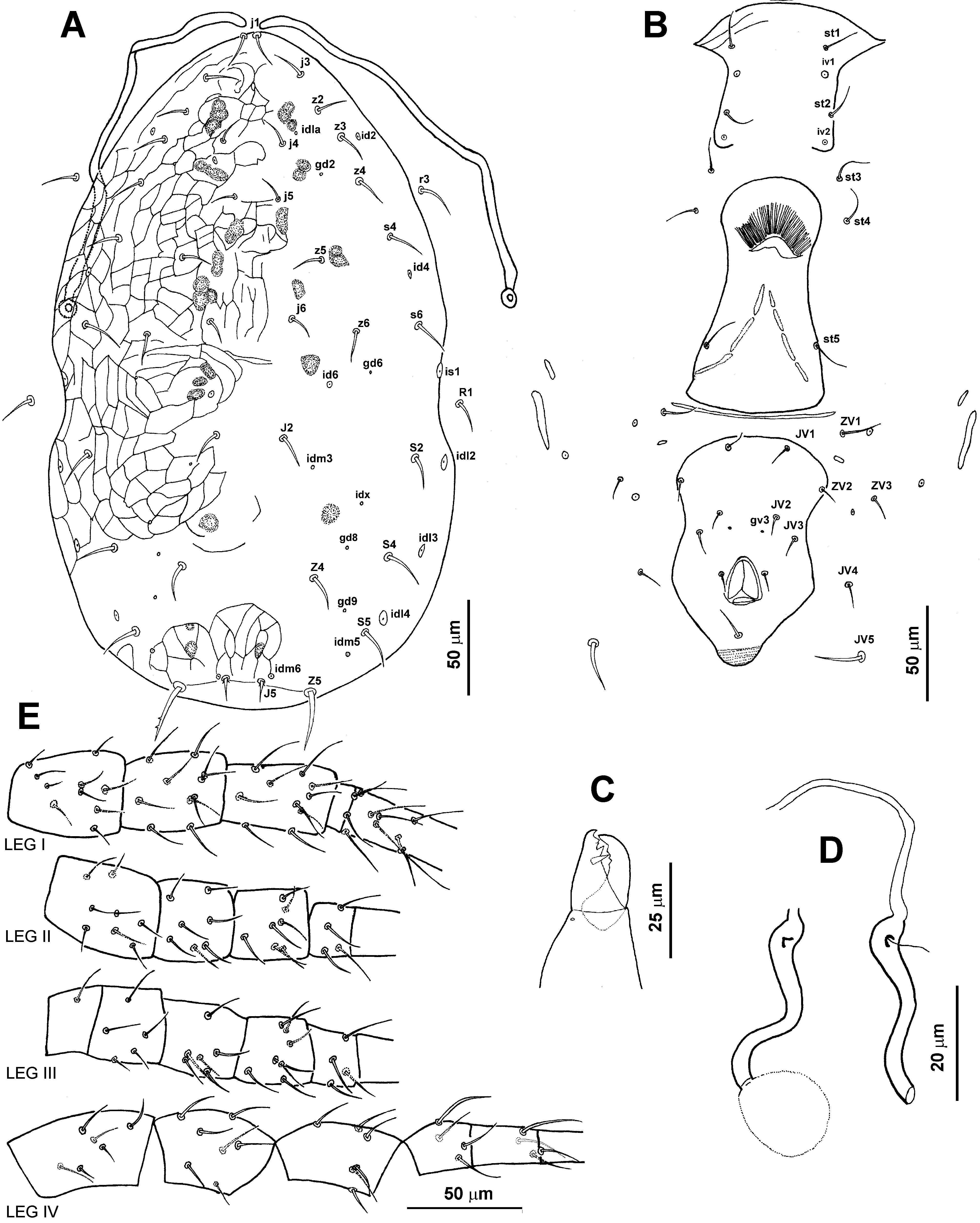
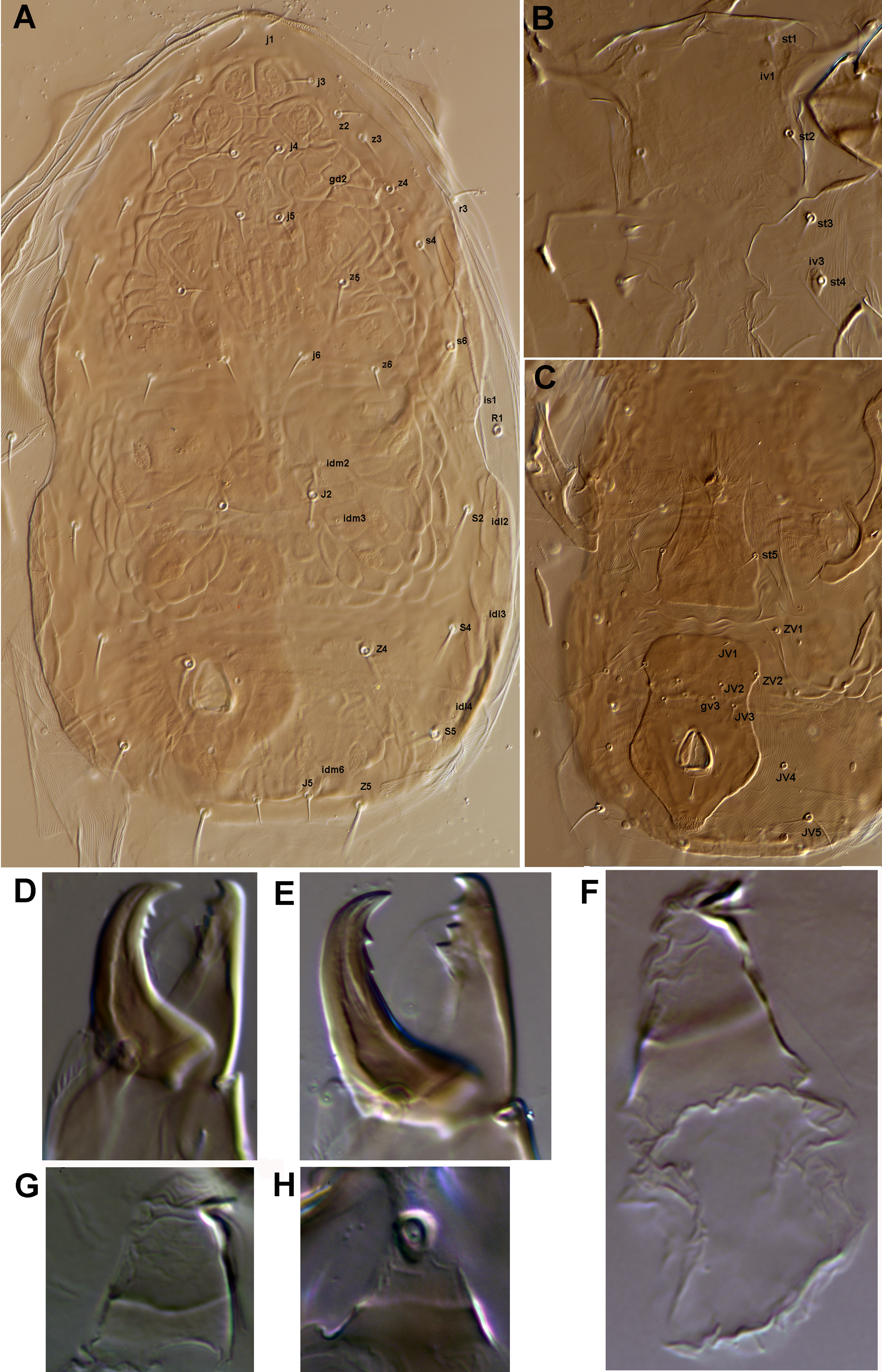
Dorsum (Figures 1A, 2A) — Dorsal shield nearly oval, incised at level of R1, reticulate throughout except area between S4 and S5; 325 (336) long, 200 (203) wide; all setae smooth except Z5 (barbed), bearing 11 pairs of discernible lyrifissures (id1a, id4, id6, idm3, idm5, idm6, is1, idl2, idl3, idl4 and idx) and 4 pairs of solenostomes (gd2, gd6, gd8 and gd9). Muscle marks discernible, mainly on podosomal areas between z2 and j4, j4 and j5, j5 and z5, s4 and z5, z5 and j6, j6 and J2, S2 and S4, and prior to J5. Lateral setae r3 and R1 smooth, on soft membranous cuticle lateral to dorsal shield, r3 at level of z4, R1 at level of shield incisions. Peritremes extending forward to bases of j1. Lengths of setae: j1 17 (17), j3 16 (17), j4 13 (12), j5 12 (12), j6 14 (14), z2 16 (16), z3 17 (16), z4 18 (19), z5 15 (16), z6 15 (15), s4 17 (19), s6 20 (20), J2 16 (16), J5 11 (12), Z4 19 (18), Z5 27 (29), S2 18 (20), S4 21 (21), S5 19 (21), r3 17 (21), R1 17 (18).
Ventral idiosoma (Figures 1B, 2B) — Sternal shield smooth (without reticulation), 56 (56–57) wide, anterior margin moderately convex, medial posterior margin vague, bearing 2 pairs of attenuate setae (st1, st2) and 2 pairs of lyrifissures (iv1, iv2), iv1 between st1 and st2, iv2 at corners of the posterior margin; setae st3 on soft cuticle, st4 on platelets. Genital shield smooth, 113 (120) long, 58 (60) wide. Lengths of setae: st1 19 (20), st2 13 (12), st3 19 (19), st4 15 (13), st5 11 (18). A thin and long transversal sclerite between genital and ventrianal shields, a pair of small plates between ZV1 and ZV2. Ventrianal shield approximately pentagonal, smooth, anterior margin convex, with prominent waist at JV2 level, 116 (118) long, 75 (76) wide, with 4 pairs of pre-anal setae ( JV1, JV2, JV3 and ZV2) and a pair of pores gv3 posteromedial to JV2; distance gv3-gv3 18 (18), 4 pairs of setae ( ZV1, ZV3, JV4 and JV5) and 3 pairs of lyrifissures on soft cuticle surrounding ventrianal shield. Primary metapodal plate 30 (32) long, 4 (4) wide, the secondary plate 8 (9) long, 2 (1) wide.
Calyx of insemination apparatus tubular (Figure 1D, 2D, 2E), 37 (39) long and 2 (2) wide, major duct long and slender, 34 long, 1 wide, minor duct thread-like, connecting to centre of atrium.
Gnathosoma — Chelicera (Figure 1C, 2C) with movable digit 25 (26) long, bearing 2 teeth, fixed digit 25 (27) long, bearing 3 discernible teeth, terminal two next to each other.
Leg lengths (I–IV): 299 (307), 232 (233), 230 (269) and 289 (344). Legs I, II and III without macrosetae. Basitarsus and telotarsus of leg IV (Figure 1E) each with a smooth macroseta, 20 (20) and 25 (31) long, respectively.


ADULT MALE. Unknown.
Holotype female, Hubei: Shiyan, Shennongjia National Nature Reserve, Pinus massoniana, 15.VIII.1981, collected by Wei-nan Wu and Zhao-quan Li. Paratype: 1 female, same data as holotype. Both specimens are deposited at the Guangdong Institute of Applied Biological Resources, Guangdong, China.
China: Fujian (Wu, 1985; Zhang and Lin, 1987), Hubei (Wu and Li, 1984), Hunan (Wu and Lan, 1992), Jiangxi (Hou, 1996).
Fragaria × ananassa (Hou, 1996), Hyllostachys heterocycla (Zhang and Lin, 1987), Pinus massoniana (Wu and Li, 1984; Wu, 1985).
In their original description Wu and Li (1984) miscounted the number of dorsal setae, overlooked z6 and incorrectly illustrated the sternal shield. Wu et al. (1997) corrected these mistakes. In 2009 Wu et al. transferred this species from Typhlodromus (Anthoseius) to Kuzinellus. We herein confirm their corrections and changes. Apart from these we have noted and illustrated the following characters which were not presented in the original and subsequent publications (Wu and Li, 1984; Wu et al., 1997; 2009): lyrifissures, gland openings and muscle marks on idiosoma; atrium, major and minor ducts of insemination apparatus, and legs I–III. The second tooth of the fixed digit of chelicera is very small and situated next to the apical tooth. The margins of the platelets bearing st4 and iv2 were not discernible in the long preserved specimens. This species was originally described as Typhlodromus (Anthoseius) cervix and listed as such in the catalog of Moraes et al. (1986) and in the Phytoseiidae database of Demite et al. (2017), which would need to be updated.
Typhlodromus trisetus Wu et al., 1992: 48; Wu et al., 1997: 173.
Amblydromella (Amblydromella) triseta; Denmark and Welbourn, 2002: 307.
Kuzinellus trisetus; Moraes et al., 2004: 274; Wu et al., 2009: 331.
ADULT FEMALE (n=5).
Dorsum (Figures 3A, 4A) — Dorsal shield nearly oval, incised at level of R1; 399 (390–414) long, greatest width 221 (211–228), 208-218 wide, reticulate throughout except area between Z4 and S5; all setae smooth except Z5 (barbed). Dorsal shield with 14 pairs of discernible lyrifissures (id1a, id2, id4, id6, idm2, idm3, idm4, idm5, idm6, idx, is1, idl2, idl3 and idl4) and 6 pairs of solenostomes (gd2, gd4, gd6, gd8 and gd9). Muscle marks discernible, mainly on dorsal shield between dorsal setae and median setae, and anterior to J5. Lateral setae r3 and R1 smooth, on soft membranous cuticle laterad of dorsal shield, r3 at level between z4 and s4, R1 at level of shield incisions. Peritremes extending forward to bases of j1. Lengths of setae: j1 18 (16–22), j3 16 (14–17), j4 14 (12–15), j5 13 (12–14), j6 14 (13–15), z2 15 (13–17), z3 16 (16–18), z4 17 (16–18), z5 15 (13–18), z6 15 (13–16), s4 18 (15–20), s6 21 (20–21), J2 19 (17–20), J5 12 (11–13), Z4 21 (20–23), Z5 27(25–30), S2 20 (20–22), S4 21 (19–24), S5 19 (18–20), r3 19 (17–21), R1 18 (17–20).
Ventral idiosoma (Figures 3B, 4B, 4C) — Sternal shield approximately as long as wide, 60 (57–65) long, 59 (56–62) wide, smooth, anterior margin straight, posterior margin have 2 corners, posterior margin medially smooth or zigzagged, bearing 2 pairs of attenuate setae (st1, st2) and 2 pair of lyrifissures (iv1, iv2), iv1 close to st1, iv2 at corners of posterior margin; setae st3 and st4 on membranous cuticle. Genital shield smooth, 128 (124–132) long, 60 (54–62) wide. Lengths of setae: st1 23 (20–24), st2 21 (20–21), st3 21 (18–23), st4 20 (16–21), st5 22 (21–23). A series of slender transversal sclerites present between genital and ventrianal shields, a pair of small plates present between ZV1 and ZV2. Ventrianal shield (Plate 1C) approximately pentagonal, smooth, anterior margin convex, with a prominent waist at level of JV2, 125 (122–130) long, 76 (64–80) wide, bearing 4 pairs of pre-anal setae ( JV1, JV2, JV3 and ZV2) and a pair of pores (gv3) posteromedial to JV2, distance gv3-gv3 21 (18–25),4 pairs of setae ( ZV1, ZV3, JV4 and JV5) and 3 pairs of lyrifissures on soft cuticle surrounding ventrianal shield. Primary metapodal plate 39 (36–42) long, 5 (4–7) wide, the secondary plate 14 (13–15) long, 2 (2–3) wide.
Calyx of insemination apparatus (Figures 3D, 4F, 4G, 4H) U- or V-shaped with basal 1/2 to 2/3 membranous and thin, and apical 1/3 to 1/2 thick.
Gnathosoma — Chelicera (Figures 3C, 4D, 4E) with movable digit 25 (24–26) long, bearing 2 or 3 teeth, fixed digit 27 (25–28) long, bearing 3 discernible teeth, pilus dentilis thorn-shaped, 8 (6–9) long, opposing distal tooth of movable digit.
Leg lengths (I–IV): 356 (341–371), 290 (287–305), 298 (290–301), 390 (380–402). All legs without obvious macrosetae (Figure 3E).


ADULT MALE. Unknown.
Holotype female, 10 paratype females, Qianshan, Liaoning, ex Larix sp.24.VII.1984, collected by Wei-nan Wu; 3 females, Ningwu, Luyashan National Nature Reserve, 38°44'34" N, 111°55'10" E, 2491 m, ex Pinus sp., 7.IX.2014, collected by Bing-Qian Su and Meng-Jiao Yin (accession no.: T14_0268); 1 female, Huguan, Taihang Mountains Grand Canyon, Purple Mass of Mountain, 35°54'47" N, 113°29'44" E, 1446 m, ex Viburnum mongolicum, 6.X.2014, collected by Min Ma (T14_0346); 1 female, same data as T14_0346 except: 35°54'47" N, 113°29'44" E, 1454 m, Spiraea trilobata, 7.X.2014, (T14_0347); 11 females, same data as T14_0268 except: Jiaocheng, Pangquangou National Nature Reserve, 37°49'35" N, 111°27'56" E, 1793 m, 28.VIII.2014, (T14_0191).
Holotype and paratypes are deposited in Guangdong Institute of Applied Biological Resources, Guangdong, China. Specimens collected from Shanxi are deposited in the Insect Ecology Laboratory, College of Agronomy, Shanxi Agriculture University, Taigu, China.
China: Henan (Lin et al., 2010), Liaoning (Wu et al., 1992), Shanxi (present paper).
Fern, Dianthus sp. (Lin et al., 2010); Larix sp. (Wu et al., 1992); Pinus sp.; Spiraea trilobata, Viburnum mongolicum (present paper).
This is the first report of Kuzienllus trisetus in Shanxi province. We have noted and illustrated the following characters which were not presented in the original publication (Wu et al., 1992): lyrifissures, gland openings and muscle marks on idiosoma, and legs I–III. The detailed structure of the insemination apparatus is given to show the unevenness of the calyx. The number of teeth on the movable digit is variable, from two to three. The apical tooth on the fixed digit is sometimes bifurcate. The areas around st4 and iv2 were not discernible in the long preserved specimens.
1. Spermatheca with calyx elongate and tubular, uniformly sclerotized (Fig. 1D)
...... Kuzinellus cervix (Wu and Li, 1984)
— Spermatheca with calyx short, U- or V-shaped, with basal 1/2 to 2/3 membranous (Fig. 2D)
...... Kuzinellus trisetus (Wu, Lan and Zhang, 1992)
We would like to thank Dr Zhi-Qiang Zhang (Landcare Research, New Zealand) for his critical comments on a preliminary version of this manuscript. We appreciate Misses Bing-Qian Su and Meng-Jiao Yin for their assistance in the collection of specimens.
Beard J.J. 2001. A review of Australian Neoseiulus Hughes and Typhlodromips De Leon (Acari: Phytoseiidae: Amblyseiinae). Invertebr. Taxon., 15: 73–158. doi:10.1071/IT99017 ![]()
Chant D.A., McMurtry J.A. 2007. Illustrated keys and diagnoses for the genera and subgenera of the Phytoseiidae of the world (Acari: Mesostigmata). West Bloomfield: Indira Publishing House, USA., 219 pp.
Demite P.R., Moraes G.J. de, McMurtry J.A., Denmark H.A., Castilho R.C. 2017. Phytoseiidae Database. [accessed on 12 Nov. 2017] Available from: www.lea.esalq.usp.br/phytoseiidae
Denmark H.A., Welbourn W.C. 2002. Revision of the genera Amblydromella Muma and Anthoseius De Leon (Acari: Phytoseiidae). Int. J. Acarol., 28(4): 291–316. doi:10.1080/01647950208684308 ![]()
Evans G.O. 1963. Observations on the chaetotaxy of the legs in the free-living Gamasina (Acari: Mesostigmata). Bull. Brit. Mus. (Nat. Hist.) Zool., 10(5): 277–303. doi:10.5962/bhl.part.20528 ![]()
Gerson U., Smiley R.L., Ochoa R. 2003. Mites (Acari) for pest control. Blackwell Science Ltd., UK, 539 pp. doi:10.1002/9780470750995 doi:10.1002/9780470750995 ![]()
Helle W., Sabelis, M. W. 1985. Spider mites: Their Biology, Natural Enemies and Control Vol. 1B. Elsevier, Amsterdam, The Netherlands, 458 pp.
Hou P.H. 1996. Investigation on species and vertical distribution of Phytoseiidae in Wuyishan Nature Reserve, Jiangxi Province. Jiangxi Plant Protection, 19(2): 12–14.
Kamran M., Basahih J.S., Alatawi F.J. 2017. A new species of Kuzinellus Wainstein, 1976 (Acari: Mesostigmata: Phytoseiidae) from Saudi Arabia, with a key to the world species. Int. J. Acarol., 43(7): 545–551. doi:10.1080/01647954.2017.1360937 ![]()
Lin J.Z., Ma L.M., Zhang Y.X., Ji J., Chen, X. 2010. Investigation of free living gamasid mite in Henan, China (IV) (Acari: Mesostigmata). Wuyi Sci. J., 26: 1–10.
Lindquist E.E., Sabelis M.W., Bruin J. 1996. Eriophyoid Mites - Their Biology, Natural Enemies and Control. World Crop Pest Series Vol. 6, Amsterdam: Elsevier Science Publishers, The Netherlands, 790 pp.
Ma M., Fan Q.-H., Li S.C. 2016. Typhlodromus Scheuten (Acari: Phytoseiidae) from Shanxi province of China. Syst. Appl. Acarol., 21(12): 1614–1630. doi:10.11158/saa.21.12.3 ![]()
McMurtry J.A., Croft B.A. 1997. Life-styles of phytoseiid mites and their roles in biological control. Ann. Rev. Entom., 42: 291–321. doi:10.1146/annurev.ento.42.1.291 ![]()
Moraes G.J. de, McMurtry J.A., Denmark H.A. 1986. A catalog of the mite family Phytoseiidae. References to taxonomy, synonymy, distribution and habitat. Brasilia: EMBRAPA - DDT, Brazil, 353 pp.
Moraes G.J. de, McMurtry J.A., Denmark H.A., Campos C.B. 2004. A revised catalog of the mite family Phytoseiidae. Zootaxa, 434: 1–494. doi:10.11646/zootaxa.434.1.1 ![]()
Sabelis M.W., Van Rijn P.C.J. 1997. Predation by insects and mites. In: Lewis T. (Ed.). Thrips as Crop Pests. London: CAB-International. p. 259–354.
Wainstein B.A. 1962. Some new predatory mites of the family Phytoseiidae (Parasitiformes) of the USSR fauna. Entomol. Obozr., 41: 230–240.
Wainstein B.A. 1976. New tribe of the family Phytoseiidae (Parasitiformes). Zool. Zh., 55: 696–700.
Wu W.N., Lan W.M. 1992. Phytoseiidae. In: Peng J., Liu Y. (Eds.). Iconography of Forest Insects in Hunan China. Changsha: Hunan Science and Technology Press. p. 1473.
Wu W.N., Li, Z.Q. 1984. Three new species of the genus Phytoseius from south China (Acarina: Phytoseiidae). Acta Entomol. Sin., 27(4): 457–461.
Wu W.N. 1985. A new species of the genus Typhlodromus, with notes on two other species from Fujian province. Wuyi Sci. J., 5: 83–87.
Wu W.N., Lan W.M., Zhang, S.Y. 1992. New species and new records of phytoseiid mites from northeast China III (Acari: Phytoseiidae). Acta Zootaxon. Sin., 17(1): 48–56.
Wu W.N., Liang L.R., Lan, W.M. 1997. Acari: Phytoseiidae. Economic Insect Fauna of China 53. Beijing: Science Press, 227 pp.
Wu W.N., Ou J.F., Huang J.L. 2009. Fauna Sinica, Invertebrata vol. 47. Arachnida Acari: Phytoseiidae. Beijing: Science Press, 511 pp.
Zhang Y., Lin J. 1987. Description of agricultural mites from Fujian province (I). Fujian J. Agri. Sci., 5(1): 51–59.


2018-01-06
Date accepted:
2018-06-19
Date published:
2018-09-10
Edited by:
Tixier, Marie-Stéphane

This work is licensed under a Creative Commons Attribution 4.0 International License
2018 Ma, Min; Fan, Qing-Hai and Li, Sheng-Cai
 Download article Download low definition
Download the citation
RIS with abstract
(Zotero, Endnote, Reference Manager, ProCite, RefWorks, Mendeley)
RIS without abstract
BIB
(Zotero, BibTeX)
TXT
(PubMed, Txt)